Mindful Wellness
  • Home
    • Start Here >
      • Look Around
      • Find Your Why >
        • Affirm Your Values
      • Identify Imbalances >
        • Shift Your Paradigm
  • Learn
    • Pages >
      • Inner World >
        • Awareness >
          • The Human >
            • Awareness Systems >
              • Mind >
                • Psychology >
                  • Biological >
                    • Sensation & Perception >
                      • Stress >
                        • Hormesis
                      • Visualization
                  • Behavioral >
                    • Emotions
                  • Cognitive >
                    • Conscious Mind
                    • Subconscious Mind
                  • Social
                  • Personality >
                    • Ego >
                      • Cognitive Dissonance >
                        • Anxiety
              • Body >
                • Anatomy >
                  • Organ Systems >
                    • Endocrine System >
                      • Pineal Gland
                    • Fascia
                • Physiology >
                  • Bioenergetics >
                    • Mitochondria
                  • Hormones >
                    • Testosterone
                    • Thyroid
                    • Pregnenalone
                    • Progesterone
                  • Bone Biomechanics
                  • Inflammation
              • Spirit >
                • Altered States >
                  • Entheogens
                • Intention >
                  • Creating Reality
                • Parapsychology
            • Core Pillars >
              • Happiness
              • Nutrition >
                • Nutrients >
                  • Macronutrients >
                    • Carbohydrates >
                      • Inositol
                    • Fat >
                      • Linoleic Acid
                    • Protein >
                      • Amino Acids >
                        • L-Theanine
                      • Collagen
                    • Water
                  • Micronutrients >
                    • Vitamins >
                      • Vitamin A
                      • Vitamin C
                      • B Vitamins
                      • Vitamin D
                    • Minerals >
                      • Copper
                      • Electrolytes
                      • Magnesium
                      • Boron
                  • Antioxidants >
                    • Glutathione
                  • Flavonoids >
                    • Greens
                    • Diosmin / Hesperidin
                  • Antinutrients >
                    • Histamine
                    • Mycotoxins
                  • Phytonutrients
                  • Therapeutics >
                    • Shilajit
                    • GABA
                    • Probiotics
                    • Enzymes
                    • Hydrochloric Acid
                    • Methylene Blue
                    • Adrenal Cocktail
                    • Diatomaceous Earth
                    • Mushrooms
                  • Energy Metabolism >
                    • Alcohol
                    • Caffeine
                    • Sugar-Sweetened Beverages
                  • Nutrient Timing >
                    • Intermittent Fasting >
                      • Calorie Restriction
                • Steps to an Optimal Diet >
                  • Optimal Diet Foundation >
                    • Digestion
                  • Create Your Own Food >
                    • Impact of Supplements >
                      • BCAAs
                      • Creatine Monohydrate
                      • Diatomaceous Earth
                      • Omega-3 Fatty Acids
                      • Probiotics
                      • Spirulina
                  • Whole Foods >
                    • Alkaline vs. Acidic >
                      • Acidic Foods
                      • Alkaline foods
                  • Plant-Based >
                    • Vegetarianism
                  • Animal Products >
                    • Dairy
                  • Organic
                  • Local & Seasonal
                  • Biochemical Individuality
                • Sample Meal Plans
                • Obesity >
                  • Cholesterol
                  • Weight Management
              • Fitness >
                • Active Warmup
                • Concurrent Training >
                  • Aerobic Exercise >
                    • HIIT
                  • Resistance Training >
                    • Push
                    • Pull
                    • Legs
                    • Abdominals
                    • Accessory Muscles
                  • Sports-Specific Training
                • Stretching
                • Exercise Adherence
              • Silence >
                • Sleep
          • Remedies & Therapies >
            • Ayurveda >
              • Detoxification >
                • Methylation
                • Basti
              • Doshas
              • Chakras
            • Functional Medicine >
              • FODMAP
              • Testing
              • Parasites
            • Physical Therapy >
              • Lumbar Spine
            • Contrast Therapy >
              • Deliberate Heat Exposure
              • Deliberate Cold Exposure
            • Herbalism >
              • Milk Thistle
              • Cannabis
              • Ginkgo biloba
              • Ginseng
            • Subtle Energy >
              • Earthing
          • Risk Factors >
            • Chemicals
            • Cigarettes
        • Lifestyle Applications >
          • Conscious Creations >
            • The Basics >
              • How to Shop
              • Kitchen Essentials
            • Food >
              • Sprouting
              • Vegetables >
                • Cruciferous Vegetables >
                  • Maca
              • Fiber
              • Fruits
              • Whole Grains >
                • Gluten
            • Recipes >
              • Appetizers >
                • Appetizers
              • Breakfast
              • Entrées >
                • Entrées
              • Sweets
              • Beverages >
                • Beverages
              • Dressings
          • Mindful Movement >
            • Breathing
            • Mobilization
            • Stretches
            • Stabilization
            • Exercises
            • Program Design
          • Personal Growth >
            • Communication
            • Effectiveness >
              • Goal Setting
            • Meditation >
              • Mindfulness
            • Motivation
            • Human Relations
            • Simple Living >
              • Self-sufficiency
              • Minimalism >
                • Quality Over Quantity
              • Basic Needs
      • Outer World >
        • Relationship Concepts
        • Community Concepts >
          • Development >
            • Decentralization
            • Infrastructure
            • Sustainability >
              • Permaculture
          • Values >
            • Do No Harm
            • Cooperation
            • Sense of Community
            • Service
          • Culture
        • World concepts >
          • Geopolitics >
            • Agriculture
            • Government >
              • Politics >
                • Systems >
                  • Agorism
                  • Democracy >
                    • Voting
              • Military Industrial Complex >
                • Declassified Projects >
                  • Bluebeam
                  • Highjump
                  • Manhattan
                  • MKUltra
                  • Mockingbird
                  • Northwoods
                  • Paperclip
                  • Popeye
                  • Stormfury
              • Healthcare >
                • Pharmaceuticals
                • Vaccines
                • Water Fluoridation
            • Infrastructure >
              • Energy >
                • Hydrocarbons
            • Economy >
              • Banks >
                • Federal Reserve >
                  • Money
              • Corporatocracy >
                • Alphabet
                • Facebook
          • Technology >
            • Biotechnology >
              • Genetic Engineering
            • EMFs >
              • Cell Phones
              • Microwaves
              • Smart Meters
            • Geoengineering
            • Internet >
              • Smart Grid >
                • Artificial Intelligence
                • Biometrics
              • Social Media
            • Zero-Point
          • Media >
            • Context >
              • Mainstream Media
            • Advertising >
              • Television
            • Propaganda >
              • Education >
                • Conditioning
                • History of Humanity
              • Psychological Operations >
                • False Flag Attacks >
                  • 9/11
              • Symbols >
                • Language
        • Cosmological Concepts >
          • Philosophy >
            • Hermetic Philosophy >
              • Mentalism >
                • Law of One
              • Correspondence
              • Vibration
              • Polarity
              • Gender
              • Rhythm
              • Cause & Effect
            • Metaphysics >
              • Cosmology
              • Ontology >
                • Hidden Messages >
                  • Numeric >
                    • Numerology
                  • Visual >
                    • Sacred Geometry >
                      • Fibonacci Sequence
            • Logic
            • Knowledge
          • Science >
            • Life >
              • Biology
            • Physical >
              • Physics >
                • Astronomy >
                  • Contents >
                    • Solar System >
                      • Earth
                      • Sun
                • General >
                  • Entropy
                  • Time
                  • Frequencies >
                    • Dimensions
                • Theorectical >
                  • Quantum Mechanics
                  • Multiverse
                  • Holographic Universe
          • Phenomena >
            • Extraterrestrials
            • Interplanetary Climate Change
    • Domains >
      • I >
        • Awareness
        • Lifestyle
      • We >
        • Relationships
        • Community
      • All >
        • World
        • Cosmos
  • Offerings
    • All Products >
      • Body Care >
        • Bath Salts >
          • Relax
        • Soap Bars >
          • Gentle Lavender Soap
        • Herbal Oils >
          • Radiance Skin Serum
        • Salves/Lotions >
          • Relief Body Butter (CBD + Magnesium)
          • CBD Body Butter - Herbal Salve
          • Dirty Chai Body Butter
          • Sweet Rose Cream
          • Golden Body Butter
          • Boo-Boo Salve
          • Loving Sister Salve
          • Sacred Moon Body Butter
      • Extracts >
        • Tinctures >
          • Mycelium Mix
          • Balance Formula
          • Respire
          • Endurance
          • Vital Roots - Detox
          • Bio Shield - Microbe Detox
          • Progesterone Balance
          • Bone Strength
        • Tonics / Syrups >
          • Fire Cider
          • Nutri-Tonic
          • Sour Cider
          • Bitter Tonic
          • Elderberry Immunity
          • Resilience
      • Herbal Tea >
        • Afternoon Delight
        • Happy Tummy
        • Homestead Garden
        • Mystic Garden
        • Nutri Tea
        • Serenity
        • Sleepy Tea
        • 5th Chakra Evolved Chai
      • Supplements >
        • BPC-157
    • Wellness Programs >
      • Movement Program
      • Nutrition Program
      • Lifestyle Program
      • Integral Wellness Program
    • Leave A Review
  • About
    • Mindful Wellness
    • Subscribe
    • How to Use This Website >
      • Research
    • Resources >
      • Sponsors >
        • Products
      • Questionnaires >
        • Nutrition & Lifestyle Questionnaire
        • Toxicity Questionnaire
      • Free Guides >
        • Kickstart Your Path To Holistic Vitality
      • Workshops >
        • Low Back Pain Workshop
      • Film & Literature
      • Quote Archive
    • Disclaimer
    • Support

Awareness

Understanding the Stages of Menopause

3/1/2025

0 Comments

 
Picture
Hormonal shifts are an inevitable part of every woman’s life, but the transition into and through menopause is often misunderstood or oversimplified. Understanding the stages--perimenopause, menopause, and postmenopause—can help women feel more informed, empowered, and proactive about their health.
​
This article breaks down each phase in detail, outlining the key signs, symptoms, and physiological changes so you can better recognize and respond to what your body may be experiencing.

​Perimenopause: The Transitional Phase

Perimenopause marks the beginning of reproductive decline. It’s the transitional phase that can start as early as a woman’s late 30s or early 40s and typically lasts 4–10 years.

What’s happening hormonally?
  • Estrogen levels begin to fluctuate unpredictably—sometimes high, sometimes low.
  • Progesterone steadily declines due to fewer ovulatory cycles.
  • FSH (Follicle Stimulating Hormone) starts to rise in response to declining ovarian function.

​Common Symptoms:
  • Irregular menstrual cycles (shorter, longer, heavier, or missed)
  • Heightened PMS (more mood swings, breast tenderness, migraines)
  • Sleep disturbances (difficulty falling or staying asleep)
  • Hot flashes and night sweats (often mild or occasional at this stage)
  • Fatigue and lower stress tolerance
  • Weight gain, especially around the belly
  • Increased anxiety, mood swings, or brain fog
  • Changes in libido
  • Increased insulin resistance and inflammation
  • Heart palpitations or joint aches

Perimenopause is largely a progesterone-deficient state with fluctuating estrogen. It’s often the most symptomatically turbulent phase.

​Menopause: The Milestone Moment

Menopause is defined as the point in time when a woman has gone 12 consecutive months without a menstrual period. The average age is 51 in the United States.
​
What’s happening hormonally?
  • According to conventional medicine, both estrogen and progesterone drop significantly and remain low. Recent literature suggests low progesterone is the main culprit.
  • FSH remains high as the ovaries cease hormone production.

​Common Symptoms:
  • More intense hot flashes and night sweats
  • Vaginal dryness, irritation, and thinning tissues
  • Painful intercourse due to decreased lubrication
  • Decreased libido
  • Mood swings, anxiety, or depression
  • Ongoing sleep issues
  • Thinning skin, hair loss, and accelerated aging
  • Loss of bone density and lean muscle
  • Increased abdominal fat and metabolic slowdown
  • Changes in cholesterol (higher LDL, lower HDL)

Menopause is not just about the absence of periods—it’s a significant hormonal shift with systemic effects on metabolism, bone health, cardiovascular function, and emotional well-being.

Postmenopause: The New Normal

Postmenopause refers to the rest of a woman’s life after the 12-month mark without a period. Some symptoms subside, but the effects of long-term low hormones become more pronounced.

What’s happening hormonally?
  • According to conventional medicine, estrogen and progesterone remain consistently low. Recent literature suggests low progesterone is the main culprit.
  • The body adjusts to a new hormonal baseline, but the risk of chronic conditions increases.

Common Symptoms:
  • Improved or subsided hot flashes for many women
  • Persistent vaginal dryness and urinary issues (urgency, infections)
  • Higher risk of osteoporosis and fractures
  • Increased risk of cardiovascular disease
  • Slower metabolism and weight maintenance challenges
  • Sarcopenia (age-related muscle loss)
  • Collagen loss and skin thinning
  • Pelvic floor weakness and possible prolapse or incontinence

While many uncomfortable symptoms may lessen, postmenopause introduces new health considerations—bone density, heart health, and muscle preservation become top priorities.

​Rethinking Estrogen Decline in Menopause: A Closer Look at Tissue vs. Serum Levels

For decades, conventional medicine has framed menopause and postmenopause as a state of estrogen deficiency, largely based on blood serum levels. This reductionist view has shaped the widespread use of estrogen replacement therapies (ERT) under the assumption that low circulating estrogens are the root cause of common menopausal symptoms. However, emerging research challenges this narrative, offering a more nuanced understanding of hormonal dynamics in the postmenopausal body.
​
A recent study measured steroid hormone concentrations not just in serum, but also in adipose tissue—where hormones are stored and act locally. This dual approach reveals a critical insight: serum hormone levels may significantly underrepresent actual tissue concentrations. The study found that estradiol (E2), estrone (E1), and estrone sulfate (E1S) levels in adipose tissue of postmenopausal women were not only detectable but remained within or even above the expected physiological range. In some cases, estrone and estrone sulfate in tissue were orders of magnitude higher than what serum testing alone would suggest.
​
Additionally, while serum progesterone appeared low and borderline deficient, tissue levels were still present—though not proportionally balanced with estrogens. This leads to a key takeaway: it's not just the absolute hormone levels that matter, but their ratios—especially the progesterone to estrogen (Pg/E2) ratio, which serves as a vital indicator of endocrine balance. Optimal ratios are generally considered to be in the 200–500 range. Alarmingly, in the study’s menopausal control group (those not receiving HRT), the Pg/E2 ratio hovered between 16 and 38 across serum and tissue samples—well below the optimal range, signifying a state of relative estrogen dominance.

In cases of estrogen dominance, a woman may have low circulating estrogen yet still have excessive estrogenic stimulation in tissues due to:
  • Aromatase activity (converting androgens into estrogens, especially in fat tissue)
  • Poor estrogen metabolism (e.g., impaired phase I/II liver detox, low methylation, sluggish COMT)
  • Estrogen receptor hypersensitivity
  • Xenoestrogen exposure (plastics, parabens, pesticides)

This estrogen dominance—despite no "deficiency" in absolute estrogen levels—has been observed in nearly all cases of ER+ breast cancer, and yet it remains overlooked in many menopausal women labeled as “healthy.” Had these hormone profiles been seen in younger women, they likely would have prompted clinical concern and intervention. Instead, the misperception of “low estrogen” leads many into estrogen replacement without addressing the underlying imbalance.

In light of these findings, a one-size-fits-all approach to hormone therapy, particularly estrogen-centric models, may be not only misguided but potentially harmful. Greater emphasis must be placed on tissue-level hormone dynamics and hormonal ratios, as well as the potential need to support progesterone rather than indiscriminately boosting estrogen.

The myth of estrogen deficiency

The claim that estrogen is protective and that its loss drives menopausal symptoms and age-related disease doesn't hold up under scrutiny. Rates of estrogen-sensitive cancers actually increase with age in both women and men, and anti-estrogenic medications remain a primary intervention in treating such cancers. Furthermore, large-scale studies like the Women’s Health Initiative (WHI) have shown that estrogen supplementation—particularly when unopposed by progesterone—actually increases the risk of cardiovascular disease (CVD), strokes, and heart attacks in women of all ages.

Perhaps even more revealing, research has shown that aging men can produce more estrogen on a daily basis than young, ovulating women. If estrogen were truly the protective, life-sustaining hormone it’s often made out to be, why then would rates of chronic disease increase alongside estrogenic load with age?
​
In contrast, recent observational and interventional studies suggest that declining levels of progesterone and androgens—not estrogen—are the true hormonal changes correlated with many hallmark symptoms and diseases of aging. One study found that low testosterone and DHEA levels (not low estrogen) were strongly linked to increased risk of CVD in aging women. This pattern mirrors a consistent decline in androgens with age, even as estrogen levels remain within or above reference range, particularly in perimenopausal women.
Learn more about testosterone
Even the classic symptoms of menopause—such as night sweats, insomnia, and mood fluctuations—appear more closely tied to low progesterone, not low estrogen. A placebo-controlled interventional trial using bioidentical progesterone showed significant improvements in sleep and night sweats—two of the most commonly reported and disruptive perimenopausal symptoms. The authors noted that previous attempts to alleviate these symptoms with estrogen (either alone or in low-dose contraceptives) were largely ineffective, further challenging the estrogen-deficiency dogma.

As Dr. Michelle Fung, endocrinologist and co-author of the study, observed:
“Although menopausal women have low hormone levels, perimenopausal women may experience heavy flow, sore breasts, and migraine headaches related to higher estrogen levels.”

Dr. Jerilynn Prior, another leading voice in the field, concluded that progesterone’s efficacy in improving sleep, reducing the severity of night sweats, and even moderating daytime vasomotor symptoms (VMS) should make it a first-line therapeutic consideration--not estrogen.

It is time we question the mainstream hormone replacement paradigm. Rather than viewing menopause as a deficiency disease of estrogen, we must recognize it as a time when the balance between hormones shifts—often toward relative estrogen dominance due to precipitous drops in progesterone and androgens. In light of these findings, restoring bioidentical progesterone and supporting androgenic tone—not flooding the system with synthetic or unopposed estrogens—may be the more physiologically sound path to supporting women through this transition and beyond.

Summary of key differences

Symptom
Peri-Menopause
Menopause
Post-Menopause
Menstrual Cycle
Irregular (heavier/longer)
Absent for 12 months
Absent
Estrogen
Often elevated and erratic; can drive symptoms like sore breasts and migraines
Fluctuates and begins to trend downward, but not consistently low
Stabilizes at a lower baseline, but still present and not truly “deficient”
Progresterone
Declines steadily, contributing to insomnia, anxiety, and night sweats
Very low due to anovulation
Remains very low unless supplemented
Androgens (Testosterone, DHEA)
Begin to decline noticeably; affects libido, mood, and metabolic function
Often lower than estrogen; imbalance contributes to fatigue
Lowest levels, strongly linked to CVD, weight gain, low libido
Hot Flashes / Night Sweats
Can begin; often due to progesterone drop + estrogen dominance
Often peak in intensity
Frequently improve, though not always
Mood Swings / Anxiety
Common; linked to hormonal volatility and dropping progesterone
Can peak or worsen
Often stabilize, especially with hormonal balance support
Vaginal Dryness
May be occasional; not always present
Becomes more common
Often persistent
Libido
May fluctuate; impacted by androgen decline
Often decreases
May stay low without androgen support
Weight Gain / Metabolism
Tends to increase, especially abdominal fat
Worsens due to lower androgens and insulin sensitivity
Often persists without lifestyle/hormonal intervention
Bone Loss Risk
Begins to rise as progesterone and androgens decline
Increasing; estrogen alone doesn't protect fully
High risk; linked more to low androgens and progesterone
Cardiovascular Risk
Rising; influenced by hormonal imbalance (esp. low progesterone & androgens)
Higher risk, often incorrectly blamed on estrogen deficiency
Highest; strong association with low androgens, not estrogen

​A Functional Approach

  • Perimenopause often responds well to support for progesterone production, adrenal health, and blood sugar balance.
  • Menopause may benefit from a personalized approach involving herbal support, mitochondrial and metabolic care.
  • Postmenopause is best supported through nutrient-dense foods, strength training, stress management, and regular health screenings.

Menopause isn’t a disease—it’s a natural biological transition. However, the symptoms and health risks it brings are real. With proper education and personalized care, women can not only survive but thrive through every phase.

If you're in the midst of these changes, you don’t have to navigate them alone. Working with a practitioner who understands both conventional and functional approaches can make a world of difference.

references

​Hetemäki N, Robciuc A, Vihma V, Haanpää M, Hämäläinen E, Tikkanen MJ, Mikkola TS, Savolainen-Peltonen H. Adipose Tissue Sex Steroids in Postmenopausal Women With and Without Menopausal Hormone Therapy. J Clin Endocrinol Metab. 2025 Jan 21;110(2):511-522. doi: 10.1210/clinem/dgae458. PMID: 38986008; PMCID: PMC11747684.

​Prior, J.C., Cameron, A., Fung, M. et al. Oral micronized progesterone for perimenopausal night sweats and hot flushes a Phase III Canada-wide randomized placebo-controlled 4 month trial. Sci Rep 13, 9082 (2023). https://doi.org/10.1038/s41598-023-35826-w

​Islam, Rakibul M et al. Safety and efficacy of testosterone for women: a systematic review and meta-analysis of randomised controlled trial data. The Lancet Diabetes & Endocrinology, Volume 7, Issue 10, 754 - 766
​
Islam, Rakibul M et al. Associations between blood sex steroid concentrations and risk of major adverse cardiovascular events in healthy older women in Australia: a prospective cohort substudy of the ASPREE trial. The Lancet Healthy Longevity, Volume 3, Issue 2, e109 - e118

https://www.monash.edu/medicine/news/latest/2019-articles/large-study-shows-beneficial-role-of-testosterone-for-postmenopausal-women
​

https://www.monash.edu/medicine/news/latest/2022-articles/low-testosterone-levels-in-women-associated-with-double-the-risk-of-cardiac-events

0 Comments

Understanding Alzheimer’s: A Holistic Perspective

12/17/2024

0 Comments

 
Picture
Alzheimer’s disease (AD) is a progressive neurological disorder that primarily affects memory, cognition, and behavior. As the most common form of dementia, AD represents a significant and growing global health challenge, with millions of individuals and families impacted each year. The rising prevalence of Alzheimer’s, particularly in aging populations, underscores the urgent need to understand this complex condition more deeply and address its far-reaching consequences.

This article aims to provide a comprehensive exploration of Alzheimer’s Disease, delving into its causes, prevalence, and innovative preventative solutions. While conventional perspectives on AD have focused heavily on amyloid beta plaques and pharmaceutical interventions, there is a pressing need to broaden our understanding of its etiology and potential treatments. By integrating insights from emerging research and holistic approaches, we can uncover new pathways to mitigate the impact of AD and enhance quality of life for those affected.

Understanding Alzheimer’s disease through a multidimensional lens is not just a matter of advancing scientific knowledge—it is a vital step toward empowering individuals, caregivers, and healthcare professionals to navigate the complexities of this condition with greater confidence.

Understanding Alzheimer's

Picture

What is alzheimers? 

Alzheimer’s disease is a progressive brain disorder that slowly destroys memory and thinking skills, and eventually, the ability to carry out simple tasks. It is the most common cause of dementia among older adults.
Key Facts:
  • Alzheimer’s disease is a physical illness that damages brain cells, leading to cognitive decline and dementia.
  • Conventional medicine has determined that age is the biggest risk factor, with most people developing symptoms after age 60.
  • Women are more likely to develop Alzheimer’s than men, with almost two-thirds of Americans with Alzheimer’s being women.
  • Early symptoms typically appear as mild memory loss, which worsens over time, affecting daily life and activities.
  • There is no known cure, but early diagnosis and treatment can help slow down the progression of the disease.
  • Alzheimer’s disease is diagnosed by ruling out other conditions with similar symptoms, allowing for a diagnosis with up to 95% accuracy.
Conventional Causes and Risk Factors:
  • Age: The biggest risk factor, with most people developing symptoms after age 60.
  • Family history: Having a first-degree relative with Alzheimer’s increases the risk.
  • Genetics: Certain genetic mutations, such as APOE-e4, increase the risk.
  • Lifestyle factors: Physical inactivity, smoking, and obesity may contribute to the risk.
  • Build-up of amyloid and tau proteins in the brain: These proteins form plaques and tangles, damaging brain cells.
Symptoms:
  • Memory loss, especially for recent events
  • Difficulty learning new information
  • Trouble with communication and language
  • Disorientation and confusion
  • Mood changes, such as depression and anxiety
  • Personality changes
  • Difficulty with daily tasks and activities

Stages of AD

A. Preclinical Phase
  • Duration: Variable, potentially (3-5) years before MCI onset
  • Disease Progression: Early dysfunction begins in the olfactory bulb and hypothalamus, affecting sensory and regulatory pathways.
Picture
Picture
Symptoms:
  • Olfactory Dysfunction (Anosmia): A reduced or complete loss of smell, often overlooked but significant as the olfactory nerve (the only monosynaptic nerve in the body) directly connects to the brain without passing through intermediary relay stations. This unique connection makes it particularly vulnerable to early neurodegenerative changes.
  • Altered Circadian Rhythms and Sleep Patterns: Early disruptions in sleep, including difficulty falling asleep, staying asleep, or experiencing restful sleep, as well as changes in daily activity levels. These alterations are linked to the hypothalamus's role in regulating the sleep-wake cycle.
1. Mild Cognitive Impairment (MCI) / Prodromal Sensory Phase
  • Duration: Approximately 7 years
  • Disease Progression: Begins in the temporal lobe, the region of the brain responsible for memory and language.
Picture
  • Symptoms:
    • Noticeable short-term memory loss, such as forgetting recent conversations or misplacing items.
    • Subtle difficulties with word recall or decision-making.
    • Slight changes in mood or behavior, often mistaken for normal aging.
    • The prevalence of clinical MCI increases with age and is higher for persons with lower levels of education
2. Mild Alzheimer’s Disease
  • Duration: Approximately 2 years
  • Disease Progression: Spreads to the parietal lobe, affecting spatial reasoning and navigation.
Picture
  • Symptoms:
    • Increased forgetfulness, including difficulty recalling familiar names or locations.
    • Challenges with organizing tasks or managing finances.
    • Confusion about time, place, or personal events.
    • Noticeable changes in personality or withdrawal from social activities.
3. Moderate Alzheimer’s Disease
  • Duration: Approximately 2 years
  • Disease Progression: Expands into the frontal lobe, impairing executive function, planning, and emotional regulation.
Picture
  • Symptoms:
    • Difficulty with complex tasks like dressing appropriately or following multi-step instructions.
    • Increased dependence on caregivers for daily activities.
    • Heightened emotional distress, including anxiety, agitation, or paranoia.
    • Frequent repetition of questions or stories.
4. â€‹Severe Alzheimer’s Disease
  • Duration: Approximately 3 years
  • Disease Progression: Impacts the occipital lobe and other areas, leading to widespread brain atrophy.
Picture
  • Symptoms:
    • Inability to recognize close family members or familiar surroundings.
    • Loss of ability to speak coherently, leading to reliance on non-verbal communication.
    • Significant physical decline, including difficulties with walking, swallowing, and maintaining posture.
    • Total dependence on caregivers for personal care and basic needs.

Conventional AD Pathophysiology

Alzheimer’s Disease (AD) is a complex neurodegenerative disorder characterized by specific pathological changes in the brain that precede clinical symptoms by decades. Two hallmark features define its pathophysiology: 
  1. senile plaques formed by aggregates of amyloid-beta (Aβ) peptides
  2. neurofibrillary tangles (NFTs) composed of hyperphosphorylated tau protein.
These processes trigger widespread neurodegeneration, leading to progressive cognitive decline and functional impairments.

Senile Plaques and Amyloid-Beta Aggregates
​Aβ is very tiny - its a 4 kDa fragment of the amyloid precursor protein (APP), a transmembrane protein broadly expressed in brain neurons, vascular and blood cells (including platelets), and astrocytes (to a lesser extent). 
1. Aβ is a tiny piece of a larger protein: It’s like a small shard broken off from a bigger protein known as amyloid precursor protein (APP). The size of Aβ is measured in "kilodaltons (kDa)," which is a unit for how big molecules are, and Aβ is very small at 4 kDa.
2. APP is found in many places in the body: The larger protein (APP) is located in brain cells (neurons), blood vessel cells, certain blood cells like platelets, and, to a lesser degree, in support cells in the brain called astrocytes.
3. APP spans the cell membrane: APP is a "transmembrane protein," meaning it stretches across the outer layer of a cell, partly inside and partly outside, where it plays various roles in normal cell functions.

​
In simpler terms, think of APP as a big brick in a wall (the cell membrane). Over time, tiny fragments like Aβ can break off from the brick. While this is normal in small amounts, in Alzheimer’s Disease, these fragments build up in the brain in harmful ways.​
Picture
The generation of Aβ involves two critical proteolytic steps involving Î˛-Secretase (BACE1) [cleaves APP at its ectodomain], and Îł-Secretase [further cleaves APP at intra-membranous sites], producing Aβ peptides. Over time, these peptides aggregate to form dense extracellular plaques. These neocortical neuritic plaques are considered a hallmark of AD, marking abnormal brain aging.

​Neuropathological and neuroimaging studies reveal that Aβ accumulation follows a spatial-temporal pattern:
  • Begins in association cortices, areas with high metabolic bioenergetic activity.
  • Progresses from the neocortex to allocortex, then to the brainstem, and eventually involves the cerebellum.
This upstream brain accumulation of Aβ species and plaques precedes the spreading of tau pathology, neuronal loss, and clinical manifestations by 20–30 years.

​
Tau Protein and Neurofibrillary Tangles (NFTs)
​
Within neurons, tau protein plays a critical role in stabilizing microtubules in axons, which serve as highways for transporting nutrients and molecules. Tau proteins have been shown to directly bind microtubules, promote nucleation and prevent disassembly, and to induce the formation of parallel arrays. In AD:
  • Tau becomes hyperphosphorylated and loses its stabilizing function.
  • Disintegration of microtubules occurs, disrupting intracellular transport.
  • Hyperphosphorylated tau aggregates into neurofibrillary tangles (NFTs), further impairing neuronal function.
This intracellular tau pathology typically correlates with the severity of cognitive decline in AD and spreads sequentially through connected brain regions.
Picture
Picture
In Alzheimer’s Disease (AD), a protein called tau—which is like scaffolding inside nerve cells—stops working properly and creates problems. Normally, tau helps stabilize structures called microtubules, which are like tiny highways that transport nutrients and signals within the cell to keep it healthy and functioning.
Here’s what happens in AD:
1. Tau gets damaged:
It becomes "hyperphosphorylated," meaning it gets too many chemical changes, making it lose its ability to do its job.

2. Cellular highways break down: Without tau's support, the microtubules fall apart. This stops the nerve cell from getting the nutrients and signals it needs, like a road collapsing and blocking traffic.
3. Clumps of tau form: Damaged tau proteins stick together into neurofibrillary tangles (NFTs) inside the nerve cells. These clumps make it even harder for the cells to function and eventually lead to their death.

​The buildup of these tau tangles happens in a predictable pattern, spreading through brain areas involved in memory, thinking, and other functions. This spread is strongly linked to how severe the symptoms of Alzheimer's become, like memory loss and confusion.

Genetic Risk Factors: APOE-e4

APOE-e4: A Major Genetic Risk Factor
The apolipoprotein E (APOE) gene is one of the most well-studied genetic markers in Alzheimer’s disease (AD), with its e4 variant (APOE-e4) recognized as a significant risk factor for late-onset AD. While the APOE gene comes in three main allelic forms--e2, e3, and e4—it is the e4 variant that stands out for its profound impact on brain health.

The Role of APOE-e4 in AD Pathophysiology
The APOE gene encodes a protein involved in the transportation of lipids, including cholesterol, within the brain. This protein plays a crucial role in maintaining neuronal health and repairing brain tissues. However, the e4 variant has unique properties that disrupt normal brain function and contribute to the development of AD.

​It’s important to note that possessing the APOE-ε4 allele is 
not deterministic of AD. Many individuals with this variant never develop the disease, while others without it may still experience AD. This suggests a complex interplay between genetics, environment, and lifestyle factors.

The APOE protein facilitates the transport of cholesterol and other lipids within the central nervous system, playing a crucial role in maintaining neuronal function and repair. Cholesterol is essential for synapse formation and stability, yet dysregulated cholesterol metabolism is a hallmark of AD pathology. 
Amyloid-beta plaques, a defining feature of AD, are known to accumulate more readily in environments of altered cholesterol homeostasis, which raises intriguing questions about the interaction between APOE, cholesterol, and the progression of AD.
​

Interestingly, cholesterol-lowering drugs such as statins have been shown in some studies to augment pathological changes in AD rather than mitigate them. This paradox may be explained by the fact that cholesterol is a vital component of neuronal membranes and synaptic function. Overly aggressive cholesterol reduction, especially within the brain, could disrupt these processes and exacerbate neurodegeneration. Additionally, statins' effects on amyloid-beta processing and neuroinflammation appear to vary depending on an individual's APOE genotype, highlighting the gene’s nuanced influence.
Learn more about cholesterol
The Interaction Between APOE-e4, Inflammation, and Lifestyle Factors
​
While genetics set the stage, environmental and lifestyle factors play a pivotal role in determining whether APOE-e4 carriers will develop AD. This gene-environment interaction provides hope for interventions.

Epigenetics and Modifiable Risk Factors
​Beyond genetics, APOE is likely influenced epigenetically, meaning its expression and function can be modulated by environmental and behavioral factors. Emerging research suggests that nutrition, physical activity, stress levels, and overall lifestyle choices play significant roles in regulating APOE activity and its downstream effects on cholesterol metabolism and brain health.

For instance, diets rich in omega-3 fatty acids, antioxidants, and low in processed sugars are associated with better cholesterol profiles and reduced inflammation, both of which may support brain health and reduce AD risk. Similarly, physical activity and stress management strategies may positively influence APOE expression by promoting vascular health and reducing oxidative stress.

This intersection of genetics, epigenetics, and lifestyle underscores a hopeful message: while APOE-ε4 may elevate the risk of AD, modifiable risk factors offer powerful tools to influence disease outcomes. Future research into personalized interventions targeting cholesterol metabolism and APOE function could unlock novel strategies for preventing and managing AD.
Picture
Empowering APOE-e4 Carriers with Preventative Strategies
The presence of APOE-e4 is not a definitive sentence but rather a call to action. By understanding the gene’s mechanisms and addressing modifiable risk factors, individuals can take proactive steps to preserve cognitive health.
​
In conclusion, while APOE-e4 poses a significant genetic risk for late-onset AD, it also underscores the importance of lifestyle in shaping health outcomes. Understanding this gene’s role not only highlights the pathophysiology of AD but also opens doors to impactful, preventative solutions.
  • Individuals carrying one copy of the APOE-e4 gene have a 3 to 4 times higher risk of developing AD compared to non-carriers.
  • Those with two copies (homozygous APOE-e4) face an 8 to 12 times higher risk.
  • However, carrying the APOE-e4 allele does not guarantee the development of AD; environmental and lifestyle factors significantly influence disease manifestation.
  1. Impact on Amyloid Beta Metabolism
    • The APOE-e4 protein is less efficient at clearing amyloid beta (Aβ), a sticky protein that forms plaques in the brain.
    • This inefficiency allows Aβ to accumulate and aggregate, leading to the formation of plaques—a hallmark of AD.
    • APOE-e4 carriers often exhibit higher levels of amyloid deposits at earlier ages compared to non-carriers.
  2. Influence on Tau Pathology
    • Beyond amyloid, APOE-e4 also interacts with tau proteins, which can become tangled inside neurons.
    • These tangles disrupt nutrient transport and contribute to neuronal death.
    • Studies suggest that APOE-e4 exacerbates tau pathology, accelerating brain degeneration in specific regions, such as the hippocampus.
  3. Increased Neuroinflammation
    • APOE-e4 is associated with heightened inflammatory responses in the brain.
    • Chronic neuroinflammation further damages neurons, impairs synaptic communication, and exacerbates amyloid and tau pathology.
  • Inflammation and Lifestyle
    • Chronic inflammation—stemming from poor diet, sedentary behavior, or exposure to environmental toxins—amplifies the detrimental effects of APOE-e4.
    • Anti-inflammatory interventions, such as a nutrient-dense diet and regular physical activity, can mitigate this risk.
  • Insulin Resistance
    • APOE-e4 carriers are particularly susceptible to insulin resistance, which has been implicated in the "Type 3 Diabetes" hypothesis of AD.
    • Strategies like intermittent fasting and reducing refined carbohydrates can help stabilize blood sugar levels and support brain health.
  • Oxidative Stress
    • The APOE-e4 variant is linked to increased oxidative stress in the brain, making neurons more vulnerable to damage.
    • Antioxidant-rich foods and therapies like red light exposure can help counteract this vulnerability.
  • Cholesterol Metabolism
    • Given the role of APOE in lipid transport, optimizing cholesterol levels through diet and supplementation (e.g., omega-3 fatty acids) is especially critical for APOE-e4 carriers.
  • Tailored Interventions
    • APOE-e4 carriers benefit most from personalized approaches that address their unique vulnerabilities to inflammation, oxidative stress, and insulin resistance.
  • Holistic Support
    • Combining holistic strategies—such as a high-quality diet, regular exercise, and stress management—with conventional care can help delay or even prevent the onset of AD.

Deterministic Genes in Alzheimer’s Disease: The Role of PSEN1, PSEN2, and APP

Among the genes linked to AD, PSEN1, PSEN2, and APP stand out as deterministic. Mutations in these genes are directly responsible for the development of early-onset familial Alzheimer’s disease (EOFAD), a rare form of the disease that typically manifests before age 65. While these genetic mutations inevitably lead to AD, emerging research suggests that epigenetic factors may influence the pathological expression of these genes, opening a window of opportunity for modifiable interventions.
Picture
Picture
The Deterministic Genes in Alzheimer’s Disease
  • PSEN1 (Presenilin 1)
    PSEN1, located on chromosome 14, encodes a protein that forms part of the gamma-secretase complex. This enzyme is crucial for processing amyloid precursor protein (APP) into amyloid-beta (Aβ) peptides. Mutations in PSEN1 alter this process, favoring the production of longer, more aggregation-prone Aβ42 peptides. These peptides are a primary component of the amyloid plaques characteristic of AD pathology.
  • PSEN2 (Presenilin 2)
    PSEN2, similar to PSEN1, encodes a component of the gamma-secretase complex and shares its role in APP processing. While mutations in PSEN2 are less common, they also result in increased production of pathogenic Aβ42 peptides. Mutations in PSEN2 have been associated with variable disease onset and progression, suggesting a possible interaction with other genetic and environmental factors.
  • APP (Amyloid Precursor Protein)
    APP, located on chromosome 21, is the precursor to amyloid-beta peptides. Mutations in the APP gene can directly lead to the overproduction or altered processing of Aβ peptides. Duplication of the APP gene, as seen in some cases of Down syndrome, also increases the risk of AD, underscoring its central role in the disease.
While mutations in PSEN1, PSEN2, and APP are rare, accounting for less than 1% of all AD cases, their penetrance is nearly 100%. Individuals who inherit these mutations are virtually guaranteed to develop AD, making these genes truly deterministic.

The Epigenetic Influence on Deterministic Genes
Despite the certainty of AD development in individuals with PSEN1, PSEN2, and APP mutations, the age of onset, severity of symptoms, and rate of progression can vary significantly, even among individuals with the same mutation. This variability points to the role of epigenetic regulation—modifications to gene expression without changes to the DNA sequence itself.
​
Epigenetic mechanisms, such as DNA methylation, histone modification, and non-coding RNAs, are influenced by environmental and lifestyle factors, including diet, physical activity, stress, and exposure to toxins. These mechanisms could modulate the activity of PSEN1, PSEN2, and APP, potentially influencing the timing and severity of pathological changes.
Learn more about methylation
For instance:
  • Inflammation: Chronic inflammation, driven by poor nutrition or stress, can exacerbate gamma-secretase dysregulation and Aβ production.
  • Oxidative Stress: High levels of oxidative stress may alter the epigenetic landscape, promoting aberrant gene expression.
  • Lifestyle Interventions: Positive lifestyle factors, such as a Mediterranean diet, regular exercise, and stress reduction, have been shown to reduce amyloid load and neuroinflammation in animal models, potentially impacting the epigenetic regulation of these genes.

A Case for Modifiable Risk Factors
​
While PSEN1, PSEN2, and APP mutations predetermine the development of AD, the possibility of epigenetic modulation suggests that environmental and behavioral factors could influence the disease trajectory. Early intervention strategies aimed at optimizing brain health may delay the onset of symptoms or reduce their severity, even in individuals with deterministic mutations.

Ongoing research is exploring therapeutic approaches that target the epigenetic mechanisms underlying AD, which includes lifestyle interventions, such as tailored diets, exercise regimens, and cognitive stimulation programs designed to create a neuroprotective environment.

PSEN1, PSEN2, and APP mutations offer a unique window into the molecular mechanisms of Alzheimer’s disease. While these genes are undeniably deterministic, epigenetic influences may shape their expression and impact the progression of AD. This emerging understanding highlights the dynamic interplay between genetics and environment, reinforcing the importance of modifiable lifestyle factors in promoting brain health.

Structural Brain Changes in AD

AD profoundly alters the brain’s structure, leading to functional decline and severe cognitive impairments. These structural changes reflect the underlying pathology and align with the progression of clinical symptoms. Key brain changes include:
  • Shrinkage of the Hippocampus: The hippocampus, a region critical for memory and learning, is one of the first areas affected in AD. Its degeneration disrupts the ability to form and retrieve new memories, which explains early symptoms such as forgetfulness and difficulty navigating familiar environments.
  • Enlargement of Ventricles: As brain tissue dies and shrinks, the ventricles—fluid-filled spaces in the brain—expand. This enlargement compensates for the loss of surrounding tissue and reflects the severity of neuronal death.
  • Cerebral Atrophy: Cerebral atrophy involves a generalized shrinking of the brain, including both the cortex (outer layer) and subcortex (deeper structures such as the hippocampus and basal ganglia). This loss of synapses and brain tissue results in reduced brain weight and is associated with the advanced stages of AD.

Cortical vs. Cerebral Atrophy: Key Differences
While cortical atrophy and cerebral atrophy describe brain shrinkage, they differ in scope and progression:
Cortical Atrophy:
  • Definition: Focused on the cortex, the outermost layer of the brain responsible for higher-level functions like memory, language, and voluntary movement.
  • Associated Symptoms: Memory loss, impaired judgment, difficulty with language, and motor planning issues.
  • Conditions: Predominantly seen in diseases like Alzheimer’s and frontotemporal dementia.
Cerebral Atrophy:
  • Definition: Refers to shrinking of the entire brain, including both cortical and subcortical regions.
  • Associated Symptoms: Broader cognitive and functional impairments, including memory loss, motor skill issues, and personality changes.
  • Conditions: Common in advanced Alzheimer’s, traumatic brain injury, multiple sclerosis, and stroke.
Key Difference: Cortical atrophy primarily affects the brain’s outer layer, while cerebral atrophy encompasses both outer and deeper structures.

Sequence of Atrophy in AD
Cortical atrophy often precedes cerebral atrophy in Alzheimer's Disease (AD). The progression of atrophy in AD reflects the pathophysiological spread of tau tangles and amyloid plaques:
  1. Cortical Atrophy in Early AD:
    • Initial damage occurs in the medial temporal lobe, including the hippocampus—a critical region for memory formation and spatial navigation.
    • Early symptoms include memory loss, difficulty planning, and organizing.
    • The atrophy spreads outward, affecting other cortical areas like the temporal and parietal lobes.
  2. Cerebral Atrophy in Advanced AD:
    • Atrophy extends to deeper subcortical structures, including the thalamus and basal ganglia.
    • The entire brain begins to shrink significantly, leading to loss of motor function, personality changes, severe cognitive and functional decline.

​Mechanisms Driving Cortical Atrophy
​1. Amyloid Beta Plaques
  • What they are: Toxic protein fragments that accumulate outside neurons, forming dense plaques.
  • Effect: Disrupt cell communication and trigger inflammation, leading to neuronal death.
2. Tau Protein and Neurofibrillary Tangles
  • Function of Tau: Normally stabilizes microtubules, which transport nutrients within neurons.
  • In AD: Tau becomes hyperphosphorylated, forming tangles that disrupt nutrient transport and cause neuronal degeneration.
3. Disruption of the Myelin Sheath
  • What is Myelin? A protective coating around nerve fibers that ensures efficient electrical signal transmission.
  • In AD: Inflammation and damage degrade the myelin sheath, slowing or blocking communication between brain regions.
Picture
Symptoms Linked to Specific Brain Regions
As cortical atrophy progresses, it affects different areas of the brain, leading to distinct symptoms:
  1. Hippocampus (Memory): Shrinkage disrupts the formation and retrieval of memories.
  2. Prefrontal Cortex (Planning and Decision-Making): Atrophy impairs reasoning, judgment, and problem-solving.
  3. Temporal Lobes (Language and Recognition): Degeneration causes difficulty understanding language, naming objects, and recognizing faces.
  4. Parietal Lobes (Spatial Awareness): Damage leads to issues navigating familiar environments.
  5. Global Atrophy: In advanced stages, widespread shrinkage results in profound cognitive, personality changes, and motor decline.

Myelin Degradation and Cortical Atrophy as Synergistic Factors
​The breakdown of the myelin sheath compounds the effects of cortical atrophy in several ways:
  • Slower Communication: Signals between brain regions slow or fail to transmit efficiently.
  • Increased Vulnerability: Neurons become more susceptible to damage and death due to disrupted communication and metabolic stress.
This cascading failure accelerates brain shrinkage, amplifying the visible symptoms of AD.

Leading Cause of Death
According to 2022 data provided by the CDC, AD is the seventh leading cause of death, with some sources citing dementia as the fifth leading cause of death. While AD can ultimately lead to death, it is typically not the direct cause of death. As the disease advances, it can lead to complications that are life-threatening, including:
  1. Infections: People with advanced AD often develop difficulties swallowing, leading to an increased risk of aspiration pneumonia (infections caused by inhaling food or liquid into the lungs). Infections like urinary tract infections (UTIs) are also common.
  2. Malnutrition and Dehydration: Difficulty eating, drinking, and swallowing can result in significant weight loss, malnutrition, and dehydration, which weaken the immune system and overall health.
  3. Falls and Injuries: Loss of coordination and balance can lead to falls, which may result in severe injuries, including fractures or head trauma, that contribute to declining health or death.
  4. Complications from Immobility: As AD progresses, individuals may become bedridden, which increases the risk of pressure ulcers (bedsores), blood clots, and respiratory issues.
  5. General Organ Failure: In advanced stages, the brain's ability to regulate essential functions like breathing, heart rate, and body temperature may deteriorate, leading to organ failure.

AD is often listed as a contributing or underlying cause of death rather than the immediate cause. For example, a death certificate may attribute the cause of death to pneumonia or sepsis, with AD noted as a significant factor that led to the person's vulnerability. In the United States and many other countries, AD ranks among the leading causes of death, particularly in older adults. Its impact underscores the importance of early detection, research into treatments, and support for those living with the disease and their caregivers.

​The conventional understanding of AD revolves around the interplay between Aβ plaques, tau tangles, degradation of the myelin sheath, and progressive neurodegeneration, collectively eroding the brain’s ability to function. These changes underscore the devastating impact of AD on memory, cognition, and motor skills, reflecting its status as a major neurodegenerative disorder. Despite these insights, significant gaps remain in addressing the triggers and upstream factors driving these pathological processes. A deeper exploration of these mechanisms is essential to pave the way for novel interventions and preventative strategies.

What is dementia?

Dementia is an broad umbrella term used to describe a collection of symptoms associated with cognitive impairment. There are more than a dozen specific types of dementia disorders. It refers to the decline in cognitive abilities severe enough to interfere with daily activities. These symptoms can affect memory, thinking, and behavior, including changes such as sleep disturbances, hallucinations, delusions or agitation.

Dementia itself is not a disease. Dementia is a general term for a decline in mental ability severe enough to interfere with daily life, while Alzheimer's is a specific disease and the most common cause of dementia. The symptoms are caused by underlying biological changes in the brain that are linked to specific diseases.
Picture
Key Characteristics of Dementia:
  1. Early clinical symptoms: difficulty remembering recent conversations, names, or events. Apathy and depression.
  2. Later symptoms: impaired communication, poor judgment, disorientation, confusion, behavior changes, and difficulty speaking, swallowing, and walking.
  3. Cognitive decline: Dementia affects memory, thinking, language, and problem-solving abilities.
  4. Progressive: Symptoms worsen over time, affecting daily life and relationships.
  5. Multi-faceted: Dementia can manifest in different ways, depending on the underlying cause.
Common Types of Dementia:
  1. Alzheimer’s disease: The most common cause of dementia, accounting for 60-80% of cases.
  2. Vascular dementia: Caused by reduced blood flow to the brain, often resulting from strokes or small vessel disease.
  3. Dementia with Lewy bodies: Characterized by abnormal protein deposits in the brain, similar to Parkinson’s disease.
  4. Frontotemporal dementia: Affects the front and temporal lobes of the brain, leading to changes in personality, behavior, and language.
  5. Mixed dementia: A combination of two or more types of dementia.
Picture
Early Signs and Symptoms of Dementia:
  1. Memory loss: Difficulty recalling recent events, conversations, or learning new information.
  2. Difficulty concentrating: Trouble with planning, organizing, and making decisions.
  3. Language and communication: Problems with word-finding, following conversations, or understanding written or spoken language.
  4. Mood changes: Increased anxiety, agitation, depression, or apathy.
  5. Personality changes: Loss of empathy, judgment, or social skills.

What is cognitive decline?

Cognitive Decline: A gradual and often irreversible deterioration of cognitive abilities, including memory, attention, language, problem-solving, and decision-making skills, typically occurring with advancing age.

Cognitive decline can manifest in various ways, such as:
  1. Memory impairment: Difficulty recalling recent events, learning new information, or remembering familiar words and names.
  2. Slowed processing speed: Taking longer to complete tasks, respond to questions, or make decisions.
  3. Language difficulties: Trouble finding the right words, following conversations, or understanding complex instructions.
  4. Executive function deficits: Impaired ability to plan, organize, and execute tasks, leading to difficulties with daily activities, such as managing finances or cooking.
  5. Visuospatial impairments: Trouble with spatial awareness, navigation, or visual perception.

​Cognitive decline can be a normal part of aging, but it can also be a symptom of various underlying conditions, including:
  1. Alzheimer’s disease: A progressive neurological disorder characterized by the buildup of beta-amyloid plaques and neurofibrillary tangles in the brain.
  2. Vascular dementia: Caused by reduced blood flow to the brain, often resulting from strokes or small vessel disease.
  3. Frontotemporal dementia: A group of disorders affecting the frontal and temporal lobes, leading to changes in personality, behavior, and language.
  4. Traumatic brain injury: A result of physical trauma, such as a head injury or concussion, which can cause widespread damage to brain tissue.
  5. Depression: A mental health condition that can affect cognitive function and contribute to feelings of fatigue, apathy, and disinterest.
It’s essential to note that cognitive decline is not an inevitable part of aging, and some individuals may experience little to no decline. Lifestyle factors, such as regular exercise, social engagement, and cognitive stimulation, can help promote cognitive health and potentially delay or mitigate cognitive decline.

What is neurodegeneration?

Neurodegeneration refers to the progressive damage or degeneration of nerve cells, which can result in the breakdown of various bodily functions, including memory and decision-making. This process is often associated with aging and can lead to the development of neurodegenerative diseases.
  • Definition: Neurodegeneration is characterized by the loss of neuronal cells and function, leading to cognitive and motor dysfunctions, depending on the brain area affected.
  • Causes and Consequences: Neurodegeneration can be caused by various factors, including age-related changes, and can result in the development of diseases such as Alzheimer’s and Parkinson’s (see more below).
  • Effects on the Brain: Neurodegeneration can lead to the destruction of parts of the nervous system, especially areas of the brain, resulting in permanent and incurable conditions, although some are now treatable with medical advances.
  • Types of Neurodegenerative Diseases: Examples of neurodegenerative diseases include Alzheimer’s disease, Parkinson’s disease, Huntington’s disease, Amyotrophic Lateral Sclerosis, Multiple Sclerosis, and frontotemporal dementia, among others.
  • Impact on Daily Life: Neurodegenerative diseases can cause a range of symptoms, including memory loss, cognitive impairment, motor dysfunction, and changes in behavior, ultimately affecting a person’s ability to move, communicate, and think properly.
Picture

Prevalence and RIsk factors

Alzheimer's prevalence

AD is a significant global health challenge, with its prevalence rising due to aging populations worldwide. Recent estimates shed light on the number of individuals affected across different stages of the AD continuum—preclinical, prodromal, and AD dementia—revealing that the disease impacts 22% of all individuals aged 50 and above, or approximately 416 million people globally with a total estimated worldwide cost of dementia being more than a trillion US dollars per year. Here’s a breakdown of the findings:

AD Dementia Prevalence
AD dementia affects an estimated 32 million people globally, representing 1.7% of individuals aged 50 and above. Among this population, two-thirds (65%) are women, and prevalence increases significantly with age. Regional differences are notable, with estimates ranging from 85% higher to 61% lower than the global mean in certain areas. â€‹
US Prevalence
In 2017, there were 3.65 million Americans living with AD. The 2020 US Census–adjusted prevalence of clinical AD was 11.3%:
  1. 10.0% among non-Hispanic Whites,
  2. 14.0% among Hispanics,
  3. 18.6% among non-Hispanic Blacks.
This number could grow to 9 to 13 million by 2060, barring medical breakthroughs to prevent or cure AD.
  1. 423% higher among Hispanics,
  2. 192% higher among Blacks,
  3. 63% higher among Whites.
Picture
​Prodromal AD
​Prodromal AD, an early stage where mild cognitive impairment (MCI) is present due to AD pathology, impacts 69 million people (3.7% of those aged 50+), with 57% being women. The prevalence starts at 2.7% at age 60, rising to 26.7% in those aged 90 and above. This group constitutes 55% of the global MCI population and represents a key target for preventative therapies.

Preclinical AD
Preclinical AD, characterized by the presence of AD pathology without noticeable symptoms, affects 315 million people globally (17% of those aged 50+). Prevalence starts at 12% at age 50, diverging by gender at advanced ages--13.6% in men and 7.5% in women aged 90 and above. Women represent 52% of the preclinical AD population overall.

Combined Impact
Across all stages, 22% of individuals aged 50 and above are on the AD continuum, with prevalence increasing steeply with age. Women constitute 54% of this population, with overrepresentation in advanced ages and disease stages. In Europe, approximately 25% of individuals aged 50+ are affected, including 6.9 million with AD dementia, 15 million with prodromal AD, and 52 million with preclinical AD.

Key Insights for Healthcare and Policy
  1. Preclinical and Prodromal Stages Dominate: The majority of individuals on the AD continuum are in early stages, offering a substantial window for prevention and early intervention.
  2. Women’s Higher Prevalence: Women are disproportionately affected, especially in advanced stages, necessitating gender-sensitive health strategies. There is also evidence suggesting that women have higher tau pathology burden, faster accumulation of tau, and faster rates of brain atrophy, even controlling for AD pathology, compared to men.
  3. Potential for Early Therapies: An estimated 86 million people with prodromal AD and mild AD dementia represent a key target group for emerging AD treatments.
  4. Regional Variations: Significant differences in prevalence across regions highlight the need for localized health and policy responses.

Challenges and Opportunities
The findings underscore the scale of AD as a public health concern, far exceeding the previously estimated 50 million individuals with dementia globally. Improved understanding of the early stages of AD can guide dementia prevention strategies, national dementia plans, and healthcare planning for emerging therapies. However, disparities in healthcare access, diagnostic procedures, and treatment eligibility remain critical challenges.

​Alzheimer’s disease, spanning preclinical, prodromal, and dementia stages, impacts a staggering number of people globally, with significant gender and age variations. As research advances, there is an urgent need for proactive healthcare policies, early intervention programs, and support systems to address this growing burden effectively.

Early- onset Alzheimer's: A Rising Concern

AD has traditionally been considered an illness of the elderly. However, a disturbing trend has emerged: a dramatic rise in Early-Onset Alzheimer’s Disease (EOAD), with diagnoses increasing by 200% among individuals in their 30s, 40s, and 50s.
​
Understanding Early-Onset Alzheimer’s
EOAD is generally defined as Alzheimer’s that develops before age 65, although there is no strict age cutoff. The condition remains relatively rare, accounting for about 5.5% of all Alzheimer’s cases worldwide. Rates vary by country, with some notable examples:
  • 6.8% in Sweden
  • 9.7% in the UK
  • 5.7% in the US
  • 4.4% in developing countries
This variability suggests that environmental, genetic, and lifestyle factors all play a role in the disease’s development. Interestingly, developed countries tend to have slightly higher rates, potentially due to better diagnostic resources or increased awareness.

Why Are Diagnoses Increasing?
The exact reasons for the rise in EOAD cases are unclear. However, factors such as family history, genetics, environmental influences, modifiable risk factors (like diet and exercise), and even epigenetic changes may also contribute. These findings emphasize the importance of investigating prevention strategies and lifestyle adjustments.

A Global Perspective
The pooled analysis of EOAD rates across different countries offers some insights:
  • Developed countries: The rate is approximately 5.9%, with a relatively consistent pattern across nations.
  • Developing countries: The rate is slightly lower, at 4.4%, but this could reflect differences in healthcare systems, diagnosis rates, or awareness.

For younger individuals, EOAD presents unique challenges, including EOAD is rare but rising, emphasizing the importance of early detection, lifestyle interventions, and advocacy for affordable treatments. With a holistic approach, we can better understand and combat this condition, improving outcomes for younger populations facing its challenges.

Fraud, Ethics, and Failures in Alzheimer’s Research and Drug Development​

​The Amyloid Beta Controversy

A significant chapter in Alzheimer’s research involves controversy surrounding the amyloid beta hypothesis. For decades, research funding prioritized studies centered on amyloid beta plaques and tau protein tangles as primary culprits of neurodegeneration. However, in 2022, investigative reports revealed that key studies supporting this hypothesis were based on deliberately falsified data.

The fraud involving Alzheimer's research primarily revolves around a groundbreaking 2006 study that heavily influenced the direction of Alzheimer’s disease research for over a decade. The study, published in Nature, claimed to have identified a specific protein called amyloid-beta, particularly a form called Aβ*56, a soluble amyloid-beta oligomer implicated in cognitive decline in Alzheimer's patients. This discovery led to an enormous amount of time, effort, and funding being directed toward developing treatments aimed at targeting this specific protein.
Read the retracted 2006 study
However, recent investigations have cast serious doubt on the legitimacy of these findings. Key issues include:
  1. Manipulated Images: In 2022, a detailed investigation revealed that several images used in the study were likely doctored to support the researchers' claims. Image manipulation is a form of scientific misconduct that involves altering data to achieve desired results, which in this case misled the scientific community into believing that Aβ*56 was responsible for cognitive decline.
  2. Misallocation of Research Funds: Because the study was so influential, it directed billions of dollars in public and private funding toward amyloid-beta research, while promising alternative hypotheses, including metabolic dysfunction and inflammation, were sidelined and received less attention. This not only wasted valuable resources but also delayed the pursuit of other promising avenues for Alzheimer’s research, possibly hindering the discovery of more effective treatments.
  3. Failed Treatments and Delayed Therapeutic Development: As pharmaceutical companies and researchers focused primarily on amyloid-targeting drugs, many of these treatments failed in clinical trials. The focus on a potentially flawed hypothesis delayed progress in understanding the true underlying causes of Alzheimer’s and developing effective therapies.
  4. Ethical Implications: Patients and their families have been affected by this fraud, as they have placed hope in treatments that were developed based on faulty research. The ethical breach goes beyond the scientific community, impacting millions of people who suffer from Alzheimer’s and their caregivers, who were waiting for breakthroughs in treatment.
The scandal has shaken trust in the scientific community, prompting a reevaluation of research standards and the peer review process.

​
Unraveling the Threads of Alzheimer’s Research: A Look into Scientific Misconduct and Misdirection
In August 2021, Dr. Matthew Schrag, a neuroscientist and physician at Vanderbilt University, received a call that thrust him into the depths of an investigation surrounding scientific integrity. A colleague connected him to an attorney probing allegations of misconduct in research tied to Simufilam, an experimental drug for Alzheimer’s disease. The drug’s developer, Cassava Sciences, claimed it improved cognition by repairing a protein that blocks amyloid beta (Aβ) deposits, a hallmark of Alzheimer’s. However, doubts arose when two prominent neuroscientists, who also happened to be short sellers, alleged fraudulent research supporting Simufilam’s efficacy.

Schrag, known for his critiques of the controversial FDA approval of another Alzheimer’s drug, Aduhelm, decided to investigate. Using his expertise in image analysis, he identified signs of altered or duplicated images in dozens of journal articles. Schrag shared his findings with the National Institutes of Health (NIH) and the FDA, casting doubt on the credibility of research funded by millions of federal dollars. While Cassava Sciences denied any misconduct, the implications extended beyond Simufilam.

The Discovery of Aβ*56: A Breakthrough or Mirage?
​Schrag’s inquiry led him to challenge a foundational piece of Alzheimer’s research published in Nature in 2006. The study, led by Sylvain LesnĂ© of the University of Minnesota, identified an Aβ subtype called Aβ*56, which was suggested to directly cause memory impairment in rats. This discovery bolstered the amyloid hypothesis, a dominant theory positing that Aβ clumps are central to Alzheimer’s progression. The study became a cornerstone of the field, cited over 2,300 times and inspiring numerous related experiments.
Picture
However, Schrag’s findings raised serious doubts. Independent image analysts and Alzheimer’s researchers reviewed his evidence, agreeing that many images in Lesné’s papers appeared tampered with. This alleged misconduct not only undermined the 2006 study but also threatened the validity of subsequent research derived from its findings.
​
The Impact of Misconduct on Alzheimer’s Research
The implications of Schrag’s revelations are profound. As mentioned, misguided research stemming from potentially fraudulent studies has likely diverted billions of dollars and years of effort. For scientists like Nobel laureate Thomas Südhof, the damage extends beyond wasted resources; it also warps the scientific understanding of Alzheimer’s. Some researchers argue that the amyloid hypothesis, though dominant, has stifled exploration of alternative theories, such as mitochondrial dysfunction or inflammation.

The controversy highlights the challenges of Alzheimer’s research, where reproducibility is notoriously difficult. Even well-intentioned experiments often lead to inconclusive results. Schrag’s role as a whistleblower underscores the need for rigorous scrutiny in a field striving to combat one of the world’s most devastating diseases.

Schrag’s Ethical Stand
Despite the gravity of his findings, Schrag refrains from labeling the disputed studies as outright fraud, emphasizing the importance of objective evidence. His meticulous approach reflects a commitment to uncovering truth while respecting the complexities of scientific inquiry. As Alzheimer’s research continues to evolve, Schrag’s work serves as a sobering reminder of the stakes involved in maintaining scientific integrity.

​
The Alleged Manipulation
Schrag flagged irregularities in images presented in the Nature paper and subsequent works. His analysis, using advanced image analysis techniques, identified anomalies in Western blot bands—specifically, evidence of duplicated bands purportedly manipulated to emphasize the presence of Aβ*56 in older mice as their symptoms progressed.​
Picture
Picture
Picture
Picture
Picture
(Graphic) C. Bickel/Science; (Data) S. Lesné et al., Nature 440, 352 (2006). https://doi.org/10.1038/nature04533
Schrag's findings were corroborated by independent image analysts Elisabeth Bik and Jana Christopher, who identified additional instances of duplication and manipulation across multiple papers.

Scientific Skepticism and Fallout
The implications of these findings extend far beyond individual experiments. The 2006 Nature paper, a cornerstone for many subsequent studies, has been cited thousands of times. Yet the existence and role of Aβ56 in Alzheimer’s remain contentious. Efforts to replicate the findings have largely failed, with prominent researchers like Harvard’s Dennis Selkoe unable to detect Aβ56 in human tissues.

Selkoe, who reviewed Schrag’s dossier, found it credible and noted that manipulation likely affected at least 15 images across Lesné’s publications. Even corrections issued for some papers replaced questionable images with others showing further anomalies. These issues cast a shadow over the reliability of data used to support hypotheses about Alzheimer's disease.

A Broader Problem in Science
The controversy has also highlighted systemic issues in scientific research:
  • Lack of Replication: Journals and funding mechanisms often prioritize novel findings over replication studies, which could have flagged inconsistencies earlier.
  • Oversight Failures: Senior researchers like Ashe, while not directly implicated in misconduct, are criticized for inadequate scrutiny of data underpinning their high-profile work.
  • Ethical Responsibilities: As co-authors and mentors, senior scientists bear the onus of ensuring the integrity of published data, particularly when such work forms the basis for significant funding and clinical research directions.

Repercussions and Next Steps
The University of Minnesota, where Lesné and Ashe conducted their work, has initiated a review of Lesné’s research. The NIH, which funded much of the research, may escalate the matter to the Department of Health and Human Services Office of Research Integrity for a formal investigation.
In the interim, journals such as Nature and Science Signaling are reassessing the flagged papers, with some issuing expressions of concern. However, the damage to the Alzheimer’s research field—both reputational and scientific—may take years to repair.

Reflections on the Future of Alzheimer's Research
The allegations surrounding Aβ*56 highlight the need for greater rigor and transparency in biomedical research. This includes:
  • Enhanced Image Analysis: Widespread adoption of techniques like Schrag’s could identify data manipulation early.
  • Replication Incentives: Funding agencies and journals must prioritize reproducibility to validate seminal findings.
  • Ethical Accountability: Institutions and senior researchers must establish systems to detect and prevent misconduct, fostering trust in scientific inquiry.
Ultimately, while the investigation into Lesné’s work continues, the broader Alzheimer's research community faces a critical opportunity to address these challenges and reaffirm its commitment to scientific integrity.

Moving Beyond Amyloid Beta: A Holistic View of Alzheimer's

The role of amyloid beta in Alzheimer’s disease (AD) has been a cornerstone of research for decades, yet the focus on targeting plaques has yielded little efficacy in prevention or treatment. While amyloid beta plaques are undeniably present in the brains of individuals with AD, their presence alone does not tell the full story. Strikingly, some individuals without AD show brains full of amyloid, challenging the notion that these plaques are the sole culprits of the disease.
​
Emerging insights from functional medicine suggest that amyloid beta may not be the root cause but rather a response to underlying metabolic or inflammatory dysfunction. This shift in perspective emphasizes addressing the triggers that drive amyloid accumulation, such as chronic inflammation, oxidative stress, and impaired metabolism.
​
Genetic predispositions also play a role, as seen in individuals with APP mutations. These mutations introduce an additional cleavage site in amyloid precursor protein, leading to an excess of misfolded amyloid. This misfolding contributes not only to plaque formation but also to the neurofibrillary tangles characteristic of AD.
​
Despite the billions of dollars and countless studies directed at anti-amyloid therapies, the results have been disappointing. The failure to demonstrate significant efficacy in halting or reversing the disease highlights a critical flaw in the amyloid beta hypothesis. The time has come to move beyond plaques and tangles and toward a comprehensive approach that addresses the root causes of Alzheimer’s. This paradigm shift offers a promising path forward in both prevention and treatment, focusing on the metabolic and inflammatory underpinnings that drive this devastating disease.

The controversy surrounding the amyloid beta hypothesis underscores a deeper issue within Alzheimer’s research: a field plagued by misdirected priorities and, in some cases, scientific misconduct. While the 2006 study’s fabricated data shifted the course of Alzheimer’s research for years, it wasn’t an isolated incident. As the scientific community wrestled with the fallout, another scandal emerged, casting doubt on some of the most respected figures in neurodegenerative disease research.
​
In 2016, the appointment of Eliezer Masliah as director of the NIA Division of Neuroscience was heralded as a turning point. With a distinguished career and an ambitious vision for Alzheimer’s research, Masliah’s leadership symbolized hope during what many viewed as a “golden era.” Yet, the revelation of research misconduct across decades of his work has not only tarnished his reputation but also raised critical questions about the integrity of scientific inquiry in a field desperate for answers.

Eliezer Masliah and the allegations of research misconduct: A Turning POINT for Neurological Research

In 2016, Eliezer Masliah, a highly esteemed neuropathologist, was appointed director of the National Institute on Aging (NIA) Division of Neuroscience. Tasked with overseeing a burgeoning $2.6 billion annual budget for Alzheimer’s research, Masliah brought decades of expertise and over 800 highly-cited publications on Alzheimer’s and Parkinson’s diseases to his role. His appointment symbolized hope and progress in neurodegenerative disease research during what he called “the golden era of Alzheimer’s research.”

However, recent revelations have cast a shadow over his legacy. A 300-page dossier compiled by forensic analysts and neuroscientists has revealed extensive concerns about manipulated images in 132 of Masliah’s published papers, spanning from 1997 to 2023. These findings led the National Institutes of Health (NIH) to conclude that Masliah engaged in research misconduct in at least two publications, citing falsification and fabrication of figure panels.

The Fallout from Misconduct Allegations
The alleged manipulations involve reused and altered images of Western blots and brain tissue micrographs, raising doubts about the validity of many studies that have shaped our understanding of neurological diseases. These questionable studies also underpin the development of experimental drugs, such as Prothena’s antibody prasinezumab for Parkinson’s disease. While prasinezumab has already failed to demonstrate efficacy in clinical trials, the dossier suggests that its preclinical foundation may be flawed, potentially impacting ongoing trials.

Experts reviewing the dossier described its contents as staggering. “Breathtaking,” said neuroscientist Christian Haass of Ludwig Maximilian University. Others, like Samuel Gandy of Mount Sinai Alzheimer’s Disease Research Center, expressed disbelief at the sheer scale of manipulated data.

Masliah’s Leadership and Influence
Before joining the NIA, Masliah led groundbreaking research at the University of California, San Diego (UCSD), where he explored synaptic damage, amyloid production, and the role of alpha-synuclein in neurodegenerative diseases. â€‹

Alpha-Synuclein: A Key Player in Neurodegenerative Diseases

What is Alpha-Synuclein?
Alpha-synuclein (aSyn) is a protein encoded by the SNCA gene in humans. It is a neuronal protein that plays a vital role in regulating synaptic vesicle trafficking and neurotransmitter release. This protein is abundant in the brain, particularly in the axon terminals of presynaptic neurons, where it interacts with phospholipids and other proteins.

In presynaptic terminals, aSyn facilitates the release of neurotransmitters from compartments known as synaptic vesicles. This process is critical for normal brain function, as it enables effective communication between neurons. Smaller amounts of aSyn are also found in the heart, muscles, and other tissues.

What Happens in Disease?
In conditions such as Parkinson’s disease, Lewy body dementia, multiple system atrophy, and even some cases of Alzheimer’s disease, aSyn undergoes pathological changes. The protein begins to misfold and clump together, forming insoluble aggregates known as Lewy bodies. These toxic aggregates accumulate inside neurons, disrupting their normal functions.
​

Imagine trying to work in a cluttered environment where the tools you need are inaccessible—this is similar to what happens in neurons overwhelmed by Lewy bodies. Over time, these aggregates damage neurons, leading to their degeneration and the progressive loss of brain function seen in neurodegenerative diseases.

The Gut-Brain Connection and Alpha-Synuclein
The role of aSyn in neurodegenerative diseases extends beyond the brain. Emerging research highlights the gastrointestinal (GI) tract as a significant area of interest in synucleinopathies. While the GI tract has already been linked to disorders such as autism spectrum disorder, depression, anxiety, and Alzheimer’s disease, protein aggregation and inflammation in the gut represent a novel area of investigation for aSyn pathology.

Some studies suggest that misfolded aSyn may originate in the gut, potentially triggered by inflammation, environmental toxins, or microbiome imbalances. From the gut, the protein may travel to the brain via the vagus nerve, forming a critical link in the gut-brain axis.

Alpha-Synuclein and Alzheimer’s Disease
Interestingly, aSyn pathology is also observed in both sporadic and familial cases of Alzheimer’s disease (AD). Although Alzheimer’s disease has traditionally been associated with amyloid beta plaques and tau tangles, recent research increasingly recognizes the involvement of aSyn.

This overlap between Alzheimer’s disease and synucleinopathies suggests that shared mechanisms may underlie these disorders. Understanding these connections could open the door to novel therapeutic approaches that target both brain and gut health.

The Need for a Holistic Approach
​
As research evolves, it becomes clear that addressing aSyn pathology requires a comprehensive understanding of its role in the brain, gut, and overall health. Advancing treatments and preventative strategies will likely depend on targeting both the central nervous system and the gut-brain axis to mitigate the effects of this protein’s misfolding and aggregation.
Picture
Bidirectional relationship between toxic αSyn species and cellular dysfunctions. Different factors are well known to contribute to the generation of misfolded toxic αSyn species. Additionally, abnormal levels or misfolded forms of αSyn impact several aspects of neuronal function, thus contributing to the pathogenesis of PD.
His efforts were instrumental in advancing imaging techniques and therapeutic approaches. His prolific output earned him global recognition as one of the top researchers in his field, ranking among the most-cited scientists in Alzheimer’s and Parkinson’s research.

At NIA, Masliah wielded significant influence over the direction of aging-related neuroscience research, including funding decisions and the setting of scientific priorities. “NIA sets the agenda worldwide for age-related diseases,” said Scott Ayton of the Florey research institute in Australia.

The Scope of the Problem
The allegations extend beyond isolated errors. Masliah’s position as a lead or corresponding author on all the suspect papers raises questions about his oversight and involvement. Neuroscientists reviewing the dossier contend that the scale and duration of the issues suggest systemic misconduct rather than accidental mistakes.

Matthew Schrag of Vanderbilt University emphasized the implications: “The volume of papers and resources involved are enormous—as is Dr. Masliah’s leadership and influence on the field.”

Broader Implications for Neuroscience
The controversy surrounding Masliah underscores the critical need for transparency and integrity in scientific research. While NIH has addressed misconduct in two publications, the dossier implicates scores more, calling for further investigation by NIH, scholarly journals, and UCSD.

This scandal not only threatens Masliah’s reputation but also undermines trust in the research underpinning therapeutic development for neurodegenerative diseases. As stakeholders grapple with these revelations, the field must reckon with the broader implications for scientific rigor and accountability.

Masliah’s case serves as a stark reminder of the ethical responsibilities that accompany scientific discovery, especially in a field where the stakes are as high as they are in Alzheimer’s and Parkinson’s research. Whether this will prompt systemic changes in research oversight remains to be seen.

Allegations of Scientific Misconduct Rock Foundations of Parkinson’s and Alzheimer’s Research
In 2023, the scientific community faced an alarming revelation when forensic image analysts began flagging potential image manipulation in numerous research papers authored by Eliezer Masliah, a prominent neuroscientist and National Institute on Aging official. The allegations, discussed on the online forum PubPeer, quickly drew widespread attention due to Masliah’s esteemed position and the implications for groundbreaking Alzheimer’s and Parkinson’s treatments tied to his work.

Unveiling the Concerns
Forensic analysts, including Columbia University neurobiologist Matthew Schrag, independent image expert Kevin Patrick (known as “Cheshire” online), and neuroscientist Mu Yang, undertook an extensive review of Masliah’s publications. Using tools like Image-twin software and their own scrutiny, they identified recurring patterns of apparent image manipulation in key experiments. The anomalies included duplicated and altered images, often “seamlessly blended,” raising suspicions of deliberate falsification.
​
Microbiologist Elisabeth Bik, renowned for her expertise in research integrity, corroborated many of the findings. Their investigation, conducted independently and voluntarily, culminated in a dossier highlighting hundreds of questionable images across Masliah’s publications. This work paralleled earlier investigations into similar misconduct by other high-profile researchers.

Making a drug look better?
National Institute on Aging neuroscience chief Eliezer Masliah was senior author of a 2015 study of the Alzheimer’s disease treatment Cerebrolysin in BMC Neuroscience. Brain-tissue images from normal mice and mice engineered to overexpress a mutant form of the tau protein might have been doctored to suggest the drug reduces damage from the tau.

​Overlapping copies
In the paper, normal (non-tg) mice show no tau damage. The mutant mice (3RTau) show tau damage unless treated with Cerebrolysin. But a merged image of the younger, normal mouse and the older, treated mutant appear unnaturally similar. Yellow sections are identical.
Picture
A series of clones
Dissimilar (red or green) areas within the merged image seem to be caused by efforts to obscure the duplication. Small “cloned” areas within each image are indicated by same-color boxes. The journal’s publisher said it would review the concerns.
Picture
(GRAPHIC) C. BICKEL/SCIENCE; (DATA) E. ROCKENSTEIN ET AL. BMC NEUROSCIENCE 16:85 (2015)/CC BY; DOI 10.1186/S12868-015-0218-7

Implications for Drug Development
Masliah’s research underpins critical developments in treatments like prasinezumab, an experimental drug targeting Parkinson’s disease. The drug aims to slow disease progression by neutralizing toxic alpha-synuclein proteins. However, the dossier raises questions about the validity of the scientific evidence supporting prasinezumab’s development.

For example, a 2015 study in BMC Neuroscience included brain-tissue images suggesting that the Alzheimer’s drug Cerebrolysin reduces tau protein damage in mice. Analysts discovered evidence of overlapping and cloned sections in these images, indicating possible manipulation. Such findings undermine the study’s conclusions and cast doubt on related clinical trials.

Four foundational papers for prasinezumab’s development, co-authored by Masliah and the late Dale Schenk, were also flagged for suspect images. Despite this, Roche and Prothena, the pharmaceutical companies involved in prasinezumab’s development, remain committed to advancing clinical trials, citing broader evidence supporting their approach.
Manipulations in key experiment?
Five images in a 2005 paper in Neuron from Masliah and Prothena founder Dale Schenk appear to have been doctored in ways that could influence paper's findings. It is among several papers cited as key to the experimental drug prasinezumab that show apparent falsifications.
Picture
(GRAPHIC) C. BICKEL/SCIENCE; (SOURCE) E. MASLIAH, E. ROCKENSTEIN, A. ADAME ET AL., NEURON, 46:6 (2005) DOI: 10.1016/J.NEURON.2005.05.010

A Pattern of Misconduct
The alleged issues extend beyond prasinezumab. The dossier scrutinized Masliah’s seminal 2000 paper in Science, which suggested alpha-synuclein might drive brain cell death and contribute to Lewy body formation in Parkinson’s. Evidence of doctored images in this study raises further doubts about its findings, which have influenced decades of research and drug development.

Neuroscientist Michael Okun called the findings “deeply troubling,” noting their potential to erode trust in clinical trial approvals. Similarly, Christian Haass of Ludwig Maximilians University Munich described the revelations as “shocking.”

Ongoing Investigations and Accountability
The National Institutes of Health (NIH), which funds much of Masliah’s research, began its investigation in mid-2023. The U.S. Food and Drug Administration (FDA) has yet to comment on the dossier’s impact on prasinezumab’s clinical program. Meanwhile, Science has pledged to address concerns raised about the implicated studies.

The controversy highlights systemic issues in research oversight and the responsibility of senior scientists to ensure integrity. It also raises questions about the roles of Masliah’s collaborators, including those who co-authored numerous papers flagged for misconduct.

A Path Forward
As pharmaceutical companies and regulatory bodies navigate these revelations, the scientific community must grapple with the broader implications. Independent replication of key findings is essential to restore trust in the affected research areas. For patients and families affected by Parkinson’s and Alzheimer’s, the situation underscores the need for rigorous, transparent science to guide therapeutic development.

The outcomes of ongoing investigations and clinical trials will determine whether therapies like prasinezumab can overcome the shadow cast by these allegations. Until then, the scientific world watches closely, seeking to balance accountability with the pursuit of promising medical advances

The Ethics and Risks Behind Alzheimer’s Drug Trials: A Closer Look at Leqembi and Kisunla

​By 2021, nearly 2,000 volunteers had stepped forward to test an experimental Alzheimer’s drug known as BAN2401, later marketed as Leqembi. For Eisai, the pharmaceutical company behind the trial, the stakes were high: a potential breakthrough for Alzheimer’s and billions of dollars in profits. However, the path to testing and approval of Leqembi has been marred by ethical controversies and safety concerns that raise serious questions about the balance between innovation and risk.
Picture
A Risky Genetic Profile Kept Secret
To evaluate Leqembi’s efficacy and safety, Eisai specifically sought volunteers with genetic profiles that made them particularly susceptible to Alzheimer’s. These individuals, however, were also at a higher risk of brain bleeding or swelling when taking the drug. Despite this, Eisai chose not to inform 274 high-risk volunteers of their genetic vulnerability, as revealed by documents obtained by The New York Times.

One such participant was 79-year-old Genevieve Lane, a resident of the Villages in Florida. She died in September 2022 after just three doses of the drug. Her autopsy revealed 51 microhemorrhages in her brain, with the drug’s side effects contributing to her death.

Dr. Matthew Schrag, a Vanderbilt University neurologist involved in Ms. Lane’s autopsy, emphasized the severity of the drug’s risks: “This is a medication that has some significant side effects, and we need to be aware of them.”

Another high-risk volunteer also died, and over 100 others experienced brain bleeding or swelling. While many injuries were mild, some were severe and life-threatening, underscoring the dangers associated with the treatment.

FDA Approval Amid Controversy
In early 2023, the FDA approved Leqembi, asserting that its modest benefit—a slight slowing of cognitive decline for about five months—outweighed its risks. A second drug, Kisunla, received FDA approval in July 2023. Similar to Eisai, Kisunla’s manufacturer, Eli Lilly, chose not to inform 289 high-risk trial participants of their genetic predisposition. Dozens of these participants suffered “severe” brain injuries, sparking additional ethical concerns.

Ethical Dilemmas and Industry Practices
The secrecy surrounding genetic risk in these trials has drawn sharp criticism from Alzheimer’s experts and bioethicists. George Perry, editor of the Journal of Alzheimer’s Disease, labeled the nondisclosure provisions “ethically fraught,” arguing, “You have to ask patients if they want to know it, but then it should be disclosed. That would be part of informed consent.”

Eisai and Eli Lilly defended their practices, with Lilly stating that participants were advised to assume they were at higher risk. Nonetheless, subsequent trials have allowed participants the option to learn their genetic results before enrollment, reflecting growing recognition of ethical concerns.

Modest Benefits, Significant Risks
Leqembi and Kisunla work by targeting beta amyloid plaques in the brain, a hallmark of Alzheimer’s. While these drugs successfully reduce plaque, their clinical benefits are limited, delaying cognitive decline by only a few months. This modest efficacy has intensified debates about the amyloid hypothesis, the dominant but increasingly questioned theory of Alzheimer’s disease.

Dr. Rudolph J. Castellani, a pathology professor at Northwestern University, warned of the drugs’ toxicity, stating that researchers have yet to fully grapple with their severity. Both the European Union and Australia recently declined to approve Leqembi, citing safety concerns and the drug’s limited effectiveness.

The Cost of Hope
For pharmaceutical companies, aging communities like the Villages in Florida are fertile ground for Alzheimer’s trials. Charter Research, which conducted the Leqembi trial, drew participants through an array of free events and services, tapping into the hopes of residents like Ms. Lane.

“Hope Starts Here,” read the Charter ad that caught Ms. Lane’s eye. For her, the trial represented a chance to combat a disease that had begun to rob her of her independence and joy. Tragically, that hope came at a devastating cost.

The Way Forward
​
The story of Leqembi and Kisunla underscores the urgent need for greater transparency and ethical rigor in clinical trials. While the pursuit of Alzheimer’s treatments is essential, it must not come at the expense of informed consent or participant safety. As researchers continue their quest for a cure, the lessons from these trials should serve as a guide for balancing innovation with compassion and accountability.

Concerns Mount Over Brain Shrinkage from Leqembi

A recent analysis has brought troubling insights into the effects of antiamyloid Alzheimer’s drugs, including lecanemab (Leqembi), an antibody granted accelerated approval in the United States earlier this year. Published in Neurology, the study highlights that these drugs, while designed to slow cognitive decline, may cause brain shrinkage—a phenomenon documented across multiple clinical trials.
Picture
Researchers observed that participants treated with antiamyloid drugs, such as lecanemab and donanemab, experienced greater brain volume loss compared to those on a placebo. Lecanemab, for instance, was linked to a 28% greater brain shrinkage over 18 months, translating to a loss of 5.2 mL of brain matter. In people taking the now-approved dose of lecanemab, brain ventricle size increased by 36% more that it did in people on placebo—or an additional 1.9 mL. Furthermore, the size of brain ventricles—fluid-filled cavities—expanded significantly, a potential marker of nearby tissue atrophy.

The findings, led by neuroscientist Scott Ayton from the Florey Institute of Neuroscience and Mental Health, suggest the drugs may trigger neurodegeneration through inflammation, as evidenced by their association with amyloid-related imaging abnormalities (ARIA). ARIA, a known side effect of these therapies, includes brain swelling and bleeding, which affected 21% of participants in lecanemab’s pivotal trial, sometimes with severe or fatal outcomes.
​
While proponents of these therapies argue that brain volume changes may stem from the drugs clearing harmful beta amyloid proteins, critics emphasize the uncertainty surrounding their implications. “We don’t know what it means,” said Lon Schneider, director of the California Alzheimer’s Disease Center, calling for further investigation into whether these changes might correlate with worse clinical outcomes.

As the FDA considers granting lecanemab full approval later this year, researchers are urging transparency and deeper analysis of trial data. Madhav Thambisetty of Johns Hopkins University expressed concern over incomplete reporting, emphasizing the need to understand how brain volume loss impacts individual patients. Ayton echoed this sentiment, urging drug developers and regulators to clarify whether these effects pose risks or are benign.
​
Despite the drugs’ modest success in slowing cognitive decline, the unresolved questions surrounding brain shrinkage highlight the complexity of balancing potential benefits with unknown long-term risks. As Ayton noted, “You have more data—tell us why we shouldn’t be concerned.”

Aduhelm: Treatment and Controversy

Aduhelm (aducanumab), an immunotherapy developed by Biogen, represents a significant yet controversial step in AD treatment. Originally created at Neurimmune using naturally occurring autoantibodies from memory B-cells of dementia-free elderly individuals, Aduhelm targets β-amyloid deposits in the brain. While its FDA approval does not extend to cognitive symptom improvement, it is intended to reduce β-amyloid levels—a surrogate biomarker for AD progression. This approval has ignited debate, as Aduhelm's annual cost of $56,000 limits accessibility and raises questions about its role in clinical practice.
Picture
The drug’s potential is best suited for secondary prevention in asymptomatic individuals with confirmed cerebral β-amyloid deposits. Evidence suggests β-amyloid accumulates decades before symptoms, initiating tauopathy and neuroinflammation that eventually lead to dementia. However, in symptomatic patients, the efficacy of anti-amyloid therapies to improve cognition remains underwhelming. Aduhelm showed mixed results in clinical trials, fueling skepticism about its approval.

Despite these limitations, Aduhelm's approval marks a paradigm shift. Historically, the FDA required AD drugs to demonstrate cognitive improvement, akin to expecting a statin trial to mimic bypass surgery outcomes in heart disease management. By acknowledging that lowering β-amyloid alone has clinical value, the FDA opens avenues for more efficient trials and innovative therapies. This decision may expedite the development of safer, affordable anti-amyloid drugs, such as oligomer-targeting immunotherapies, microglial clearance agents, or γ-secretase modulators.
​
Looking ahead, Aduhelm and similar therapies could become actionable treatments following diagnostic tests for β-amyloid, offering new hope for reducing AD risk presymptomatically. While the high cost and controversies surrounding Aduhelm temper enthusiasm, its approval may ultimately lay the groundwork for a multifaceted approach to AD prevention, benefiting millions at risk of developing dementia.

Resignations Spark Controversy Over FDA’s Approval of Alzheimer’s Drug

Picture
The recent FDA approval of Aduhelm, an Alzheimer’s drug with contested benefits, has led to the resignation of three experts from an FDA advisory committee. The drug, also known as aducanumab, was approved despite a nearly unanimous vote against it by the committee, raising concerns about the agency’s decision-making process.

Dr. Aaron Kesselheim, a professor at Harvard Medical School and a long-serving member of the FDA advisory panel, was the latest to step down. In a strongly worded resignation letter, Kesselheim called the approval “probably the worst drug approval decision in recent U.S. history.” He criticized the agency for shifting its evaluation criteria at the last minute to grant accelerated approval, a move he argued lacked sufficient evidence of the drug’s effectiveness.
​
Neurologists Dr. David Knopman and Dr. Joel Perlmutter also resigned earlier in the week, highlighting broader dissatisfaction within the medical community. With these departures, the advisory panel has lost a third of its external members, underscoring the controversy surrounding Aduhelm’s approval.
​
The Debate Over Aduhelm’s Effectiveness and Cost
Aduhelm is the first treatment approved since 2003 that targets Alzheimer’s underlying disease process by reducing amyloid plaque buildup in the brain. However, conflicting study results leave its ability to prevent memory loss and cognitive decline in question. While one study suggested benefits, another did not, creating uncertainty about the drug’s actual impact.

Biogen, Aduhelm’s manufacturer, has set a list price of $56,000 annually, with patient copayments estimated at $11,500 for those with Medicare. Critics argue that this high cost, combined with inconclusive benefits, may undermine trust in the FDA and the healthcare system’s affordability.

Broader Implications for Drug Approval
Kesselheim warned that Aduhelm’s approval sets a concerning precedent, potentially lowering the threshold for approving future drugs without robust evidence. He also drew parallels to the controversial approval of eteplirsen, a drug for Duchenne muscular dystrophy, suggesting a pattern of prioritizing accelerated approvals over scientific rigor.

Mixed Reactions from Advocates and PatientsWhile some patient advocacy groups, such as the ALS Association, celebrate the decision as a sign of the FDA’s responsiveness to urgent medical needs, others fear it may erode confidence in the agency’s standards. This debate highlights the tension between accelerating access to treatments and ensuring their efficacy and safety.
​
The Aduhelm controversy reflects broader challenges in balancing innovation, patient needs, and scientific evidence within the healthcare system.

Unpacking the Aluminum-Alzheimer’s Debate: Conflicts of Interest and Scientific Integrity

The claim that aluminum plays no role in the development of Alzheimer’s disease (AD) has been widely circulated by major organizations, including the Alzheimer’s Association (Alz.org) and researchers affiliated with the National Institutes of Health (NIH). According to these sources, aging remains the greatest risk factor for AD, while beta-amyloid plaques and tau tangles are considered the primary culprits. However, these assertions raise several concerns, particularly when examining the financial entanglements of these institutions and the growing body of research suggesting that aluminum is neurotoxic and may play a role in neurodegenerative diseases.
Aluminum is neurotoxic: Comb through the literature yourself
The Alzheimer’s Association’s Stance and Omission of Key Data
The Alzheimer’s Association insists that aluminum does not contribute to AD, yet it continues to promote the beta-amyloid hypothesis, a theory that has come under significant scrutiny. This hypothesis was initially reinforced by a now-retracted 2006 study that provided fraudulent data supporting the amyloid-plaque theory. Despite the retraction, Alz.org has failed to acknowledge its misleading foundation while continuing to advocate for treatments targeting beta-amyloid plaques.

Additionally, financial conflicts of interest cloud the Alzheimer’s Association’s credibility. The organization receives substantial funding from pharmaceutical companies, raising concerns about potential bias. In the fiscal year 2023 alone, the association received $4.9 million from pharmaceutical, biotech, and clinical research firms. It notably supported the FDA’s controversial approval of Aduhelm (aducanumab), a drug from Biogen, despite substantial scientific opposition. Critics question whether the Alzheimer’s Association’s support was influenced by its receipt of at least $1.4 million from Biogen and its partner, Eisai, since 2018. Although the organization has implemented conflict-of-interest policies, public skepticism remains regarding the integrity of its research advocacy.

The NIH and Its Financial Ties to the Pharmaceutical Industry
Another significant player in Alzheimer’s research is the National Institutes of Health (NIH), which funds studies and influences medical consensus. Dr. Jonathan Hollander, a program director for the National Institute of Environmental Health Services (NIEHS), a branch of the NIH, has stated that aluminum is not a known cause of dementia. Similarly, Dr. Allison B. Reiss dismisses the notion that aluminum is a leading factor in AD as an “exaggeration and distortion.” However, such definitive statements fail to acknowledge the substantial body of evidence demonstrating aluminum’s neurotoxicity.

Complicating matters, NIH scientists have received substantial financial compensation from pharmaceutical companies. Between late 2021 and 2023, NIH and its researchers collected approximately $710 million in royalties from the industry, with much of the funding directed to the National Institute of Allergy and Infectious Diseases (NIAID). Although these payments were disclosed following lawsuits advocating for transparency, they raise serious concerns about potential biases in medical research and policy recommendations. While NIH has established conflict-of-interest policies, the extent to which industry funding influences research outcomes remains an open question.

The Scientific Debate on Aluminum’s Role in Alzheimer’s Disease
While it is true that correlation does not imply causation, dismissing aluminum’s potential role in AD outright is scientifically irresponsible. Research has consistently demonstrated that aluminum is a neurotoxin capable of accumulating in brain tissue. Some researchers argue that this accumulation may be a consequence rather than a cause of AD, as a damaged blood-brain barrier in Alzheimer’s patients could allow increased aluminum deposition. However, others suggest aluminum exposure may contribute to blood-brain barrier dysfunction in the first place, exacerbating neurodegeneration.

The complexity of AD—likely influenced by multiple risk factors including genetics, environmental toxins, and metabolic dysfunction—makes it difficult to isolate any single cause. However, it is disingenuous for institutions like the Alzheimer’s Association and the NIH to claim with certainty that aluminum plays no role, especially when substantial research supports its neurotoxic effects.

Transparency, Scientific Rigor, and Public Trust
The Alzheimer’s Association and NIH should prioritize transparency and scientific integrity over industry-aligned narratives. Given the millions of dollars in funding from pharmaceutical companies, it is reasonable to question whether these organizations’ public stances are influenced by financial incentives. Rather than dismissing aluminum’s role in AD outright, a more balanced approach would be to acknowledge the existing evidence and encourage further investigation.

Scientific discourse should be rooted in objective analysis rather than corporate influence. The public deserves accurate, unbiased information about potential risks, and institutions tasked with leading Alzheimer’s research must be held accountable for any conflicts of interest that could compromise their findings. Until then, the debate over aluminum’s role in Alzheimer’s disease remains far from settled.

integrative Pathophysiology

Common Causes of Neurodegenerative Decline from a Holistic Perspective

1. Chronic Inflammation
  • Persistent low-grade inflammation due to infections, autoimmune conditions, or inflammatory dietary triggers (e.g., processed foods, high sugar intake, and omega-6 fatty acid imbalance).
  • Systemic inflammation contributes to neuroinflammation, a key driver of neuronal damage.
  • Persistent inflammation damages neural tissue, disrupts neurotransmitter balance, and exacerbates plaque formation.
  • Glycotoxins, primarily Advanced Glycation End Products (AGEs), are harmful compounds formed when proteins or fats react with sugar (glycation).
  • High dietary intake of AGEs (e.g., processed foods, grilled meats, fried foods, and baked goods) increases systemic inflammation.
  • AGEs activate the Receptor for Advanced Glycation End Products (RAGE), which triggers chronic neuroinflammation and oxidative stress in the brain.
2. Oxidative Stress
  • Imbalance between free radicals and antioxidants, leading to cellular damage.
  • Caused by environmental toxins, poor diet, smoking, excessive alcohol, and lack of antioxidant-rich foods.
  • Sustained stress triggers excessive cortisol production, which damages hippocampal neurons.
  • Unresolved emotional trauma contributes to dysregulated brain function.
Learn more about stress
3. Metabolic Dysfunction, Insulin Resistance and Type 3 Diabetes
  • Dysregulated blood sugar and insulin resistance impair brain energy metabolism.
  • Sometimes referred to as "Type 3 Diabetes" in relation to Alzheimer's.
  • Insulin Resistance: The brain’s inability to effectively utilize glucose is a hallmark of Alzheimer’s, often referred to as “type 3 diabetes.” Chronic hyperinsulinemia leads to inflammation, oxidative stress, and reduced synaptic plasticity.
  • Mitochondrial Dysfunction: Damaged mitochondria impair energy production in neurons, accelerating cognitive decline. Often linked to poor diet, lack of exercise, and toxin exposure.
learn more about mitochondria
4. Toxin Accumulation and Heavy Metal Toxicity
  • Aluminum Accumulation: Found in many household products and vaccines, aluminum has been linked to neurotoxicity. It contributes to amyloid beta plaque formation and disrupts cellular communication in the brain.
  • Other Metals: Mercury, lead, arsenic, and cadmium may also contribute to oxidative stress and neuronal damage.
  • Environmental Toxins: Pesticides, herbicides, industrial chemicals, drugs like alcohol, and air pollution. Persistent organic pollutants (POPs) can disrupt hormonal signaling and promote neuroinflammation.
  • Mycotoxins: Mold exposure and related fungal toxins.
learn more about detoxification
learn more about alcohol
5. Endotoxin-Induced Neuroinflammation
  • Lipopolysaccharides (LPS), a component of Gram-negative bacterial endotoxins, trigger systemic inflammation.
  • LPS can cross the blood-brain barrier, activating microglia and astrocytes, leading to chronic neuroinflammation.
  • Persistent activation of immune cells in the brain contributes to neuronal damage and cognitive decline.
6. Blood-Brain Barrier Disruption
  • Chronic endotoxin exposure weakens the integrity of the blood-brain barrier (BBB).
  • Increased permeability allows inflammatory mediators, toxins, and pathogens to infiltrate the brain, accelerating neurodegeneration.
  • This breach in the BBB exacerbates oxidative stress and promotes amyloid-beta accumulation.
7. Amyloid-Beta and Tau Pathology
  • Endotoxins stimulate amyloid-beta (Aβ) production as an antimicrobial response.
  • Prolonged Aβ accumulation leads to plaque formation, synaptic dysfunction, and neuronal loss.
  • LPS also promotes tau hyperphosphorylation, contributing to neurofibrillary tangle formation.
8. Dysbiosis and Gut-Brain Axis Dysfunction
  • The gut microbiome significantly influences brain health. Imbalances in gut bacteria can disrupt the gut-brain connection.
  • Dysbiosis can lead to increased intestinal permeability (“leaky gut”), allowing inflammatory cytokines to enter systemic circulation and affect the brain
learn more about digestion
9. Nutritional Deficiencies
  • B Vitamins: Especially B6, B12, and folate, which are critical for methylation and neuroprotection. Deficiencies impair methylation, increasing homocysteine levels and neurodegenerative risk.
  • Omega-3 Fatty Acids: Deficiency in DHA, a structural component of brain cells.
  • Magnesium: Important for cognitive function and neuroprotection.
  • Antioxidants: Vitamin C, vitamin E, and glutathione.
learn more about micronutrients
learn more about glutathione
10. Hormonal Imbalances
  • Declines in estrogen, testosterone, and thyroid hormones can negatively impact brain health.
  • High cortisol levels due to chronic stress accelerate neurodegeneration.
learn more about testosterone
11. Sleep Disorders
  • Sleep apnea and poor sleep quality reduce the brain’s ability to clear waste (via the glymphatic system), increasing risk of amyloid-beta buildup.
learn more about sleep
12. Electromagnetic Field (EMF) Exposure
  • Chronic exposure to high levels of EMFs from devices like phones, Wi-Fi, and other electronics may contribute to oxidative stress and neuroinflammation.
learn more about EMFs
13. Poor Diet and Lifestyle
  • Diets high in processed foods, refined sugars, and trans fats.
  • Sedentary lifestyle and lack of physical exercise reduce neurogenesis and cognitive resilience.
Steps to build an optimal diet
learn more about movement
14. Viral and Bacterial Infections
  • Herpes Simplex Virus (HSV-1): Linked to Alzheimer's Disease.
  • Lyme Disease and Co-Infections: Can cause neuroinflammation.
  • Epstein-Barr Virus (EBV): Associated with autoimmune and neurological disorders.
15. Autoimmune Processes
  • Immune system attacks neural tissues due to cross-reactivity (e.g., anti-neuronal antibodies).
  • Examples include multiple sclerosis and other neuroinflammatory conditions.
learn more about immune function
16. Genetic Predispositions and Epigenetic Factors
  • APOE-e4 allele increases risk for Alzheimer's.
  • Epigenetic modifications, influenced by lifestyle and environment, modulate gene expression.
17. Impaired Detoxification Pathways
  • Inefficient liver and cellular detoxification systems lead to toxin buildup.
  • Often tied to poor glutathione recycling and methylation issues.
18. Sedentary Behavior and Lack of Cognitive Stimulation
  • Physical inactivity reduces brain-derived neurotrophic factor (BDNF), critical for neuroplasticity.
  • Insufficient mental challenges fail to keep neural networks active and robust.
Is Sitting the New Smoking?
19. Head Trauma and Concussions
  • Even mild traumatic brain injuries can trigger long-term neurodegenerative processes.
20. Smoking and Substance Abuse
  • Smoking and excessive alcohol intake contribute to oxidative stress, inflammation, and reduced neurogenesis.​
21. Aging-Related Factors
  • Natural declines in mitochondrial function, immune surveillance, and regenerative capacity due to cellular senescence.

Glial Cells: Protection Against Oxidative Stress

Picture
The human brain is a powerhouse of activity, constantly processing information, regulating bodily functions, and adapting to new experiences. However, this high level of function also generates significant oxidative stress, particularly in the form of reactive oxygen species (ROS)—unstable molecules that can cause cellular damage. To combat this threat, the brain relies on a fascinating and highly efficient protective mechanism involving glial cells and tau protein.
​
While neurons are often considered the stars of the nervous system, glial cells play an equally crucial role in maintaining cognitive health. One of their essential functions is safeguarding neurons from oxidative damage by managing toxic byproducts known as peroxidated lipids (LPOs).

LPOs are the damaged versions of polyunsaturated fatty acids (PUFAs) that result from oxidative stress. Since PUFAs are an integral part of neuronal membranes, they are particularly vulnerable to ROS attacks. When neurons experience excessive ROS exposure, they undergo a self-preservation strategy—they release these harmful LPOs instead of keeping them inside, reducing their own oxidative burden.

The Role of Glial Cells in Cleaning Up LPOs
This is where glial cells step in as the brain’s clean-up crew. They take up the toxic LPOs from neurons and store them in lipid droplets—small fat-containing structures that prevent these toxic molecules from causing further harm. This process functions like a cellular recycling program, allowing the brain to safely contain and break down damaging lipids before they accumulate to dangerous levels.
Picture
The Importance of Tau Protein in Lipid Droplet Formation
Recent research has uncovered a crucial factor in this protective mechanism: tau protein. Tau is best known for its role in stabilizing microtubules within neurons, but it also plays a vital function in enabling glial cells to form lipid droplets.

What Happens When Tau Levels Are Too Low?
Without sufficient tau protein, glial cells struggle to form lipid droplets, which means that toxic LPOs remain uncontained and can spread damage to surrounding neurons. This leaves the brain more vulnerable to oxidative stress, potentially leading to neurodegenerative conditions.

The Delicate Balance of Tau
While too little tau disrupts this protective mechanism, too much tau can also be harmful. Excessive tau accumulation is linked to neurodegenerative diseases such as Alzheimer’s disease, where abnormal tau tangles interfere with cellular function. Maintaining a balanced level of tau is critical for cognitive health and brain resilience.

Implications for Brain Health and Neurodegeneration
This discovery sheds light on how oxidative stress contributes to neurodegenerative diseases and highlights the importance of glial cell function and tau protein balance in protecting the brain. It also suggests potential strategies for promoting brain health:
  1. Supporting Glial Cell Function
    Ensuring that glial cells can effectively perform their protective role may help reduce oxidative damage and support cognitive longevity. Strategies for optimizing glial function include:
    âś” Consuming brain-protective nutrients such as omega-3s, phosphatidylserine, and choline.
    âś” Engaging in regular exercise, which has been shown to enhance glial cell activity.
    âś” Prioritizing quality sleep, as glial cells are especially active in clearing toxins during deep sleep.

  2. Managing Oxidative Stress
    Since oxidative stress accelerates neuronal damage, reducing ROS levels is key to preventing LPO accumulation. This can be achieved through:
    âś” Antioxidant-rich diets, including vitamins A, C, E, and polyphenols from fruits and vegetables.
    âś” Reducing exposure to environmental toxins such as heavy metals, pollutants, and processed seed oils that increase oxidative stress.
    âś” Minimizing PUFA consumption, as excessive PUFAs increase the likelihood of LPO formation when exposed to oxidative stress.

  3. Supporting Healthy Tau Levels
    Keeping tau protein in the optimal range is crucial for both neuroprotection and cognitive function. Ways to support healthy tau metabolism include:
    ​✔ Avoiding excessive glucose spikes, as insulin resistance has been linked to tau-related neurodegeneration.
    âś” Ensuring proper sleep and circadian rhythm regulation, since sleep disturbances can promote tau buildup.
    âś” Using targeted nutrients such as magnesium, DHA, and curcumin, which may help maintain tau homeostasis.

The ability of glial cells to detoxify the brain by managing peroxidated lipids represents an incredible natural defense mechanism against oxidative stress. However, this system relies on the proper function of tau protein, making it essential to maintain a delicate balance of tau levels for long-term brain health. Understanding these mechanisms provides valuable insights into neuroprotection and cognitive longevity. By supporting glial function, reducing oxidative stress, and maintaining tau balance, we can take proactive steps to protect our brains from premature aging and neurodegenerative diseases.

accumulation of Beta-Amyloid is protective

For decades, the prevailing theory of Alzheimer’s disease has revolved around amyloid beta (Aβ) plaques as the primary driver of cognitive decline. This belief has fueled aggressive pharmaceutical efforts to develop drugs that remove Aβ from the brain, despite limited success. However, growing evidence suggests that Aβ may not be the villain it was once thought to be. Instead, Aβ could play a protective role in the brain, accumulating in response to underlying damage rather than causing it.

Aβ and Glucose Metabolism: A New Perspective
A study published in Alzheimer’s & Dementia found a surprising correlation between Aβ accumulation and glucose metabolism in the brains of Alzheimer’s patients. Contrary to the belief that Aβ deposition is simply a toxic buildup, researchers discovered that areas with higher glucose metabolism—regions of the brain with greater energy demand—also showed increased Aβ accumulation.

This suggests that Aβ may serve a functional purpose, potentially acting as a protective measure in metabolically active regions of the brain. However, this protective mechanism appears to have limits. As Alzheimer’s progresses, individual brain regions with higher Aβ levels also exhibit lower glucose metabolism, indicating that excessive accumulation may eventually impair brain function.

The Protective Role of Aβ and Tau Proteins
Rather than being the primary causes of neurodegeneration, Aβ and tau proteins appear to be responses to underlying cellular stress.
  • Amyloid Beta (Aβ): Emerging research suggests Aβ may act as an antioxidant, helping to neutralize oxidative stress and protect neurons. It may also play a role in sealing damaged blood vessels, preventing further harm to surrounding brain tissue.
  • Tau Protein: Tau helps stabilize microtubules within neurons, supporting structural integrity and transport within brain cells. It may also assist in managing oxidative stress, much like Aβ.
This means that Aβ and tau, while often viewed as harmful in Alzheimer’s, could be the brain’s attempt to repair and defend itself. Their accumulation, therefore, is not the root cause of Alzheimer’s but a symptom of deeper metabolic dysfunction and inflammation.

Why Alzheimer’s Drugs Keep Failing
Despite mounting evidence that Aβ is not the main culprit, the U.S. Food and Drug Administration (FDA) continues to approve Alzheimer’s drugs that target Aβ, such as Leqembi. These drugs attempt to reduce Aβ in the brain, yet they have not shown meaningful improvements in cognitive function and may even cause harm.

If Aβ is part of the brain’s natural defense system, removing it without addressing the underlying damage could leave neurons even more vulnerable to stress, leading to further deterioration. This could explain why many anti-amyloid drugs fail in clinical trials or produce only marginal benefits with serious risks.

A Holistic Approach to Alzheimer’s Prevention and Treatment
If Aβ and tau are protective responses rather than direct causes of Alzheimer’s, then treatments should focus on addressing the root causes of neurodegeneration rather than simply removing these proteins.

Key factors that contribute to Alzheimer’s include:
  1. Metabolic Dysfunction: Insulin resistance, impaired glucose metabolism, and mitochondrial dysfunction are strongly linked to neurodegenerative diseases. Supporting metabolic health through diet, exercise, and mitochondrial function may be more effective than targeting Aβ.
  2. Chronic Inflammation: Systemic and neuroinflammation can drive oxidative stress and contribute to brain damage. Managing inflammation through diet, lifestyle, and anti-inflammatory compounds (e.g., omega-3 fatty acids, curcumin, and resveratrol) can support brain health.
  3. Oxidative Stress: Aβ may accumulate as a response to oxidative damage. Antioxidant-rich foods, proper sleep, and reducing exposure to environmental toxins can help mitigate oxidative stress.
  4. Vascular Health: Poor circulation and damaged blood vessels can contribute to Alzheimer’s risk. Supporting cardiovascular health through exercise, nitric oxide-boosting foods, and proper hydration is crucial.
  5. Glymphatic System Function: The brain’s waste clearance system, the glymphatic system, is vital for removing toxins, including Aβ. Sleep, hydration, and lymphatic support (such as sauna therapy) can enhance this natural detoxification process.
Picture
learn more about the glymphatic system
The outdated amyloid hypothesis has led to misguided treatment approaches that fail to address the root causes of Alzheimer’s disease. Rather than being the enemy, Aβ and tau proteins may be protective mechanisms against underlying damage. This paradigm shift highlights the need for a more holistic approach—one that prioritizes metabolic health, inflammation control, oxidative stress reduction, and overall brain resilience.
​
Instead of focusing solely on removing Aβ, we should be asking: Why is the brain producing Aβ in the first place? Addressing the root causes of neurodegeneration may offer the most effective path to preventing and managing Alzheimer’s disease.

type 3 diabetes

Alzheimer’s disease, often referred to as "Type 3 Diabetes," is increasingly being recognized as a condition deeply connected to insulin resistance. Just as type 2 diabetes results from the body’s inability to respond effectively to insulin, leading to elevated blood sugar levels, Alzheimer’s disease appears to follow a similar pathway within the brain. This link highlights the importance of metabolic health and its role in cognitive function.

One key but often overlooked factor in insulin regulation is magnesium, a mineral essential for glucose metabolism and insulin function. Research has shown that magnesium deficiency is common among individuals with insulin resistance and metabolic disorders, including type 2 diabetes. Given the strong association between metabolic dysfunction and Alzheimer's, addressing magnesium intake may be an essential step in prevention.
​
Alzheimer’s as Type 3 Diabetes: The Role of Insulin in Brain Function
In 2005, researchers discovered that the brain not only utilizes insulin but also produces it, making insulin critical for brain cell survival. A decline in insulin production in the brain has been linked to neurodegeneration, memory loss, and cognitive decline—all hallmark symptoms of Alzheimer's disease.

Beyond regulating blood sugar, insulin supports brain function by:
  • Facilitating glucose uptake in neurons
  • Regulating neurotransmitters such as acetylcholine, essential for learning and memory
  • Protecting against cognitive impairment

When insulin signaling in the brain becomes impaired—due to high sugar consumption, chronic inflammation, or metabolic dysfunction—neuronal function declines, increasing the risk of Alzheimer's. Research indicates that people with diabetes are significantly more likely to develop Alzheimer's, reinforcing the idea that metabolic health is a key driver of cognitive decline.
Picture
Insulin signaling pathway in healthy brain.
Picture
Changes in insulin signaling pathway due to insulin resistance in brain.
Magnesium: A Crucial Player in Insulin Sensitivity
Magnesium is required to activate tyrosine kinase, an enzyme necessary for proper insulin receptor function. Without sufficient magnesium, insulin signaling is impaired, contributing to insulin resistance, poor glucose uptake, and metabolic dysfunction. Studies suggest that individuals with higher magnesium intake significantly reduce their risk of developing blood sugar imbalances and metabolic disorders.
​

A 2013 study on pre-diabetics found that those with the highest magnesium intake reduced their risk of blood sugar-related problems by 71%. Despite government recommendations of 300–420 mg per day, research suggests that many people would benefit from a higher intake—around 700 mg daily.
Learn more about magnesium
The Dangers of Sugar and Processed Foods in Cognitive Decline
One of the biggest contributors to insulin resistance, both in the body and the brain, is the overconsumption of refined sugars, processed carbohydrates, and fructose. Chronic exposure to high glucose and insulin levels blunts insulin signaling, leading to inflammation and oxidative stress, which contribute to neuronal damage and brain atrophy.

Additionally, excess fructose consumption forces the liver to prioritize fat production over cholesterol synthesis. Since cholesterol is vital for brain function, this imbalance further impairs cognitive health. Reducing sugar intake—particularly fructose—and focusing on whole, nutrient-dense foods is a fundamental strategy for preventing both metabolic dysfunction and cognitive decline.

Nutritional and Lifestyle Strategies to Reduce Alzheimer’s Risk
​
Preventing and managing insulin resistance is key to protecting brain health. The following strategies can help:
  1. Monitor Fasting Insulin Levels â€“ Aim for a fasting insulin level between 2 and 4 to maintain insulin sensitivity.
  2. Eliminate Processed Foods and Sugars â€“ Remove refined sugars, grains, and processed foods, replacing them with whole, fresh foods.
  3. Optimize Protein Intake â€“ Balance protein consumption, aiming for 0.5 grams per pound of lean body mass. Choose organic, grass-fed meats, eggs, and dairy for optimal quality.
  4. Consume Healthy Fats â€“ Include saturated and monounsaturated fats from sources like coconut oil, avocados, butter, and animal fats while avoiding trans fats and vegetable oils.
  5. Increase Magnesium Intake â€“ Aim for at least 700 mg per day from sources such as nuts, seeds, leafy greens, and mineral-rich water.
  6. Exercise Regularly â€“ High-intensity interval training (HIIT) and strength training improve insulin sensitivity and promote brain-derived neurotrophic factor (BDNF), supporting cognitive health.
  7. Balance Omega-3 and Omega-6 Intake â€“ Reduce inflammatory omega-6 fats from processed vegetable oils and increase omega-3s from sources like krill oil, wild-caught fish, flaxseeds, and walnuts.
  8. Maintain Optimal Vitamin D Levels â€“ Vitamin D plays a crucial role in insulin function and brain health. Aim for 50–70 ng/ml through sun exposure, supplementation, or a combination of both.
  9. Prioritize Quality Sleep â€“ Insufficient sleep increases insulin resistance and brain inflammation, contributing to cognitive decline. Aim for 7–8 hours of quality sleep per night.
  10. Practice Intermittent Fasting â€“ Fasting helps reset insulin sensitivity, reduces oxidative stress, and promotes ketone production, which serves as an alternative brain fuel, protecting against neurodegeneration.

Aluminum

Aluminum has long been recognized as a neurotoxin, with mounting evidence suggesting that chronic exposure may be a contributing factor in various neurological disorders, including Alzheimer's disease, autism, and Parkinson's disease. Despite the resistance from industries that utilize aluminum in their products, accumulating scientific research strongly supports the connection between aluminum exposure and neurodegenerative conditions.
Aluminum is neurotoxic: examine the literature
Aluminum and the Brain: A Silent Invader
Scientists widely agree that toxic metals like aluminum damage brain tissue and contribute to oxidative stress, which accelerates neurodegeneration. Once aluminum enters the body, it can cross the blood-brain barrier, accumulate in neural tissues, and impair cognitive function. Unlike other metals that serve biological functions, aluminum has no known beneficial role in the human body. The cumulative effects of long-term exposure are particularly concerning for vulnerable populations, including children and the elderly.

​The Aluminum-Alzheimer's Connection

Recent research has brought new attention to the role of aluminum in Alzheimer's disease. A case study from Keele University in the UK provided compelling evidence by identifying high aluminum levels in the brain of an individual who had significant occupational exposure to aluminum and later died from Alzheimer's disease. This case marks what is claimed to be the first direct link between elevated aluminum levels and Alzheimer's following occupational exposure.

A 66-year-old man developed an aggressive form of early-onset Alzheimer's after eight years of exposure to aluminum dust. Researchers concluded that this finding suggests a key role of the olfactory system and lungs in aluminum accumulation in the brain. This is not an isolated case—previous studies have also found elevated aluminum levels in the brains of Alzheimer's patients. For example, in 2004, a British woman who died of early-onset Alzheimer's had high aluminum levels in her tissues after an industrial accident released 20 metric tons of aluminum sulfate into her drinking water. These findings contribute to a growing body of research linking aluminum to neurodegenerative diseases.
​
This growing body of research suggests that aluminum accumulation in brain tissue is a critical factor in the development of Alzheimer’s disease, with new findings published in the Journal of Alzheimer’s Disease Reports placing this hypothesis on firmer ground. The research posits that without significant deposits of aluminum in the brain, Alzheimer’s disease would not occur within a typical human lifespan. This assertion is supported by evidence of exceedingly high aluminum levels in the brains of individuals diagnosed with familial Alzheimer’s disease, a condition linked to genetic predispositions affecting amyloid precursor protein (APP) metabolism.
Picture
These genetic mutations not only influence APP processing but may also predispose individuals to retain aluminum in their brain tissue. Environmental or occupational aluminum exposure further compounds this risk, potentially accelerating the onset of sporadic Alzheimer’s disease. While the exact mechanisms of aluminum retention are not fully understood, increased absorption of aluminum has been observed in conditions like late-onset Alzheimer’s and Down’s syndrome.

The idea that aluminum is a necessary catalyst for Alzheimer’s disease challenges traditional theories centered solely on amyloid plaques and tau proteins. Historically, Alzheimer’s disease emerged shortly after the widespread use of aluminum in industrial and consumer products. The first documented case, which occurred less than 20 years after the “aluminum age” began, was a woman in her fifties later identified as having familial Alzheimer’s disease—further supporting the aluminum hypothesis.
​
This research underscores the importance of exploring environmental and dietary factors alongside genetic predispositions in Alzheimer’s prevention and treatment strategies. It reframes the disease as potentially preventable by addressing aluminum exposure, making it a key area for future preventative approaches.

​
The Broader Impact of Aluminum on Health and the Environment
Aluminum is pervasive in modern life, found not only in the environment but also in food, pharmaceuticals, vaccines, cosmetics, and consumer products. A documentary titled The Age of Aluminum explores the devastating effects of aluminum exposure, including its association with breast cancer, Alzheimer's, and environmental disasters linked to aluminum mining and manufacturing.

Neuroscientist Christopher Shaw notes that researchers are increasingly acknowledging aluminum’s role in neurodegenerative diseases, particularly Alzheimer's. He states that the accumulation of aluminum from various sources (including geoengineering) is a significant contributor to the disease.
​Other rarer, but more toxic metals, such as mercury and lead, have been shown to have negative effects on the brain which may lead to Alzheimer’s. This has particularly raised concerns over links between air pollution and dementia, as pollution (environmental contaminates) from some sources can contain elevated levels of these metals.

Aluminum in Everyday Life: A Hidden Threat
Aluminum contamination is more prevalent than many realize. While aluminum is naturally present in soil, water, and air, human activities such as mining, processing, and industrial emissions have significantly increased exposure levels. Rainwater carries aluminum particles into water supplies, where they accumulate over time. According to the CDC, the average American adult consumes seven to nine milligrams of aluminum daily, primarily through food. While most ingested aluminum is excreted, some is absorbed and stored in body tissues, including the brain.
Picture
Common sources of aluminum exposure include:
  • Food: Baking powder, self-rising flour, salt, processed foods, baby formula, coffee creamers, and food additives
  • Pharmaceuticals: Antacids, analgesics, anti-diarrheals, and magnesium stearate additives
  • Vaccines: Hepatitis A and B, DTaP, pneumococcal vaccine, and Gardasil (HPV)
  • Cosmetics and Personal Care Products: Antiperspirants, deodorants, lotions, sunscreens, and shampoos
  • Consumer Products: Aluminum foil, cans, juice pouches, tins, and water bottles

Aluminum in Food: A Growing Concern
A study published in Environmental Sciences Europe analyzed over 1,400 food and beverage samples, revealing that:
  • 77.8% had aluminum concentrations of up to 10 mg/kg
  • 17.5% had levels between 10 and 100 mg/kg
  • 4.6% exceeded 100 mg/kg
Aluminum exposure can occur through food additives or when acidic and alkaline foods interact with aluminum cookware and storage containers. Cooking with aluminum foil is a notable source of contamination, with studies showing that baking or grilling meats in aluminum foil increases aluminum content by 76% to 378%, depending on cooking time and temperature.

Aluminum Exposure as an Occupational Hazard
Many workers in industries such as mining, welding, agriculture, and manufacturing are at high risk of aluminum exposure. Inhalation of aluminum dust or vapors leads to its absorption into the lungs, bloodstream, and ultimately the brain. Studies indicate that aluminum powder can cause pulmonary fibrosis and that aluminum factory workers are more susceptible to asthma and other respiratory conditions. Additionally, prolonged exposure to aluminum vapors has been shown to have neurotoxic effects, raising concerns about its role in the development of neurological diseases.

Reducing Aluminum Exposure
​While complete avoidance of aluminum may be impractical, steps can be taken to minimize exposure:
  • Avoid cooking with aluminum foil and aluminum cookware. Opt for glass, stainless steel, or cast iron alternatives.
  • Choose aluminum-free personal care products. Look for deodorants, lotions, and sunscreens that do not contain aluminum-based compounds.
  • Limit consumption of processed and packaged foods. Many contain hidden sources of aluminum due to food additives and packaging materials.
  • Filter drinking water. Consider using a high-quality water filter that removes heavy metals.
  • Be mindful of occupational exposure. Use protective gear if working in high-risk industries and follow workplace safety guidelines.

Vegetable and seed oils

The rise in Alzheimer’s disease (AD) parallels significant dietary shifts over the last century—particularly the increased consumption of polyunsaturated fats (PUFAs), such as linoleic acid found in seed oils. While once promoted as heart-healthy, emerging research suggests that excessive PUFA intake contributes to gut dysfunction, inflammation, and metabolic disturbances, all of which drive neurodegenerative diseases like AD.

Seed Oils, PUFAs, and Their Role in Brain Dysfunction
PUFAs, especially linoleic acid, are highly unstable and prone to oxidation. When oxidized, they produce toxic lipid peroxides (LPOs), which generate reactive oxygen species (ROS) and trigger widespread oxidative stress. This is particularly harmful to brain cells, as neurons are highly susceptible to oxidative damage.

A study published in Nature Neuroscience suggests that excessive lipid accumulation—similar to what occurs in diabetes—contributes to ROS production in Alzheimer’s. Unlike stable saturated fats, PUFAs are more likely to accumulate in tissues, leading to a pro-inflammatory state that sets the stage for cognitive decline.

How Seed Oils Destroy Gut Health and Brain Function
The gut-brain axis plays a crucial role in cognitive health. A thriving gut ecosystem fosters beneficial bacteria that produce short-chain fatty acids (SCFAs), particularly butyrate, which strengthens the intestinal barrier and reduces systemic inflammation. However, seed oils disrupt this balance in multiple ways:
  1. Increased Gut Permeability (Leaky Gut): PUFAs damage the gut lining, making it more permeable. This allows harmful endotoxins, such as lipopolysaccharides (LPS), to enter the bloodstream and trigger widespread inflammation, including in the brain.
  2. Impaired Energy Production: Maintaining a healthy gut environment requires adequate cellular energy, but seed oils impair mitochondrial function, making it harder for the gut to support beneficial oxygen-intolerant bacteria.
  3. Altered Gut Microbiome: Beneficial bacteria like Akkermansia and butyrate-producing species (Eubacterium, Eisenbergiella) help reduce Alzheimer’s risk. However, PUFA consumption shifts the microbiome toward a more inflammatory state, promoting the growth of harmful bacteria that release neurotoxic endotoxins.

PUFAs, Lipid Peroxidation, and the Alzheimer’s Connection
One of the key findings in Alzheimer’s research is the role of tau protein in protecting against toxic lipids. When PUFAs are damaged, they produce lipid peroxides, which tau helps neutralize. However, excessive PUFA consumption overwhelms this protective mechanism, leading to tau tangles and neurodegeneration.

Additionally, amyloid beta (Aβ) accumulation—a hallmark of AD—may not be the root cause of the disease but rather a protective response against oxidative stress. Removing Aβ without addressing the underlying metabolic dysfunction caused by PUFAs and inflammation may worsen neurodegeneration rather than improve cognitive function.

Breaking the Cycle: How to Protect Your Brain from PUFA-Induced Damage
To reduce Alzheimer’s risk, it is essential to limit PUFA intake and support brain and gut health through dietary and lifestyle changes:
  • Eliminate Industrial Seed Oils: Avoid oils high in linoleic acid, including soybean, canola, corn, sunflower, and safflower oils.
  • Choose Stable Fats: Prioritize saturated and monounsaturated fats such as grass-fed butter, coconut oil, and olive oil.
  • Support the Gut Microbiome: Eat fermented foods, fiber-rich vegetables, and prebiotics to nourish beneficial bacteria.
  • Enhance Mitochondrial Function: Engage in regular exercise, sunlight exposure, and cold therapy to improve cellular energy production.
Picture
learn more about the harms of vegetable oils
​Rather than being the brain’s primary enemy, amyloid beta and tau proteins may actually be protective responses to damage caused by chronic inflammation, oxidative stress, and PUFA-induced metabolic dysfunction. Addressing the root causes—particularly dietary PUFA intake—offers a more effective strategy for Alzheimer’s prevention than simply targeting amyloid plaques.

Endotoxins

Alzheimer’s disease (AD) is often thought of as a brain disorder, but growing evidence suggests its origins may lie in the gut. The gut-brain axis plays a crucial role in cognitive health, and disruptions in the gut microbiome can trigger chronic inflammation that drives neurodegeneration. One of the most dangerous culprits? Endotoxins—also known as lipopolysaccharides (LPS)—which can enter the bloodstream through a compromised gut barrier and wreak havoc on mitochondrial function.

What Are Endotoxins, and How Do They Drive Brain Inflammation?
​
Endotoxins are toxic components of the outer membrane of Gram-negative bacteria. In a healthy gut, they remain contained within the intestines. However, when the gut barrier is compromised—a condition known as leaky gut—these endotoxins escape into circulation, triggering systemic inflammation and mitochondrial dysfunction.

Endotoxemia, or the presence of endotoxins in the bloodstream, has been linked to a range of chronic diseases, including Alzheimer’s. This process contributes to neuroinflammation, metabolic dysfunction, and oxidative stress, all of which accelerate cognitive decline.
The Concept of "Mighty Shock"—How Endotoxins Poison Your Mitochondria
A new term, “Mighty Shock,” describes how endotoxins shut down mitochondrial function, leading to widespread cellular dysfunction. Since mitochondria are the powerhouses of brain cells, their impairment results in reduced energy production, making neurons more vulnerable to oxidative stress and damage.

Key mechanisms by which endotoxins contribute to Alzheimer’s:
  1. Triggering Chronic Inflammation: Endotoxins activate the immune system, causing excessive inflammation that leads to brain cell damage.
  2. Disrupting Glucose Metabolism: Mitochondrial dysfunction impairs glucose metabolism, a key feature observed in Alzheimer’s patients.
  3. Inducing Oxidative Stress: Endotoxins generate excessive ROS, which damage neurons and lead to amyloid beta accumulation as a protective response.

How a Disturbed Microbiome Fuels Endotoxemia and Alzheimer’s
A well-balanced gut microbiome produces beneficial short-chain fatty acids (SCFAs), particularly butyrate, which strengthen the gut lining and prevent endotoxin leakage. However, factors like poor diet, exposure to endocrine-disrupting chemicals (EDCs), and electromagnetic fields (EMFs) impair this process, making it easier for endotoxins to enter the bloodstream.

Additionally, the overgrowth of facultative anaerobes—oxygen-tolerant bacteria—can worsen endotoxemia. These pathogenic microbes release highly virulent endotoxins that poison the mitochondria, further accelerating Alzheimer’s progression.

Reversing Endotoxemia to Protect Brain Health
To prevent endotoxin-driven neurodegeneration, it is crucial to restore gut integrity and support mitochondrial health:
  • Support Beneficial Bacteria: Consume fermented foods, fiber-rich vegetables, and prebiotic-rich foods to encourage butyrate production.
  • Strengthen the Gut Barrier: Avoid processed foods, reduce PUFA intake, and consume gut-healing nutrients such as collagen, glycine, and glutamine.
  • Improve Mitochondrial Function: Prioritize sunlight exposure, exercise, red light therapy, and cold therapy to enhance cellular energy production.
  • Limit Exposure to Endocrine Disruptors: Reduce plastic use, avoid EMFs, and minimize exposure to environmental toxins.

​Alzheimer’s is not simply a disease of the brain—it is deeply connected to gut health and mitochondrial function. The concept of “Mighty Shock” highlights how endotoxins poison mitochondria, contributing to the metabolic dysfunction that precedes neurodegeneration. Instead of focusing solely on amyloid plaques, a root-cause approach that addresses leaky gut, microbiome imbalances, and mitochondrial health offers a more effective strategy for preventing Alzheimer’s disease.

Glycotoxins

While amyloid plaque formation and tau tangles have been widely studied, a deeper understanding of the root physiological imbalances contributing to AD reveals the significant role of persistent inflammation, oxidative stress, and metabolic dysfunction. One of the key drivers of these imbalances is glycotoxins, particularly Advanced Glycation End Products (AGEs). The accumulation of AGEs in neural tissues contributes to inflammation, oxidative damage, and disruptions in neurotransmitter signaling, all of which accelerate cognitive decline.

Glycotoxins and Their Role in Neurodegeneration
Glycotoxins, primarily AGEs, are harmful compounds that form when proteins or fats undergo glycation, a process in which they react with sugar molecules. This reaction occurs naturally in the body but is significantly exacerbated by dietary intake of AGEs. High levels of AGEs are found in processed foods, grilled meats, fried foods, and baked goods—foods that have undergone high-temperature cooking methods.
​
AGEs pose a significant threat to neural health as they accumulate in brain tissues over time, triggering a cascade of inflammatory and oxidative processes that damage neurons. This damage is particularly pronounced in aging populations, where the body’s ability to detoxify AGEs declines, leaving the brain vulnerable to their harmful effects.

Persistent Inflammation and Neural Damage
Chronic inflammation is a hallmark of Alzheimer’s disease, and AGEs are potent inducers of this inflammatory state. AGEs activate the Receptor for Advanced Glycation End Products (RAGE), a key mediator of inflammation and oxidative stress in the brain. When AGEs bind to RAGE, they initiate a pro-inflammatory response that leads to:
  • Increased production of cytokines and inflammatory mediators (e.g., TNF-α, IL-6, IL-1β)
  • Disruption of blood-brain barrier integrity, allowing harmful substances to enter the brain
  • Activation of microglia, the brain’s immune cells, which become overactive and contribute to neuronal damage
This persistent inflammatory response creates an environment conducive to amyloid-beta plaque accumulation, one of the defining pathological features of AD.
Disruptions in Neurotransmitter Balance
Inflammation and oxidative stress caused by AGEs also interfere with neurotransmitter function, further exacerbating cognitive decline. Acetylcholine, a key neurotransmitter involved in memory and learning, is particularly vulnerable to damage from AGEs. Chronic inflammation reduces acetylcholine synthesis and impairs cholinergic signaling, which is a major contributing factor to the cognitive deficits seen in Alzheimer’s patients.

Additionally, oxidative stress from AGEs impairs dopamine and serotonin pathways, both of which play roles in mood regulation and cognitive function. This can explain why many Alzheimer’s patients experience depression, anxiety, and behavioral changes as part of their disease progression.

Exacerbation of Plaque Formation
The accumulation of amyloid-beta plaques in the brain is a well-known feature of AD, but emerging evidence suggests that AGEs contribute directly to this process. AGEs:
  • Promote the misfolding and aggregation of amyloid-beta proteins, leading to plaque formation
  • Induce tau hyperphosphorylation, which leads to neurofibrillary tangle formation
  • Inhibit the natural clearance mechanisms of amyloid-beta, allowing toxic buildup
This suggests that AGEs not only initiate inflammatory and oxidative damage but also accelerate the very processes that define Alzheimer’s pathology.

Reducing Glycotoxin Exposure to Support Brain Health
Given the strong association between AGEs and Alzheimer’s disease, reducing dietary intake of glycotoxins is a crucial strategy for neuroprotection. Key dietary and lifestyle modifications include:
  • Choosing Low-AGE Foods: Prioritizing whole, minimally processed foods and using gentle cooking methods like steaming, boiling, or slow-cooking can significantly reduce AGE consumption.
  • Increasing Antioxidants: Antioxidant-rich foods such as berries, green leafy vegetables, and turmeric help neutralize oxidative damage caused by AGEs.
  • Supporting Detoxification: Nutrients like alpha-lipoic acid, N-acetylcysteine (NAC), and glutathione can enhance the body's ability to detoxify AGEs.
  • Regulating Blood Sugar Levels: Reducing high-glycemic foods and maintaining stable blood sugar levels can slow endogenous AGE formation.

The link between glycotoxins and Alzheimer’s disease highlights the profound impact of diet and metabolic health on brain function. By understanding the role of AGEs in persistent inflammation, neurotransmitter disruption, and plaque formation, we can adopt strategies to mitigate these effects and promote cognitive longevity. Reducing glycotoxin exposure through dietary and lifestyle interventions may be a powerful tool in preventing and slowing the progression of Alzheimer’s disease, ultimately contributing to better brain health and quality of life.

pesticides

Pesticides such as DDT, atrazine, and glyphosate have been widely used in agriculture for decades, but mounting evidence suggests their role in accelerating brain aging and contributing to neurodegenerative diseases like Alzheimer's and Parkinson's. These chemicals, particularly atrazine and glyphosate, may disrupt physiological processes fundamental to brain health, leading to imbalances that underlie neurodegeneration.
learn more about pesticides
From Farm to Fork: How We Are Exposed to Atrazine
​Atrazine is one of the most commonly used herbicides in the U.S., particularly in corn, sorghum, and sugarcane farming. It contaminates water sources through agricultural runoff, accumulating in surface and groundwater. The Environmental Protection Agency (EPA) regulates its presence in drinking water, but concerns persist about the long-term effects of chronic low-level exposure.

Individuals are exposed to atrazine primarily through:
  • Drinking Water: Contaminated water supplies in agricultural areas.
  • Food Consumption: Residues in conventionally grown crops.
  • Airborne Particles: Atrazine disperses through the air, increasing exposure even in non-agricultural regions.
  • Occupational Contact: Farmers and pesticide applicators face higher exposure levels through skin contact and inhalation.
The European Union has banned atrazine due to its health and environmental risks, whereas the U.S. continues to allow its use.
​
Atrazine and Brain Aging
​Recent studies suggest that atrazine exposure accelerates brain aging through oxidative stress and inflammation. Animal research has demonstrated that atrazine disrupts key neurotransmitters like dopamine and acetylcholine, which are critical for movement, memory, and learning. In mice exposed to atrazine, researchers observed:
  • Cognitive decline affecting memory and spatial navigation.
  • Motor impairments similar to those in Parkinson’s disease.
  • Neurotransmitter imbalances disrupting brain cell communication.
  • Increased markers of oxidative stress and inflammation.
Oxidative stress, akin to rusting in metals, damages brain cells by increasing free radicals. Chronic inflammation, a response to persistent toxic exposure, further depletes cellular function and contributes to neurodegeneration.

Atrazine, Autophagy, and Mitochondrial Dysfunction
Brain health depends on two vital cellular processes:
  • Autophagy: The removal of damaged cellular components.
  • Mitochondrial Function: The production of energy required for brain activity.
Atrazine disrupts both processes, leading to an accumulation of cellular waste and diminished energy supply to neurons. This dysfunction compromises the ability of brain cells to repair and regenerate, further exacerbating cognitive decline.

The Link Between Atrazine and Parkinson's Disease
Emerging research connects atrazine exposure with an increased risk of Parkinson’s disease. A 2024 study found that individuals in high-atrazine regions had a 31% higher risk of Parkinson’s. The suspected mechanism involves atrazine-induced dopamine depletion, a hallmark of the disease. Similar findings have been reported for other pesticides like simazine and lindane, which also correlate with increased Parkinson’s rates.

Atrazine and Alzheimer’s Disease
Alzheimer’s disease, characterized by amyloid plaques and neurofibrillary tangles, is increasingly linked to environmental toxins. Atrazine exposure has been found to:
  • Impair cognitive function in animal studies.
  • Induce oxidative damage, a key driver of Alzheimer’s pathology.
  • Disrupt neurotransmitter balance, exacerbating dementia symptoms.
  • Increase inflammatory responses in brain tissue.
Those most vulnerable to atrazine’s effects include pregnant women, young children, and individuals with genetic predispositions to neurodegenerative diseases.

The Role of Glyphosate in Neurodegeneration
Like atrazine, glyphosate is a widely used herbicide with potential neurotoxic effects. Glyphosate disrupts gut microbiota, reducing beneficial bacteria that support brain health. Key concerns include:
  • Glutamate Excitotoxicity: Glyphosate may contribute to excess glutamate, which overstimulates neurons and leads to cell death.
  • Disruption of Amino Acid Pathways: Glyphosate impairs the shikimate pathway, affecting the production of neuroprotective compounds.
  • Inflammation and Gut-Brain Axis Dysregulation: Chronic exposure to glyphosate may fuel systemic inflammation that extends to the brain.

Reducing Exposure to Pesticides

Minimizing pesticide exposure is crucial for brain health. Practical steps include:
  • Filtering Drinking Water: Use activated carbon or reverse osmosis systems to remove atrazine.
  • Eating Organic: Choose organic produce to reduce pesticide intake.
  • Supporting Detoxification: Enhance liver function with foods like cruciferous vegetables, turmeric, and antioxidants.
  • Avoiding Agricultural Exposure: Use protective gear when handling pesticides.

​The widespread use of atrazine, glyphosate, and other pesticides presents a growing concern for neurological health. Research strongly suggests that these chemicals contribute to oxidative stress, inflammation, neurotransmitter imbalances, and mitochondrial dysfunction—all key factors in Alzheimer’s and Parkinson’s disease. Regulatory action, increased awareness, and proactive lifestyle changes can help mitigate the risks posed by these harmful compounds.

Solutions

Methylene blue

​Methylene Blue: A Powerful Agent for Cellular and Brain Health
Methylene blue, a quinone-like molecule, plays a pivotal role in cellular metabolism and mitochondrial function. Its unique ability to accept and donate electrons allows it to enhance mitochondrial efficiency, addressing metabolic challenges such as reductive stress. Unlike traditional antioxidants, methylene blue cycles indefinitely between oxidized and reduced forms, ensuring sustained mitochondrial performance and energy production.
Key Benefits of Methylene Blue
  1. Mitochondrial Support: By integrating into the electron transport chain (ETC), methylene blue maintains electron flow, preventing cellular dysfunction.
  2. Brain Health: Studies demonstrate its potential to treat depression, psychosis, and neurodegenerative diseases like Alzheimer’s by improving mitochondrial function and reducing oxidative stress.
  3. Antiaging Properties: Methylene blue combats aging by maintaining mitochondrial health, reducing oxidative damage, and synergizing with red light therapy for cellular rejuvenation.
  4. Metabolic Health: It may serve as a marker for metabolic efficiency through the "Methylene Blue Test of Health," which gauges reductive stress levels.
Picture
learn more about bioenergetics
Therapeutic Applications in Alzheimer’s
​
A stabilized form, hydromethylthionine (LMTM), shows promise in treating mild to moderate Alzheimer’s disease. Clinical trials indicate LMTM reduces cognitive decline and brain atrophy at optimal doses (16 mg daily), offering hope for reversing cognitive symptoms.

Safety and Dosage Considerations
  • Recommended Dosages: Lower doses (5-15 mg daily) are sufficient for metabolic benefits. High doses (50 mg) are reserved for specific therapeutic uses under medical supervision.
  • Potential Risks: Overdosing may cause serotonin syndrome, hemolytic anemia (in G6PD deficiency), or transient side effects like nausea and confusion. It is contraindicated with certain medications and health conditions.
  • Quality Assurance: Only pharmaceutical-grade methylene blue should be used. Aquarium-grade products contain harmful contaminants (heavy metals) unsuitable for human or pet use.

​Methylene blue is a versatile compound with profound implications for mitochondrial health, brain function, and aging. However, it must be used responsibly, with proper guidance from a healthcare professional, to avoid adverse effects (including effects on microbiome) and maximize its therapeutic potential.
​

gastrointenstinal & Metabolic health

Understanding the protective roles of Aβ and tau, along with the importance of gut health, opens up new avenues for Alzheimer's prevention and treatment. Instead of targeting these proteins directly, future therapies may focus on supporting your brain's natural protective mechanisms while addressing underlying causes of neurodegeneration.
​
This includes strategies to reduce inflammation and optimize brain metabolism through gut health interventions. By nurturing a healthy gut microbiome and addressing factors that disrupt the balance of oxygen-intolerant and oxygen-tolerant bacteria, you may be able to maintain the protective effects of Aβ and tau while preventing their excessive accumulation.

Armed with this new understanding, you can take proactive steps to support your brain health and reduce your risk of Alzheimer's disease. By adopting a holistic approach to brain health that addresses these underlying factors, you support your brain's natural protective mechanisms and maintain cognitive function as you age. In addition to optimizing your mitochondrial function, the strategies below may also help reduce your Alzheimer’s risk.

Avoid gluten and casein (primarily wheat and pasteurized dairy, but not dairy fat, such as butter) -- Research suggests there's a link between gluten and neurodegenerative disease.9 Gluten also makes your gut more permeable, which allows proteins to get into your bloodstream, where they don't belong. This sensitizes your immune system and promotes inflammation and autoimmunity, both of which play a role in the development of Alzheimer's.

Optimize your gut flora by regularly eating fermented foods and lowering your LA intake, including from processed foods. High LA consumption impairs energy production, resulting in the proliferation of pathogenic gut bacteria that produce endotoxin.

Optimize your vitamin D level with safe sun exposure — Low levels of vitamin D in Alzheimer's patients are linked with poor outcomes on cognitive tests. In one study, vitamin D reduced dementia risk by 40%.10

Keep your fasting insulin levels below 3.

Eat a nutritious diet, rich in folate -- Vegetables are your best form of folate, which you can get by eating plenty of vegetables every day. Avoid supplements like folic acid, which is the inferior synthetic version of folate.

Avoid and eliminate mercury and aluminum from your body -- Dental amalgam fillings, which are 50% mercury by weight, are one of the major sources of heavy metal toxicity. Make sure you use a biological dentist to have your amalgams removed. Sources of aluminum include antiperspirants, cookware and vaccine adjuvants.

Make sure your iron level isn't elevated, and donate blood if it is — Iron accumulations in the brain tend to concentrate in areas most affected by Alzheimer's, namely the frontal cortex and hippocampus. Magnetic resonance imaging tests have revealed elevated iron in brains affected by Alzheimer's.

Eat blueberries and other antioxidant-rich foods -- Wild blueberries, which have high anthocyanin and antioxidant content, are known to guard against neurological diseases.

Avoid anticholinergics and statin drugs -- Drugs that block acetylcholine, a nervous system neurotransmitter, increase your risk of dementia. These drugs include certain nighttime pain relievers, antihistamines, sleep aids, certain antidepressants, medications to control incontinence and certain narcotic pain relievers.
​
Statin drugs are particularly problematic because they suppress the synthesis of cholesterol, deplete your brain of CoQ10 and neurotransmitter precursors, and prevent adequate delivery of essential fatty acids and fat-soluble antioxidants to your brain by inhibiting the production of the indispensable carrier biomolecule known as low-density lipoprotein.
Learn more about cholesterol

Summary of solutions

1. Optimize Metabolic Health
  • Intermittent Ketogenic Diet: Enhancing ketone availability as an alternative fuel source for neurons.
  • Intermittent Fasting: Promotes autophagy and reduces insulin resistance.
  • Blood Sugar Control: Avoid refined sugars and processed carbohydrates to stabilize glucose levels
2. Detoxify the Brain
  • Reduce Aluminum Exposure: Avoid aluminum-containing cookware, antiperspirants, and certain processed foods.
  • Chelation Therapy: Under medical supervision, remove heavy metals from the body.
  • Support Natural Detox Pathways: Utilize glutathione, activated charcoal, and sauna therapy to enhance toxin clearance.
3. Address Inflammation
  • Anti-Inflammatory Diet: Incorporate turmeric, ginger, green leafy vegetables, and omega-3-rich foods.
  • Manage Chronic Infections: Address infections like Lyme disease or chronic viral loads that contribute to systemic inflammation.
4. Support Mitochondrial Function
  • Nutritional Support: CoQ10, PQQ, magnesium, and acetyl-L-carnitine enhance mitochondrial efficiency.
  • Low-Level Laser Therapy (LLLT): Promotes cellular energy production and neuroprotection.
5. Enhance Neuroplasticity
  • Physical Activity: Regular exercise improves synaptic plasticity and promotes brain-derived neurotrophic factor (BDNF).
  • Cognitive Stimulation: Activities like puzzles, learning new skills, and social engagement help maintain neural networks.
6. Target the Gut-Brain Axis
  • Probiotics and Prebiotics: Restore gut microbiota balance.
  • Eliminate Food Sensitivities: Reduce systemic inflammation by identifying and removing trigger foods.
7. Prioritize Sleep
  • Sleep is crucial for glymphatic clearance, the brain’s waste removal system.
  • Address sleep apnea, insomnia, and poor sleep hygiene to enhance restorative processes.​
8. Stress Management
  • Chronic stress accelerates neurodegeneration. Practices such as meditation, yoga, and mindfulness can reduce cortisol levels and promote brain health.

Alzheimer’s, dementia, and neurodegenerative decline are complex conditions requiring a paradigm shift in understanding and treatment. Functional medicine’s focus on root causes—such as metabolic dysfunction, inflammation, and toxin exposure—offers a promising path forward. By addressing these underlying factors and rejecting fraudulent narratives like the amyloid beta hypothesis, we can pave the way for more effective prevention and management strategies. Empowering patients with lifestyle interventions, targeted supplementation, and integrative therapies provides hope for a future where neurodegeneration is no longer an inevitability but a preventable condition.

references

​https://www.wsoctv.com/news/local/more-americans-are-being-diagnosed-with-early-onset-alzheimers/ESWD4QLZ75GLBKNCGQ2ZJGRJCI/

​https://www.alzheimersresearchuk.org/news/aluminium-and-alzheimers/

​https://www.hippocraticpost.com/ageing/no-aluminium-no-alzheimers-disease/#:~:text=Putting%20the%20headline%20in%20context,lifespan%20of%20say%20100%20years.

​https://www.science.org/content/article/researchers-plan-retract-landmark-alzheimers-paper-containing-doctored-images

​https://www.nature.com/articles/nature04533

​https://www.science.org/content/article/potential-fabrication-research-images-threatens-key-theory-alzheimers-disease

​https://www.science.org/content/article/promising-alzheimer-s-therapy-and-related-drugs-shrink-brains

​https://www.nature.com/articles/s41380-021-01249-0

​https://www.sciencedirect.com/science/article/abs/pii/S155252601733813X

​https://alz-journals.onlinelibrary.wiley.com/doi/abs/10.1002/alz.12362

​https://alz-journals.onlinelibrary.wiley.com/doi/full/10.1002/alz.12694

​https://www.cdc.gov/nchs/fastats/leading-causes-of-death.htm

​https://www.mdpi.com/1422-0067/22/18/9987

​https://www.science.org/content/article/potential-fabrication-research-images-threatens-key-theory-alzheimers-disease

​https://www.science.org/content/article/research-misconduct-finding-neuroscientist-eliezer-masliah-papers-under-suspicion

https://www.nytimes.com/2024/10/23/health/alzheimers-drug-brain-bleeding.html#:~:text=This%20past%20July%2C%20the%20agency,as%20%E2%80%9Csevere%E2%80%9D%20brain%20bleeding.

​https://www.frontiersin.org/journals/aging-neuroscience/articles/10.3389/fnagi.2019.00186/full

​https://pubs.acs.org/doi/10.1021/acschemneuro.1c00394

Youtube, Dr. Mercola, Crucial Facts About Your Metabolism, Part 2 – Interview with Georgi Dinkov

Haidut.me October 22, 2020 (Archived)

​J Alzheimers Dis. 2019 Nov 26;72(3):931–946

https://www.mdpi.com/2073-4409/13/15/1265​

https://www.neurology.org/doi/abs/10.1212/wnl.0000000000207268

https://www.thelancet.com/article/S1474-4422(24)00335-1/abstract

https://www.neurology.org/doi/abs/10.1212/wnl.0000000000207156

​https://www.researchgate.net/profile/Safayet-Jamil/publication/380165915_Lecanemab_Unveiled_Exploring_Alzheimer's_Treatment_Advancements_Assessing_Strengths_Limitations_and_Its_Therapeutic_Landscape_Position/links/662e930208aa54017acab0cb/Lecanemab-Unveiled-Exploring-Alzheimers-Treatment-Advancements-Assessing-Strengths-Limitations-and-Its-Therapeutic-Landscape-Position.pdf

​https://www.npr.org/2021/06/11/1005567149/3-experts-have-resigned-from-an-fda-committee-over-alzheimers-drug-approval

​https://www.j-alz.com/content/time-look-upstream-and-hold-us-alzheimers-association-accountable-its-actions?

​https://web.archive.org/web/20240715060659/https://www.alz.org/media/Documents/2023-Grant-Program-Conflict-of-Interest_Alzheimers-Association_FINAL.pdf

​https://www.mdpi.com/2075-1729/14/2/196

​https://link.springer.com/article/10.1007/s11064-015-1581-6

​https://www.cell.com/iscience/fulltext/S2589-0042(22)01259-7

JAMA Internal Medicine, 30 June 2014

Medical News Today July 1, 2014

Diabetologia December 2014 [Epub ahead of print]

Medicinenet.com December 2, 2014

North American Journal of Medicine and Science June 2013

ADA Diabetes Care October 2, 2013

Diabetic Medicine December 2013

J Am Coll Nutr December 2006

Nutrients September 27, 2013

Medical News Today March 11, 2014

Journal of Medical Case Reports February 10, 2104

Medical News Today February 13, 2014

Global Healing Center July 17, 2013

Global Healing Center July 17, 2013

Voice of America April 29, 2013

CDC ATSDR Public Health Statement on Aluminum September 2005

Environmental Sciences Europe November 28, 2011

Environmental Sciences Europe November 28, 2011

​Environmental Sciences Europe November 28, 2011 Table 3

Meat Sci December 2006

NVIC.org Vaccination Pamphlet (PDF)

MSDS Aluminum

NBC News

ABC News March 9, 2014

Sci Transl Med March 5, 2014

Rev Belge Med Dent (1984). 1990;45(2):53-8; discussion 58-9.

Bottledwaterweb.com, Fiji Water

Journal of Alzheimer’s Disease 2013;33(2):423-30

Neuro Endocrinology Letters 2005 Oct;26(5):609-16.

Journal of Pineal Research 1998 Jan;24(1):15-21

Journal of Pineal Research 2005 Sep;39(2):129-36

UCLA, Curcumin for Alzheimer’s

Biogerontology. 2009 Aug;10(4):489-502.

​Ann Indian Acad Neurol. 2008 Jan-Mar; 11(1): 13–19

Ann Indian Acad Neurol. 2008 Jan-Mar; 11(1): 13–19

https://pmc.ncbi.nlm.nih.gov/articles/PMC4392553/​

U.S. EPA, Assessment of Potential Mitigation Measures for Atrazine

WHO, Atrazine in Drinking-Water


Limnol. Oceanogr., 2020, 65, 2966-2983

National Pesticide Information Center, Atrazine Fact Sheet


U.S. CDC, Agency for Toxic Substances and Disease Registry, Public Health Statement for Atrazine


Toxicol Sci. 2014 Sep 12;141(1):90-102

Public Health Reports, 2012;127(1):72-80


International Journal of Occupational and Environmental Health, July 2006, 12(3):260-7

Research, December 13, 2024, Vol. 7, Article ID: 0547

Int J Mol Sci. 2021 Jul 26;22(15):7938


J Biochem Mol Toxicol. 2023 Jan;37(1):e23232
Science Daily, February 28, 2024

PR Newswire, February 27, 2024


Journal of Exploratory Research in Pharmacology 2024;9(3):169-179

Int J Mol Sci. 2018 Jul 31;19(8):2241

Psychiatry Research Volume 337, July 2024, 115932

0 Comments

The Hidden Dangers of Vegetable Oils

10/13/2024

0 Comments

 
This documentary sheds light on the shocking truth behind the consumption of vegetable oils, revealing how they may be far more harmful to our health than previously thought. The film follows the history of these oils, their rise to ubiquity in our diets, and the serious implications for our long-term health. Let’s break down the key moments and insights covered in the documentary.

0:00 - The Switcheroo
The film opens by describing what the creators call "The Switcheroo"—a shift in public perception that occurred in the mid-20th century. Animal fats, which had been a dietary staple for centuries, were suddenly demonized, while vegetable oils were promoted as a heart-healthy alternative. This switch, the documentary argues, was based on questionable science and driven by powerful food and medical organizations.

1:52 - History of Vegetable Oils
The documentary delves into the origins of vegetable oils, originally introduced as cheap by-products of the industrial revolution. These oils, including canola, soybean, and corn oil, were never part of the human diet until modern processing techniques made them readily available. Despite their industrial beginnings, they quickly found their way into the food supply as replacements for butter and lard.

3:50 - Enter the American Heart Association
The American Heart Association (AHA) played a pivotal role in promoting vegetable oils as a healthier alternative to saturated fats. Backed by commercial interests, the AHA endorsed vegetable oils to help reduce cholesterol and prevent heart disease. The film points out, however, that this was based on flawed studies that failed to address the negative long-term effects of these oils.

5:27 - The Massive Increase in Vegetable Oil Consumption
Since the mid-20th century, the consumption of vegetable oils has skyrocketed. The documentary highlights data showing a dramatic increase in the use of vegetable oils in processed foods, leading to widespread exposure to their harmful effects.

6:06 - Is Vegetable Oil Bad or Benign?
While some argue that vegetable oils are neutral or even beneficial, the documentary challenges this notion. It raises questions about whether these oils are truly benign, pointing to emerging evidence of their links to chronic diseases and oxidative stress in the body.

6:55 - Why do some animals live longer than others?
In this segment, the film explores how the types of fats consumed by different animals affect their longevity. Animals that consume saturated fats tend to live longer, while those that rely on polyunsaturated fats—like those found in vegetable oils—show shorter lifespans. This raises concerns about how human health might be impacted by the widespread use of these oils.

7:51 - Vegetable Oil Stays in Your Body for Years
The documentary provides startling information about how vegetable oils accumulate in our tissues and remain in the body for years. These oils are stored in fat cells and can lead to inflammation and oxidative damage over time, contributing to the development of various diseases.

9:11 - Hidden Data
Throughout the documentary, the creators expose hidden data that was either ignored or suppressed by the food and medical industries. This includes studies that revealed the harmful effects of vegetable oils but were never given public attention due to commercial interests.

12:08 - Vegetable Oils are in EVERYTHING
One of the most alarming points is just how pervasive vegetable oils have become. They are found in nearly all processed foods, from snacks to salad dressings. The documentary emphasizes how difficult it is to avoid these oils, making it almost impossible for consumers to make informed choices about their health.

13:07 - Why Vegetable Oils are Bad for Health
The film explains that vegetable oils are high in omega-6 fatty acids, which promote inflammation in the body when consumed in excess. Chronic inflammation is a known contributor to heart disease, cancer, and autoimmune conditions. The imbalance between omega-6 and omega-3 fatty acids in modern diets is a key factor in the negative health outcomes associated with vegetable oil consumption.

15:04 - The Toxic Oxidation Products
When vegetable oils are heated, they produce toxic oxidation products, including aldehydes and other harmful compounds. These substances can damage cells, tissues, and DNA, increasing the risk of diseases such as cancer and neurodegenerative conditions. The film underscores the importance of avoiding fried foods cooked in vegetable oils.

16:28 - How Vegetable Oils are Made
The documentary offers a detailed look at the industrial process used to create vegetable oils, which involves high heat, chemical solvents, and deodorizers. This process strips the oils of any beneficial nutrients and creates harmful by-products. The film suggests that these oils are far from the "natural" products they are often marketed as.

18:33 - Are Vegetable Oils Linked to Alzheimer’s?
Emerging research is exploring the potential link between vegetable oils and Alzheimer’s disease. The documentary discusses how the inflammatory and oxidative properties of these oils may contribute to the development of neurodegenerative diseases by damaging brain cells over time.

20:06 - Mitochondria, The Powerhouse of the Cell
The film highlights the role of mitochondria—the cell’s energy producers—in relation to vegetable oil consumption. It argues that the toxic by-products of these oils can impair mitochondrial function, leading to reduced energy production, fatigue, and an increased risk of chronic illness.

24:35 - Most Studies on Vegetable Oils Aren’t Long Enough
Many studies that claim vegetable oils are safe are often too short to capture the long-term effects of consumption. The documentary argues that because these oils accumulate in the body over time, their health impacts may not become apparent until much later in life. Longer studies are needed to truly understand the risks.

26:04 - Why Aren’t More People Talking About This?
The documentary concludes by exploring why the dangers of vegetable oils are not more widely discussed. It points to conflicts of interest in the food and pharmaceutical industries, where financial incentives often overshadow public health concerns. Despite mounting evidence, the widespread promotion of vegetable oils continues, leaving consumers unaware of the potential harm.


The documentary presents a compelling case against the consumption of vegetable oils, revealing the hidden dangers of these seemingly harmless products. From their inflammatory effects to their role in chronic disease, the film makes a strong argument for rethinking the way we approach fats in our diet. With vegetable oils found in nearly every processed food, it’s important for consumers to be aware of the potential risks and seek healthier alternatives.
0 Comments

Repeated COVID-19 Vaccinations May Trigger Immune Tolerance to the Virus's Spike Protein: How IgG4 Antibodies Play a Role

9/14/2024

0 Comments

 
Picture
The COVID-19 pandemic led to a unforeseen advancement in medical product technology with the emergency use authorization of the first-ever mRNA gene technology ("COVID vaccines")—BNT162b2 (Pfizer-BioNTech) and mRNA-1273 (Moderna). These "vaccines" utilized synthetic mRNA molecules that encode the SARS-CoV-2 Spike protein, encapsulated in synthetic lipid nanoparticles (LNPs), allowing for the widespread delivery of mRNA into virtually all human cells. This technology is intended to mimic a natural infection, enabling the host cells to produce the viral spike protein (which happens to induce the well-known cytokine storm, and lasts for months), which then triggers a hyperactive immune response that can lead to severe inflammation and damage to tissues. 

One of the more concerning observations is that mRNA vaccines may stimulate the production of IgG4 antibodies, otherwise known as immunoglobulin G, subtype 4. More specifically, ​IgG4 antibodies induced by repeated vaccination appear to generate immune tolerance to the SARS-CoV-2 spike protein. ​Immune tolerance means the immune system is less likely to mount a strong inflammatory response to the antigen (in this case, the Spike protein of SARS-CoV-2), which can lead to a host of various immune problems, including but not limited to cancer.
"Emerging evidence suggests that the reported increase in IgG4 levels detected after repeated vaccination with the mRNA vaccines may not be a protective mechanism; rather, it constitutes an immune tolerance mechanism to the spike protein that could promote unopposed SARS-CoV2 infection and replication by suppressing natural antiviral responses." 

What are immunoglobulins?

Picture
​Immunoglobulins (antibodies) play a vital role in the human immune system, acting as the primary defense against infections and foreign substances. Among the various immunoglobulin classes (IgA, IgE, IgM, and IgG), IgG is the most abundant and is subdivided into four subclasses: IgG1, IgG2, IgG3, and IgG4. These subclasses differ in their structure, physiological roles, and interaction with immune cells. While IgG1 is the most common, accounting for the majority of serum immunoglobulins (66%), IgG4 represents a smaller fraction (4%) but has gained significant interest due to its unique properties and effects on the immune system.

Structure and Unusual Behavior of IgG4

Picture
Picture
IgG4 differs from other subclasses due to its limited ability to activate the complement system, a key immune mechanism responsible for destroying infected cells. This characteristic has led to IgG4 being classified as an unusual antibody. One of its most intriguing features is Fab arm exchange, a process in which the two halves of an IgG4 antibody can dissociate and recombine with halves from other IgG4 antibodies. This creates bi-specific antibodies with two distinct Fab arms, reducing their ability to form immune complexes and stimulate immune responses.

IgG4 antibodies have low affinity for C1q and Fc receptors, which limits their capacity to initiate effector responses. However, this bi-specific structure enables IgG4 to potentially block the inflammatory effects of other antibody classes, such as IgG1 or IgE, by displacing their antigen-binding capabilities. This property contributes to IgG4's role as a "blocking antibody", known for its anti-inflammatory effects.​
Think of C1q as a "signal booster" in the immune system. When an antibody (like other IgGs) sticks to a virus or bacteria, C1q can attach to the antibody and kickstart a bigger immune response. This response helps destroy the invader by activating a defense system called the complement system, which can puncture the bad cell or attract more immune cells to the area to help fight. However, IgG4 doesn’t do this as effectively as other types of IgG antibodies. It has a much weaker ability to call on C1q, meaning it doesn't start this complement attack as strongly. This makes IgG4 more of a "quiet observer" in some situations, which can be helpful in avoiding too much inflammation.

Fc receptors are like "docking stations" found on the surface of immune cells. These docking stations grab onto the "tail" end of antibodies (this part of the antibody is called the Fc region) and help immune cells, like phagocytes (cells that eat invaders), detect and destroy harmful germs or infected cells. IgG4 doesn’t bind well to these Fc receptors, so it doesn’t trigger a strong immune attack like other antibodies (such as IgG1). 
​

In Other Words:
  • C1q: IgG4 isn’t very good at activating C1q, so it doesn’t trigger a big immune response through the complement system.
  • Fc receptors: IgG4 also doesn’t bind tightly to Fc receptors on immune cells, so it doesn’t tell those cells to attack as strongly.

IgG4-Related Systemic Disease

IgG4 is linked to a broad range of clinical conditions, collectively referred to as IgG4-related systemic disease. This condition involves multiple organs, characterized by significant fibrosis, infiltration of IgG4-positive plasma cells, and dispersed immune cell infiltrates. Although the disease can affect different organs, common histological (anatomical tissue) features are observed, such as tissue fibrosis and immune cell infiltration.

IgG4: Friend or Foe?

The role of IgG4 in human health is complex, with evidence pointing to both protective and pathogenic effects. In allergy immunotherapy, for example, IgG4 is often viewed as a protective antibody due to its ability to reduce inflammation and prevent IgE-mediated allergic reactions. Elevated levels of antigen-specific IgG4 are associated with successful desensitization to allergens, allowing for the development of immune tolerance. During prolonged exposure to allergens, IgG4 competes with IgE for antigen binding, effectively neutralizing allergic responses without activating immune effector cells.
​
However, IgG4 can also be involved in pathogenic processes. In some autoimmune diseases, such as pemphigus vulgaris (autoimmune disease that causes blistering of the skin and mucous membranes, such as the mouth, throat, and genitals), IgG4 plays a role in the disease’s progression by contributing to tissue damage. Additionally, IgG4 has been linked to the suppression of immune responses in certain types of cancer, where its blocking ability may inhibit the immune system's capacity to fight tumor cells.

Protective Role in Allergy Immunotherapy

In the context of allergy immunotherapy, IgG4’s lack of effector function and its capacity for half-antibody exchange raise questions about its role in modulating immune responses. Several studies have demonstrated that high levels of antigen-specific IgG4 are associated with successful outcomes in allergen-specific immunotherapy. This is because IgG4 can inhibit the effects of IgE, the antibody responsible for allergic reactions.
​
Through allergen-specific memory T- and B-cell responses, the immune system adapts to tolerate allergens, reducing chronic inflammation and allergic responses. This process is crucial for building a more robust and balanced immune system.

IgG4-Related Disease and Its Pathogenesis

IgG4-related disease (IgG4-RD) is a condition that causes inflammation and tissue damage in various parts of the body. It is marked by high levels of a specific type of immune cell called IgG4 plasma cells, which are found in the affected tissues, and higher-than-normal levels of IgG4 antibodies in the blood.

IgG4-RD includes a wide variety of diseases:
  • Mikulicz’s disease (MD)
  • autoimmune pancreatitis (AIP)
  • Riedel thyroiditis
  • interstitial pneumonitis
  • interstitial nephritis
  • prostatitis
  • lymphadenopathy
  • retroperitoneal fibrosis (RPF)
  • inflammatory aortic aneurysm
It also plays a significant role in the pathogenesis of at least 13 autoimmune disorders, including rheumatoid arthritis, and myasthenia gravis.

The clinical manifestations of IgG4-RD are usually tumor-like masses or organ enlargement, which result from dense tissue infiltration by immune cells and expansion of the extra-cellular matrix. One or more organs are affected; the 11 organs considered typical of IgG4-RD including the:
  • pancreas
  • bile ducts
  • lacrimal glands
  • orbital tissues
  • salivary glands
  • lungs
  • kidneys
  • retroperitoneal tissues
  • aorta
  • meninges
  • thyroid gland
In certain autoimmune diseases, IgG4 levels correlate with disease severity, and in experimental models, IgG4 has been shown to cause disease manifestations when injected into animals. This indicates the pathogenic role of IgG4 in these disorders.

IgG4’s Role in Cancer

Recent studies suggest that IgG4 antibodies may play a role in immune evasion by cancer cells, contributing to cancer progression. Immune checkpoint inhibitors (ICBs) are commonly used in cancer immunotherapy to block proteins like PD-1 (programmed cell death protein 1) and allow the immune system to attack cancer cells. However, IgG4 antibodies, including PD-1 antibodies, have been linked to cases of rapid disease progression, known as hyper-progressive disease (HPD).
Picture
IgG4 antibodies were found to interfere with anti-tumor immune responses, blocking the ability of other antibodies (such as IgG1) to target and destroy cancer cells. This was demonstrated in studies of cancers like malignant melanoma, where locally produced IgG4 antibodies hindered the immune system's response, allowing tumors to grow unchecked.

Studies using animal models confirmed that IgG4 promotes tumor growth by blocking local immune responses. For example, in a breast cancer model, the local administration of IgG4 significantly accelerated tumor growth compared to controls. These findings suggest that IgG4 antibodies assist cancer cells in escaping immune detection, facilitating tumor progression.

​Although immune checkpoint inhibitors targeting PD-1 are effective in some cancers, their IgG4 subclass raises concerns about potential side effects, including autoimmune reactions and rapid tumor progression. IgG4's role in blocking immune responses may explain the occurrence of hyper-progressive disease in some patients undergoing cancer immunotherapy.

Studies have shown that certain cancers, including malignant melanoma, extrahepatic cholangiocarcinoma, and pancreatic cancer, often have an abundance of IgG4-positive plasma cells in and around the tumor. 

A groundbreaking study by Karagiannis et al. revealed that cancer-specific IgG4 antibodies, unlike their IgG1 counterparts, do not activate the immune processes required to destroy cancer cells. Instead, IgG4 appears to interfere with IgG1's ability to promote tumor cell death, allowing the cancer to evade immune attack. This mechanism of immune escape enables the tumor to grow unchecked, making IgG4 a key player in cancer progression.

IgG4's Impact on Tumor Immune Evasion
Karagiannis and colleagues demonstrated that tumors producing IgG4 can actively inhibit the immune system’s ability to kill cancer cells. In their study, they found that IL-4 and IL-10, cytokines associated with immune regulation, were elevated in cancerous tissues, leading to increased IgG4 production. The research showed that IgG4 not only failed to fight tumors but also blocked other immune antibodies, like IgG1, from effectively doing so.

This was further supported by experiments using immunocompetent mice models, where the introduction of IgG4 antibodies sped up tumor growth. These findings suggest that IgG4 plays a direct role in helping cancers evade the immune system.

IgG4 and Cancer Immunotherapy
IgG4’s interference with the immune system extends to cancer immunotherapy. Specifically, nivolumab, a PD-1 blocking antibody used in cancer treatment, belongs to the IgG4 class. While effective in some cases, its IgG4 nature may also contribute to the rapid progression of cancer in others. In mouse studies, treatment with nivolumab led to faster tumor growth when compared to controls, suggesting that IgG4’s role in cancer therapy may have unintended negative effects.

The connection between IgG4 and cancer highlights the challenges of using ICIs. On the one hand, PD-1 inhibitors can stimulate the immune system to fight cancer, but on the other hand, they may inadvertently promote immune evasion and tumor growth when IgG4 is involved.

Cancers Appearing in Ways Never Before Seen After COVID Vaccinations: Dr. Harvey Risch

Dr. Harvey Risch, professor emeritus of epidemiology at the Yale School of Public Health and Yale School of Medicine, has voiced concerns about a potential rise in cancer cases following COVID-19 vaccinations. Dr. Risch, whose research focuses on cancer causes and prevention, recently shared his observations in an interview with EpochTV’s American Thought Leaders. According to him, oncology clinics are facing significant delays in appointment availability, especially in New York, where patients are now waiting months instead of weeks for cancer-related consultations.

One of the key points Dr. Risch highlights is the appearance of unusual cancer cases, particularly in younger individuals. For instance, he cites cases of 25-year-olds developing colon cancer despite having no family history, a rare occurrence under normal circumstances. He believes that something must be triggering these early-onset cancers, which don't align with traditional understandings of cancer development, which can take years or even decades to manifest.

Dr. Risch explained that a healthy immune system plays a crucial role in detecting and neutralizing cancerous cells before they can multiply. However, if the immune system is weakened or compromised, it may fail to perform this function effectively, allowing cancerous cells to grow unchecked. He believes that in some people, the COVID-19 vaccines have impaired their immune systems to varying degrees, potentially leading to an increased risk of developing cancer, recurring infections, or other serious health conditions.

Dr. Risch used the term “turbo cancers” to describe aggressive forms of cancer that seem to develop and progress at an unusually fast rate. He mentioned cases where cancers, such as breast cancer, are reappearing in vaccinated women much sooner than expected. Typically, breast cancer that returns after surgery takes around two decades to reappear, but in some of these cases, it has resurfaced in much shorter timeframes. He also reported instances where tumors grew dramatically in the short period between initial diagnosis and follow-up appointments, surprising oncologists who are accustomed to slower cancer progression.
Picture
In light of these findings, Dr. Risch encourages individuals to be particularly attentive to any new or unusual symptoms in their bodies. Being proactive and aware of potential warning signs could help detect issues earlier.

Dr. Risch also discussed the way medical agencies track adverse events following vaccination. Officially, a person is not considered "vaccinated" until two weeks after their shot, meaning any negative reactions occurring before then are often counted as happening to unvaccinated individuals. However, Dr. Risch emphasized that a significant portion of adverse reactions, including serious health issues, can occur within the first few days of vaccination, yet they are being incorrectly attributed.

Dr. Risch criticized how public health policies were handled during the pandemic, saying key principles of public health were abandoned early on. He cited the denial of early treatments for COVID-19 and what he believes were unnecessary vaccinations, calling the approach a “colossal failure.” In his view, a lot of the current fear surrounding new COVID variants is being fueled by propaganda designed to promote more vaccinations, rather than genuine concern for public health.

While Dr. Risch acknowledges that the individual risk of a severe adverse reaction to the vaccine is relatively low, when millions of people are vaccinated, even small risks can translate to large numbers of individuals experiencing serious health consequences. According to him, these reactions can sometimes be worse than the virus itself, leaving hundreds of thousands of people with injuries or long-term health problems.

Given the mild nature of current COVID-19 variants, Dr. Risch strongly advises against receiving more mRNA vaccines. He believes that most people already have some immunity from previous infections and that the new variants are not life-threatening. For Dr. Risch, the focus should be on managing these illnesses as we do with other common infections, like the flu, without resorting to unnecessary vaccinations.
​
In summary, Dr. Risch’s concerns reflect growing skepticism over the long-term effects of COVID-19 vaccines, particularly their potential link to an increase in cancer cases. His call for awareness, both from individuals and the medical community, underscores the need for further research into the vaccines’ impact on the immune system and overall health.

Hyper-Progressive Disease and Immune Escape

​The phenomenon of HPD—where patients experience accelerated cancer progression during treatment—may be partly explained by IgG4’s involvement. As tumors produce more IgG4 antibodies, they hinder immune responses and facilitate tumor survival and growth. This could explain why a subset of patients receiving PD-1 inhibitors experience HPD rather than remission.

IgG4 and Autoimmunity

Interestingly, while IgG4 can contribute to immune suppression in cancer, it may also lead to autoimmune reactions. In some cases, the use of PD-1 inhibitors has been associated with the development of acute myocarditis, a severe inflammation of the heart muscle. This potentially life-threatening condition highlights the delicate balance ICIs strike between immune stimulation and suppression.

antibody class switching

Antibody class switching is like your immune system changing the type of weapon it uses to fight an infection. When you get vaccinated, your immune system makes antibodies—proteins that help protect you by recognizing and neutralizing harmful invaders like viruses.
​
At first, your body might make certain types of antibodies, like IgG1 and IgG3, which are really good at attacking and destroying a virus. But after repeated exposure to the same vaccine or virus (like with the repeated doses of the COVID-19 mRNA vaccines), your body may shift gears and start producing a different type of antibody, called IgG4.

IgG4 antibodies act more like peacekeepers than attackers. Instead of creating a strong inflammatory response to destroy invaders, IgG4 helps calm the immune system down. This happens after the immune system has been repeatedly exposed to the same thing over time, which is why it can occur after multiple mRNA vaccine doses.
​
While this shift to IgG4 antibodies can help prevent excessive inflammation, it also means the immune response might be less aggressive, particularly in detecting and neutralizing immune threats. In the case of the COVID-19 mRNA vaccines, scientists have noticed that repeated doses can lead to higher levels of IgG4, and it appears to leading to higher rates of cancer and other IgG4-related diseases. The body might be learning to tolerate the spike protein (the part of the virus the vaccine targets), but the effects of this shift on long-term immunity are arguably worse than the disease itself.

The Impact of Antigen Dose and Repeated Vaccination on IgG4 Antibody Production

Vaccines have long been claimed to be a cornerstone in disease prevention, but recent research has shown that certain vaccines can induce the production of IgG4 antibodies. While the mRNA COVID-19 "vaccines" have brought this response into focus, it's not unique to them—vaccines for diseases like HIV, malaria, pertussis, tetanus toxoid (TT) vaccine and the respiratory syncytial virus (RSV) have also been associated with IgG4 production. This antibody class switch is influenced by three key factors: antigen concentration, repeated vaccination, and the type of vaccine used.
​
  1. Excessive Antigen Concentration
The concentration of antigen in vaccines plays a crucial role in determining the body's immune response. Comparing the two prominent mRNA COVID-19 vaccines, mRNA-1273 (Moderna) and BNT162b2 (Pfizer), the higher dose of mRNA in the Moderna vaccine (100 µg vs. 30 µg in Pfizer) leads to a more prolonged IgG4 response. This is significant because the amount of antigen influences how long the spike protein is produced, potentially altering the body’s defense mechanisms. Notably, COVID-19-uninfected individuals who received Moderna’s vaccine showed elevated anti-S1 IgG4 concentrations, the long-term effects of which remain uncertain.

In contrast, adenovirus-based vaccines like AstraZeneca's did not elicit such a long-lasting IgG4 response (despite them being suspended in 18 countries for adverse events). The link between higher antigen doses and immune tolerance is well-documented: too much antigen can lead to T-cell exhaustion and immune tolerance, weakening the immune system’s ability to fight infections.

While the traditional view has supported the idea that “more is better” when it comes to antigen doses for vaccines, especially in cases like HIV or tuberculosis where there are no clear immune predictors of protection, this approach is not without drawbacks. The following key concerns arise from excessive antigen dosing:
  • Clonal deletion: High antigen doses can lead to T-cell death, reducing the immune system's capacity to respond effectively.
  • Immune tolerance: Prolonged exposure to large amounts of antigen can cause T-cell desensitization, where the immune system no longer reacts to infections, potentially leading to persistent infections or autoimmune disorders, including cancer.
  • Terminal differentiation: Excessive antigen exposure can cause T cells to become highly specialized and lose their ability to proliferate, leading to immune exhaustion and reducing future immune responses.
  • Avidity reduction: High antigen doses can lower the strength of the immune response by reducing the avidity (binding strength) between antigens and immune cells, making it harder for the body to mount an effective defense.

Studies have also shown that in some cases, lower vaccine doses can result in better T-cell responses. This has led experts to reconsider vaccine dosing strategies, suggesting that smaller doses may sometimes be more effective, especially in boosting immunity.

      2. Repeated Vaccination
Repeated exposure to vaccines, especially mRNA-based vaccines, can significantly influence the type of antibodies produced. After the initial two doses of COVID-19 mRNA vaccines, most individuals develop IgG1 and IgG3 antibodies, which are typically pro-inflammatory and play a key role in fighting infections. However, as more doses are administered, a shift occurs, with IgG4 levels increasing significantly, particularly after a third dose or subsequent infection with a SARS-CoV-2 variant.
Picture
A group of 29 people got three doses of the Pfizer mRNA vaccine, Comirnaty. Blood samples were taken from them at different points: after each dose, and later during two follow-up visits, one about 7 months after the second shot and the other about 6 months after the third. During this time, 10 people got infected despite being vaccinated. Researchers measured specific immune responses (different types of antibodies) in their blood using special tests. Results below a certain threshold were marked as very low. The graphs show individual results and averages for the group. Only certain comparisons between the time points are shown in the data.
This IgG4 response was not observed in those who received adenovirus-based vaccines. In one study, only recipients of the Pfizer mRNA vaccine exhibited this significant increase in IgG4 levels, particularly 5–6 months after the second vaccination. In contrast, other vaccine schedules, such as mixing Pfizer with AstraZeneca, did not show a similar IgG4 rise, emphasizing the unique nature of the mRNA vaccines in inducing this response.

     3. The Consequences of Over-Vaccination
Recent studies have raised concerns about the potential negative effects of over-vaccination with mRNA boosters (as of September 2024, the CDC has recommended the American public to administer nine doses of COVID "vaccines" since the onset of the pandemic). In mouse models, extended booster vaccination schedules diminished the effectiveness of the immune system against new infections, particularly for Delta and Omicron variants. The findings showed that excessive boosting resulted in:
  • T-cell exhaustion: Repeated antigen stimulation weakened both CD4+ and CD8+ T-cell responses.
  • Elevated immune checkpoints: There was an increase in PD-1 and LAG-3 production in T cells, markers typically associated with immune suppression.
  • Reduced germinal center activity: This affected the body’s ability to produce memory B cells, which are crucial for long-term immunity.
  • Increased immunosuppressive cytokines: Higher levels of IL-10, an immunosuppressive cytokine, were observed alongside an increase in regulatory T cells (CD25+Foxp3+CD4+ Tregs).

This suggests that repeated vaccination may diminish the immune system's ability to respond to new infections or reinfections, potentially leading to more severe disease outcomes for those who become infected again after multiple booster doses.

Interestingly, the increase in IgG4 antibodies after mRNA COVID-19 vaccinations does not appear to be caused by genetic predisposition. Around 50% of individuals showed a significant increase in IgG4 after their second mRNA vaccination, and this was consistent across different populations, indicating that repeated exposure to the antigen was the primary cause.

This finding contradicts the traditional paradigm of vaccinology, where low antigen doses are generally recommended for booster shots. Both Pfizer and Moderna vaccines used the same antigen doses for primary and booster shots, leading to elevated IgG4 levels.

The production of IgG4 antibodies following vaccination is influenced by several factors, including antigen dose, repeated exposure, and the type of vaccine. The unique ability of the mRNA vaccines to induce IgG4 antibody production—especially after multiple doses—raises important questions about long-term immunity and potential immune tolerance. As research continues, striking a balance between sufficient immune response and avoiding immune exhaustion will be crucial in optimizing vaccination strategies for future diseases.

REferences

Uversky, Vladimir N, et al. “IgG4 Antibodies Induced by Repeated Vaccination May Generate Immune Tolerance to the SARS-CoV-2 Spike Protein.” Vaccines, vol. 11, no. 5, 17 May 2023, pp. 991–991, www.ncbi.nlm.nih.gov/pmc/articles/PMC10222767/, https://doi.org/10.3390/vaccines11050991.

Koneczny, Inga. “Update on IgG4-Mediated Autoimmune Diseases: New Insights and New Family Members.” Autoimmunity Reviews, vol. 19, no. 10, Oct. 2020, p. 102646, https://doi.org/10.1016/j.autrev.2020.102646. 

Boretti, Alberto. “MRNA Vaccine Boosters and Impaired Immune System Response in Immune Compromised Individuals: A Narrative Review.” Clinical and Experimental Medicine, vol. 24, no. 1, 27 Jan. 2024, www.ncbi.nlm.nih.gov/pmc/articles/PMC10821957/#:~:text=Immunocompromised%20individuals%20may%20not%20mount, https://doi.org/10.1007/s10238-023-01264-1.

Perugino, Cory. “IgG4-Related Disease - Musculoskeletal and Connective Tissue Disorders.” Merck Manuals Professional Edition, Aug. 2023, www.merckmanuals.com/professional/musculoskeletal-and-connective-tissue-disorders/igg4-related-disease/igg4-related-disease.

​Rispens, Theo, and Maartje G Huijbers. “The Unique Properties of IgG4 and Its Roles in Health and Disease.” Nature Reviews Immunology, 24 Apr. 2023, https://doi.org/10.1038/s41577-023-00871-z.
​
​Brogna, Carlo, et al. “Detection of Recombinant Spike Protein in the Blood of Individuals Vaccinated against SARS‐CoV‐2: Possible Molecular Mechanisms.” PROTEOMICS - Clinical Applications, 31 Aug. 2023, https://doi.org/10.1002/prca.202300048.

​Irrgang, Pascal, et al. “Class Switch toward Noninflammatory, Spike-Specific IgG4 Antibodies after Repeated SARS-CoV-2 MRNA Vaccination.” Science Immunology, vol. 8, no. 79, 27 Jan. 2023, https://doi.org/10.1126/sciimmunol.ade2798.

Efthymis Oraiopoulos, and Jan Jekielek. “Cancers Appearing in Ways Never before Seen after COVID Vaccinations: Dr. Harvey Risch.” The Epoch Times, 20 Sept. 2023, web.archive.org/web/20230923113807/www.theepochtimes.com/health/cancers-appearing-in-ways-never-before-seen-after-covid-vaccinations-dr-harvey-risch-5495364. Accessed 4 Oct. 2024.

​Goldman, Serge, et al. “Rapid Progression of Angioimmunoblastic T Cell Lymphoma Following BNT162b2 MRNA Vaccine Booster Shot: A Case Report.” Frontiers in Medicine, vol. 8, 25 Nov. 2021, www.ncbi.nlm.nih.gov/pmc/articles/PMC8656165/, https://doi.org/10.3389/fmed.2021.798095. Accessed 15 May 2024.

0 Comments

The Profound Impact of Light on Human Health and Metabolism

8/18/2024

0 Comments

 
Picture
Light has played a crucial role in shaping the behavior and physiology of most species on Earth. For diurnal animals, including humans, natural daylight has been a key regulator of wakefulness, while the onset of darkness signals the time for sleep. This light–dark cycle has been a constant throughout most of our evolutionary history. However, the advent of artificial light has dramatically altered these natural patterns, extending human activity into the night and giving us control over when and how we engage with our environment. While these advancements have brought about numerous benefits, they have also introduced significant challenges to our health.
​
One of the most critical issues arising from the use of artificial light is its impact on sleep and circadian rhythms. The human circadian rhythm, which regulates our sleep-wake cycle, is highly sensitive to light. Disruptions in this rhythm due to irregular light exposure have been linked to various health problems, including sleep disturbances and an increased risk for obesity and metabolic disorders. These conditions have been on the rise globally, partly due to our altered light exposure patterns.

Light is detected by specialized photoreceptors in the retina, including rods, cones, and intrinsically photosensitive retinal ganglion cells (ipRGCs). While rods and cones are primarily responsible for image formation, ipRGCs play a crucial role in non-visual responses to light, such as circadian entrainment and sleep regulation. These cells are particularly sensitive to light at around 480 nm (blue light), which is known to have strong effects on circadian rhythms. The ipRGCs transmit light information to the brain's central circadian pacemaker, the suprachiasmatic nucleus (SCN), which regulates the release of melatonin from the pineal gland. Melatonin is a key hormone that promotes sleep and helps maintain the circadian rhythm by signaling the body when it’s time to sleep.

Beyond sleep regulation, light exposure also influences energy metabolism. During the deepest stage of non-rapid eye movement (NREM) sleep, known as slow-wave sleep (SWS), energy expenditure is at its lowest, indicating that sleep serves as a period of energy conservation. The SCN also drives daily rhythms in the concentrations of various hormones linked to metabolism, such as insulin, glucagon, and corticosterone, which in turn influence energy balance and substrate utilization in the body.
​
The disruption of natural light cycles, particularly through exposure to artificial light at night, can therefore have wide-ranging effects on both sleep and metabolic health. Understanding these mechanisms is critical as we navigate the modern world, where artificial light is ubiquitous and often unavoidable. Balancing our exposure to natural and artificial light may be key to maintaining optimal health and well-being.

Light Intensity on Sleep and Metabolism

The central circadian clock in the SCN is highly sensitive to external light cues, with characteristics such as intensity, duration, timing, and wavelength playing critical roles in regulating sleep and circadian rhythms. Each of these light properties has specific effects on human physiology, with varying degrees of influence depending on their combination and context.

Natural light intensities can range dramatically, from the intense midday sunlight of 20,000 to 100,000 lux to the much dimmer artificial indoor light, which typically ranges between 14 and 430 lux. Human exposure to these varying light levels can significantly influence physiological processes, especially when these exposures occur at night.

Studies have shown that increased light intensity at night can disrupt sleep and shift circadian rhythms. For example, continuous illumination during the dark phase in mice has been linked to increased body mass and reduced glucose processing, even without changes in food intake or activity levels. Similarly, in humans, increasing light intensity at night can lead to melatonin suppression and increased alertness, indicating a phase shift in the circadian rhythm. Epidemiological studies have also documented a relationship between light intensity at night and metabolic health. For instance, brighter bedroom environments have been associated with a higher risk of obesity and metabolic disorders, such as diabetes.

Moreover, even low levels of light at night (as low as 5 lux) can disrupt sleep architecture, leading to changes in sleep stages and a decrease in total sleep time. This disruption in sleep has secondary effects on metabolism, potentially increasing the risk for insulin resistance and other metabolic issues.

In addition to intensity, the duration and timing of light exposure are crucial. Prolonged exposure to artificial light at night can exacerbate its disruptive effects on sleep and circadian rhythms. The timing of light exposure is equally important, with evening exposure being particularly detrimental. Late-day light exposure can delay the onset of melatonin secretion, shifting the circadian rhythm and leading to difficulties in falling asleep and maintaining sleep.

Furthermore, light exposure in the evening has been shown to increase the risk of metabolic disorders. For instance, studies have found that individuals exposed to light in the evening are more likely to develop insulin resistance, which can lead to type 2 diabetes. This effect is partly mediated by the impact of light on the sympathetic nervous system, which regulates glucose metabolism.

While intensity and duration are significant, the wavelength or spectral composition of light also plays a critical role. Blue light, with a wavelength around 480 nm, is particularly effective at suppressing melatonin and disrupting sleep. This has implications for the use of screens and devices that emit blue light, especially in the hours leading up to bedtime.
​
Other characteristics, such as the color temperature of light, flickering, and the type of light source (e.g., LED vs. OLED), also influence physiological responses. For instance, light-emitting diodes (LEDs) often have higher blue light content compared to organic light-emitting diodes (OLEDs), making them more likely to disrupt sleep and circadian rhythms.

Duration and timing of Light Exposure

The duration of light exposure significantly affects sleep and circadian rhythms in a dose-dependent manner. Research has shown that even a single session of high-intensity light exposure can have profound effects. In a study involving 39 healthy young adults, exposure to 10,000 lux light for varying durations (ranging from 0.2 to 4.0 hours) led to dose-dependent suppression of melatonin and shifts in circadian rhythm. This finding underscores the sensitivity of the circadian system to prolonged light exposure, particularly at high intensities.

The effects of light exposure duration extend beyond circadian disruption and impact metabolic health as well. For instance, in a study of 48 young children, prolonged exposure to light above 200 lux was associated with increased body mass index (BMI), even when controlling for sleep duration, timing, and activity levels. Additionally, observational studies have linked extended screen time, a form of prolonged light exposure, to an increased risk of overweight and obesity in children and adolescents. A meta-analysis further supported this association, highlighting the metabolic consequences of extended exposure to light from electronic devices.

The nature of light exposure, however, is complex. Circadian phase shifts can occur even with brief, intermittent light exposure. In one study, as little as 60 minutes of 2-millisecond light pulses in the evening led to significant phase delays in the circadian rhythm of participants. This finding suggests that not only continuous light exposure but also intermittent light patterns can disrupt circadian timing and potentially affect metabolism.

The timing of light exposure plays a crucial role in determining its impact on the circadian system. Depending on whether light exposure occurs in the early or late evening, the circadian rhythm may either advance or delay. This has important implications for sleep quality and metabolic regulation. For example, exposure to light in the late evening is more likely to phase delay the circadian system, leading to difficulties in falling asleep and potential metabolic disruptions.
​
In summary, both the duration and timing of light exposure are critical determinants of their effects on sleep and metabolism. Prolonged and mistimed light exposure, whether continuous or intermittent, can lead to circadian misalignment and adverse metabolic outcomes. Understanding these dynamics is essential for mitigating the potential negative health effects of artificial light in modern environments.

Morning light exposure

​Morning bright light exposure is widely recognized as an effective treatment for individuals with Seasonal Affective Disorder (SAD) and winter depression, helping to shift the circadian rhythm and potentially improve metabolic states. Research has shown that such exposure can lower the resting metabolic rate (RMR) in SAD patients and may lead to reductions in body weight and depressive symptoms. Studies also indicate a potential influence on glycemic control, as observed in diabetic patients with winter depression, though the effects on metabolism in individuals without depression are less consistent. The combination of morning light therapy and exercise has shown promise in reducing body fat among overweight individuals. Additionally, morning bright light is effective in improving sleep, making it a viable non-invasive treatment for circadian rhythm and metabolic disorders. Further research is needed to explore these benefits across different populations.

daytime light exposure

Daytime light exposure in animals is often considered a “dead zone” in the circadian phase-response curve, where light does not significantly reset the circadian rhythm. The presence of this "dead zone" in humans remains uncertain due to differences in activity patterns between diurnal and nocturnal species. Few studies have explored how daytime light exposure impacts human metabolism.

Research shows mixed results: 14 hours of daytime light exposure did not significantly affect 24-hour energy expenditure or fat and carbohydrate oxidation in healthy individuals. However, dim daytime light combined with bright evening light reduced the usual rise in postprandial glucose in insulin-resistant older adults. Additionally, daytime light treatment in individuals with SAD led to weight loss and increased oxygen consumption. Dim light during the day also affected digestion, leading to signs of carbohydrate malabsorption and reduced gastric activity.
​
Daytime light exposure can also influence metabolism during sleep, with studies indicating that dim daytime light and bright evening light may decrease the sleeping metabolic rate. These findings highlight the significant role of daytime light conditions in sleep and overall energy metabolism, warranting further investigation into their long-term health implications.

evening light exposure

Extended light exposure during the dark phase can significantly disrupt metabolism. In animals, constant light exposure reduces the amplitude of the circadian rhythm in the suprachiasmatic nucleus (SCN), increases food intake, decreases energy expenditure, and leads to weight gain and reduced insulin sensitivity. This exposure disrupts the regular circadian rhythm and peripheral clocks, contributing to metabolic imbalances.
​
In humans, evening and prolonged light exposure are linked to higher body weight, increased BMI, and a greater risk of obesity. Actigraphy studies have shown a positive correlation between mean light exposure timing, BMI, and sleep midpoint, indicating the role of light in metabolic regulation.
Shift workers, who are exposed to light during atypical hours, consistently show higher risks of metabolic disorders, including overweight, obesity, diabetes, and metabolic syndrome. This population often faces circadian disruption, sleep deprivation, and irregular eating patterns due to their work schedules, further complicating metabolic health.

Evening light exposure also affects energy metabolism during sleep. Studies have shown that exposure to bright light before sleep increases respiratory quotient and decreases fat oxidation, suggesting a shift towards carbohydrate metabolism. This is accompanied by a suppression of melatonin, a hormone crucial for regulating sleep and metabolic processes.

Furthermore, evening light exposure can impair carbohydrate digestion and increase glucose intolerance and insulin insensitivity, especially when combined with disrupted sleep and circadian misalignment. Shift workers and individuals with night eating syndrome are particularly vulnerable to these metabolic disturbances, often showing a preference for high-fat foods and altered dietary intake patterns.

Understanding the timing and intensity of light exposure, along with dietary habits, is essential for minimizing metabolic consequences, particularly in populations with atypical light exposure, such as shift workers. Proper management of light exposure and food intake timing can help maintain circadian rhythm and support healthier metabolic outcomes.

Role of melatonin

In humans, the activity of the suprachiasmatic nucleus (SCN) is often gauged through endogenous melatonin levels, with nighttime production typically ranging from 10 to 80 μg in young adults. Peak melatonin concentrations vary widely, with one study reporting levels between 2 and 84 pg/mL among a group of 170 individuals. Although melatonin secretion is primarily driven by photic input, its receptors are distributed throughout the body in areas beyond the pineal gland, such as the retina, gastrointestinal tract, bone marrow, skin, and lymphocytes. This widespread distribution means that melatonin’s influence extends beyond regulating sleep and circadian rhythms, playing significant roles in thermoregulation and energy metabolism.
​
Research has demonstrated the critical role of melatonin in metabolic processes. Animal studies involving pinealectomy, which removes the source of melatonin, have shown that the absence of melatonin leads to metabolic abnormalities, including diminished glucose tolerance, reduced glycogen storage in the liver and muscles, and insulin resistance—conditions that mirror those found in diabetogenic syndrome. In humans, similar findings have been observed, with reduced melatonin amplitude and blunted rhythms reported in individuals with type 2 diabetes. Additionally, melatonin interacts with insulin, particularly in individuals with metabolic syndrome, highlighting its vital role in maintaining energy balance and metabolic health.

Melatonin Supplementation

Exogenous melatonin has been widely studied for its effectiveness in managing sleep disorders and circadian rhythm disruptions, particularly in individuals experiencing jet lag, shift work, or visual impairments. Earlier research has established its role in improving sleep quality and aligning circadian rhythms in these populations. Beyond its influence on sleep, melatonin has garnered attention for its potential effects on human metabolism, particularly in the regulation of lipid and glucose metabolism.

In women with obesity, studies have highlighted a negative correlation between melatonin supplementation and BMI, suggesting potential benefits for weight management. For instance, a three-week randomized crossover trial involving individuals with type 2 diabetes and insomnia revealed that melatonin treatment improved sleep efficiency and reduced wakefulness after sleep onset, though it did not significantly impact glucose or lipid metabolism. Another study focusing on normolipidemic postmenopausal women found that a two-week course of melatonin (6 mg nightly) led to an increase in plasma triglyceride and VLDL cholesterol levels, underscoring the hormone's complex metabolic effects.

The benefits of melatonin extend to the shift-working population, where its administration has been shown to alleviate circadian misalignment and enhance sleep quality, alertness, and energy intake. Notably, a randomized crossover trial demonstrated that melatonin (3 mg) reduced body weight, BMI, waist circumference, and hip circumference in shift workers without altering caloric intake, alongside a significant reduction in circadian misalignment. Another trial over 12 weeks in female shift workers with elevated BMI reported that melatonin administration did not significantly affect energy intake or food choices, indicating that melatonin's effects on weight may be independent of dietary factors.

Interestingly, the interaction between melatonin and light exposure has also been explored, revealing varying effects on metabolism. For example, in a study involving healthy males, nighttime melatonin administration under bright light conditions increased leptin levels and reduced hunger, along with improvements in glucose tolerance and insulin sensitivity. However, contrasting findings were observed in healthy females, where melatonin impaired glucose tolerance, suggesting a potential decrease in insulin sensitivity. These mixed results, likely influenced by differing light conditions across studies, highlight the need for future research to clarify the interplay between melatonin and environmental light.

In summary, while exogenous melatonin shows promise in improving sleep and potentially influencing metabolic outcomes, its effects are nuanced and may vary based on individual factors such as light exposure and underlying metabolic conditions.

Natural sources of melatonin

Melatonin, a hormone that follows a daily rhythm in vertebrates, also exists in various non-animal sources, such as unicellular algae, food plants, and medicinal herbs. This naturally occurring melatonin can be found in fruits, vegetables, grains, and beverages like coffee, tea, beer, and wine. Some foods, particularly cranberries, coffee, and certain herbs, are known to contain high levels of melatonin. Additionally, melatonin is present in meats like lamb, beef, pork, chicken, and fish. However, the impact of melatonin from these food sources on human physiology, particularly sleep, remains an area of ongoing research, as the concentration of melatonin in foods can vary significantly.

Fruits such as sour cherry, also known as Montmorency cherry, are particularly noted for their high melatonin and tryptophan content, both of which are linked to improved sleep. In studies involving healthy adults, consuming tart cherry juice for a week significantly increased melatonin levels and improved sleep parameters, including total sleep time and sleep efficiency. The sleep-promoting effects of cherry-based products have been further explored in older adults and individuals with insomnia, where improvements in sleep duration, sleep quality, and reduced wakefulness after sleep onset were observed. Moreover, studies on fruits like pineapple, oranges, and bananas have demonstrated elevated serum melatonin concentrations following their consumption, suggesting a potential role in sleep enhancement.

Milk, which naturally contains both tryptophan and melatonin, has also been investigated for its potential to improve sleep. However, the melatonin content in milk can vary widely, making it challenging to measure its exact impact on sleep. Research involving melatonin-enriched milk has shown promising results, particularly in young adults, where significant improvements in sleep satisfaction and reductions in daytime sleepiness were noted. In children, while milk-based evening drinks did not significantly affect overall sleep time, they did reduce nocturnal awakenings and improved memory recall, indicating some benefits. The use of fermented milk with probiotics, such as Lactobacillus casei strain Shirota (LcS), has also shown to improve subjective sleep quality and reduce sleep latency under stressful conditions.

​Grains, including rice, corn, barley, and whole grains, have been identified as high in melatonin, and their consumption has been associated with better sleep quality. For example, a study on cereal enriched with tryptophan demonstrated improvements in sleep efficiency and total sleep time in older adults. The potential hypnotic effects of food-derived tryptophan have also been explored, with evidence suggesting that tryptophan-rich foods, such as de-oiled gourd seeds, can improve insomnia when combined with carbohydrates. Additionally, tryptophan supplementation, particularly when paired with daytime light exposure, has been shown to promote evening melatonin secretion and enhance sleep.

In summary, while melatonin is present in a wide variety of foods, its effectiveness in improving sleep and metabolism requires further investigation. The interaction between melatonin and other nutrients within these foods, as well as the role of environmental factors like light exposure, is complex and necessitates well-controlled studies to fully understand its impact on human health.

References

Ishihara, Asuka, et al. “The Complex Effects of Light on Metabolism in Humans.” Nutrients, vol. 15, no. 6, 14 Mar. 2023, pp. 1391–1391, https://doi.org/10.3390/nu15061391. 
0 Comments

The Hidden Dangers of Microplastics

8/11/2024

0 Comments

 
Picture
For years, drinking from water bottles and using plastic containers seemed like a health-conscious choice. Plastic was seen as convenient, durable, and safe. However, recent research has begun to challenge this perception, especially when it comes to microplastics, or even worse nanoplastics—tiny particles that can enter our bodies through ingestion, inhalation, and even skin contact.

Difference Between Microplastics and Nanoplastics

Microplastics and nanoplastics are small plastic particles that have significant environmental and health impacts, but they differ in size and behavior.
  • Microplastics: There is no officially published definition of microplastics, but in general they are plastic particles that range in size from 1 micrometer (µm) to 5 millimeters (mm). They can be primary microplastics, like microbeads used in cosmetics, or secondary microplastics, which result from the breakdown of larger plastic items. Microplastics are large enough to be seen with the naked eye and can accumulate in various ecosystems, leading to potential ingestion by marine life and humans.
  • Nanoplastics: Nanoplastics are much smaller, typically less than 1 µm in size, often in the nanometer scale (1 nm = 0.001 µm). Due to their tiny size, they can penetrate biological membranes more easily, potentially causing more significant health effects. Nanoplastics are challenging to detect and study, making their impact less understood but potentially more hazardous than microplastics.
Picture
Picture
Both microplastics and nanoplastics can carry harmful chemicals and disrupt biological processes, but their difference in size affects how they interact with the environment and the body. Microplastics tend to accumulate in larger quantities in the digestive systems, while nanoplastics can penetrate tissues and organs more deeply.

Sources of Microplastics and Nanoplastics: Primary vs. Secondary

Microplastics and nanoplastics are pervasive in the environment, originating from a variety of sources that are broadly categorized as primary or secondary.
  • Primary Sources: These are plastics intentionally manufactured at a small size. Common examples include microbeads used in cosmetics and personal care products, microfibers from synthetic clothing, and plastic pellets or "nurdles" used in industrial manufacturing. These particles are released directly into the environment through product use or industrial processes.
  • Secondary Sources: These originate from the breakdown of larger plastic items such as bottles, bags, and fishing nets. Over time, exposure to sunlight, wind, and water causes these larger plastics to fragment into smaller particles. Secondary microplastics are not intentionally manufactured at their small size but result from the degradation of larger plastic debris.
Picture
Both primary and secondary sources contribute significantly to the environmental burden of microplastics and nanoplastics, with secondary sources often accounting for the majority due to the widespread use and disposal of plastic products.

The journey of discarded plastic

The journey of microplastics from production to human consumption is complex and concerning. Over 80% of microplastics originate on land, with less than 20% coming from marine sources. Due to their light and durable nature, these particles can travel vast distances across the globe, contributing to widespread environmental contamination. Processes such as thermal degradation, photodegradation, and hydrolysis ensure that microplastics persist in the environment, breaking down into even smaller nanoplastics. A single microplastic particle can fragment into billions of nanoplastic particles, suggesting a ubiquitous presence of nanoplastic pollution worldwide.

It is estimated that unless practices change, the amount of plastic entering the ocean by 2025 could be as high as 26 million metric tons per year. According to environmental advocacy group Ocean Conservancy, some plastics resist degradation so long they may be in a recognizable shape for up to 400 years.
Picture

​GREAT PACIFIC GARBAGE PATCH

Heavily polluted areas of the ocean are referred to as garbage patches, and now cover nearly 40% of the world's ocean surfaces. The Great Pacific Garbage Patch (GPGP) stands as a stark testament to the widespread pollution caused by discarded plastic, both large and microscopic. This massive accumulation of plastic debris is located in the North Pacific Subtropical Gyre, an area often described as "a gyre within a gyre," where ocean currents converge, trapping floating debris. The GPGP has grown to an estimated size of approximately 1.6 million square kilometers—about twice the size of Texas—and is so vast that it is now visible from space.
Picture
​Plastic waste, which comprises over 60% of less dense material than seawater, floats on the ocean's surface, driven by currents and winds. As these plastics travel across the globe, they encounter various environmental factors such as sunlight, temperature fluctuations, waves, and marine life, which gradually degrade them into smaller pieces known as microplastics. These microplastics are then transported offshore and become trapped within the circulating currents of oceanic gyres, particularly in the North Pacific.
Picture
The Great Pacific Garbage Patch has formed through the convergence of these buoyant plastics, accumulating in a vast area within the North Pacific Subtropical Gyre. This region, with its circular ocean currents, acts as a sink for plastic debris, drawing in and concentrating floating plastic waste. The result is a massive, swirling mass of plastic pollution that not only threatens marine ecosystems but also infiltrates our food supply.

​As plastics degrade in the ocean, they break down into microplastics, which are then mistaken for food by marine life. Fish and other sea creatures ingest these tiny particles, which then enter the food chain. When we consume seafood, we are also ingesting these microplastics, which can accumulate in our bodies over time. The presence of microplastics in the food we eat is a direct consequence of the plastic pollution in our oceans, particularly in regions like the Great Pacific Garbage Patch.

Picture

The Environmental and Health Impact

The Great Pacific Garbage Patch is not just an environmental disaster; it is a growing health concern. As plastics degrade and release toxic chemicals, they pose a threat to marine life and humans alike. These microplastics and nanoplastics do not simply pass through our bodies; they can accumulate in our organs, leading to long-term health effects. The vastness of the GPGP, combined with its persistent growth, highlights the urgent need to address plastic pollution on a global scale.

​Microplastics and nanoplastics are emerging as significant environmental contaminants with profound ecotoxicological effects on aquatic wildlife. These tiny plastic particles, which can result from the breakdown of larger plastic debris or be intentionally manufactured, have been shown to cause harm to marine organisms through a variety of mechanisms. The impacts are far-reaching, affecting everything from individual cellular functions to entire ecosystems.

Studies have demonstrated that plastics are so ingrained in the ocean food chain they have contaminated the bodies of living creatures from zooplankton to lobster, crab and fish — all creatures eaten by other animals further up the food chain. 
Picture
Mechanisms of Harm to Aquatic Wildlife
  1. Physical Impacts:
    • Ingestion and Blockage: Marine animals often mistake microplastics for food, leading to ingestion. Once ingested, these plastics can cause physical blockages in the digestive systems of animals, reducing their ability to absorb nutrients and leading to starvation or malnutrition. For example, sea turtles, fish, and seabirds have been found with stomachs full of plastic, unable to process actual food.
    • Bioaccumulation: Microplastics can accumulate within the bodies of aquatic organisms over time. As these plastics move up the food chain, from prey to predator, they can reach concentrations that are harmful to larger species, including humans who consume seafood.
  2. Biological Impacts:
    • Genotoxicity and Cytotoxicity: Microplastics have been shown to cause genetic damage (genotoxicity) and cellular toxicity (cytotoxicity) in aquatic organisms. The physical presence of these particles can disrupt normal cell functions, leading to mutations, cancer, or cell death. Studies have documented DNA damage in fish and invertebrates exposed to microplastics.
    • Oxidative Damage: Microplastics can induce oxidative stress by generating reactive oxygen species (ROS) within cells. This oxidative damage can lead to inflammation, impaired cellular functions, and even apoptosis (programmed cell death). Oxidative stress is a common pathway for toxicity in aquatic organisms exposed to environmental pollutants, including microplastics.
  3. Chemical Impacts:
    • Toxicity Pathways: Microplastics often carry harmful chemical additives, such as plasticizers, flame retardants, and heavy metals, which can leach out and enter the tissues of marine animals. These chemicals can disrupt lipid metabolism, leading to energy imbalances and affecting growth and reproduction.
    • Neurotoxicity: The chemicals associated with microplastics can have neurotoxic effects, impairing the nervous system functions of marine animals. This can lead to behavioral changes, such as altered feeding habits, impaired predator-prey responses, and disorientation.
Specific Effects on Aquatic Wildlife
  • Reduction of Feeding Activity: Ingestion of microplastics can reduce the feeding activity of marine animals, as their digestive systems become clogged with indigestible particles. This can lead to a decline in energy levels, growth rates, and reproductive success.
  • Intestinal Damage: The sharp edges of microplastics can cause physical damage to the intestinal linings of marine organisms, leading to internal injuries, infections, and impaired nutrient absorption.
  • Growth Delay: The energy required to process and expel ingested microplastics can detract from the energy available for growth. This results in stunted growth and developmental delays in juvenile marine species.
  • Embryotoxicity: Microplastics have been found to impact the development of embryos in marine animals, leading to deformities, reduced hatching success, and impaired survival rates. Embryotoxicity is a critical concern for species with vulnerable early life stages.
  • Behavioral Changes: Exposure to microplastics can lead to behavioral changes in marine animals, such as reduced feeding efficiency, altered swimming behavior, and impaired predator avoidance. These changes can have cascading effects on survival and reproductive success.
  • Diseases in Marine Animals: Microplastics can serve as vectors for pathogens, carrying harmful bacteria, viruses, and parasites into marine organisms. This can lead to increased incidences of disease, further compromising the health of affected species.

The ecotoxicological effects of microplastics and nanoplastics on aquatic wildlife are not limited to individual organisms. The accumulation of these particles in marine environments can lead to broader ecological disruptions. For example, reduced feeding activity and growth delays in key species can affect the entire food web, leading to declines in predator populations and altering ecosystem dynamics.

Moreover, the persistence of microplastics in the environment means that these impacts can accumulate and intensify over time, potentially leading to long-term declines in biodiversity and the health of marine ecosystems.

The Great Pacific Garbage Patch is a glaring example of how our discarded plastic waste has come to dominate the world's oceans, creating a cycle of pollution that impacts both the environment and human health. As this floating mass of debris continues to grow, so does the urgency to find solutions to the plastic pollution crisis.
​
Microplastics and nanoplastics are not just passive pollutants; they actively harm aquatic wildlife through a variety of mechanisms, including genotoxicity, cytotoxicity, oxidative damage, and neurotoxicity. These impacts, coupled with the physical presence of microplastics in the digestive systems of marine animals, can lead to significant ecological and biological disruptions, underscoring the urgent need for action to reduce plastic pollution in our oceans.

Microplastics in sea salt

In recent years, microplastics have become an increasingly concerning contaminant, even infiltrating the very salt we consume. A 2015 study published in Environmental Science and Technology revealed alarming findings: salt sold and consumed in China contained microsized particles of plastics derived from disposable bottles, polyethylene, cellophane, and other materials. Notably, the highest concentrations of these plastic particles were found in salt harvested from seawater.
​
To put this into perspective, the study identified over 250 particles of plastic in just one pound of sea salt. Sherri Mason, Ph.D., a professor of chemistry at State University New York Fredonia, highlighted the ubiquity of plastic contamination, suggesting that it doesn’t matter whether you purchase sea salt from Chinese or American supermarkets—the issue persists globally. In fact, Mason went on to lead another study in 2017 that demonstrated Americans could be ingesting up to 660 microparticles of plastic annually if they adhere to the recommended daily intake of 2.3 grams of salt. Given that nearly 90% of Americans consume more salt than this, the actual intake of microplastics is likely higher.
Picture
Mason's research, conducted in collaboration with the University of Minnesota, analyzed plastics found in various consumer products including beer, tap water, and salt. They discovered that sea salt is particularly susceptible to plastic contamination due to its production process, which involves evaporating saltwater and leaving behind the solid salt—along with any microplastics present in the water.

Mason emphasized that this contamination is not unique to any one region, stating, "It's not that sea salt in China is worse than sea salt in America, it's that all sea salt—because it's coming from the same origins—is going to have a consistent problem." She urged consumers to reconsider their plastic usage and its pervasive role in our society, suggesting that addressing the flow of plastic into the environment is essential to curbing this widespread contamination.
​
As consumers become more aware of the hidden dangers in everyday products, the need for alternative materials and reduced plastic consumption becomes increasingly critical for both environmental and public health.
A Word on the Benefits of Consuming Salt
Contrary to popular belief, consuming high amounts of salt does not necessarily lead to increased thirst or elevated blood pressure. In fact, studies have consistently failed to support these common assumptions. Instead, elevated insulin levels are the real culprit behind salt retention, which can lead to increased blood pressure. What drives up insulin? Refined sugars and carbohydrates. So, rather than blaming salt, it's more accurate to point the finger at sugar for these issues. Your body requires both sodium and chloride ions, the main components of salt, and it cannot produce them on its own. Therefore, it's essential to obtain these ions through your diet.

If you decide to follow a low-carb diet or engage in fasting, your insulin levels will naturally drop, leading to increased salt excretion through urine. This can cause dizziness, a common symptom when your body lacks adequate salt. The solution? Increase your salt intake. Feeling low on energy? Take salt. Experiencing headaches, brain fog, or difficulty focusing? Salt could be the answer.

Salt is an essential hydration mineral, and not getting enough can negatively impact your quality of life. Unlike some other nutrients, if you consume too much salt, your body simply excretes it through urine. In fact, drinking salt water has been associated with anti-aging properties. Maintaining healthy salt levels can boost your energy, improve sleep quality, reduce muscle cramps, and enhance exercise performance.
​
Starting your day with 16 ounces of water mixed with salt can set you on the right path for maintaining optimal hydration and overall well-being. So, rather than avoiding salt, recognize its vital role in your health and use it wisely to improve your quality of life.

​However, it's important to note that not all salt is created equal. Refined table salt is almost entirely sodium chloride, often with added man-made chemicals. In contrast, unprocessed salts, like pink Himalayan salt, offer a more balanced mix of sodium and chloride, along with other essential minerals such as calcium, potassium, and magnesium. These minerals not only contribute to the pink hue of Himalayan salt but also provide additional health benefits.
LEarn more about the benefits of salt
Himalayan salt is mined from ancient salt beds that were formed long before the advent of plastic and other toxic chemicals. These salt deposits, once part of ancient ocean beds, were lifted during the formation of the Himalayan mountains and have since been protected by layers of lava, snow, and ice for thousands of years.

In comparison to salt harvested from modern oceans, which are increasingly contaminated with persistent organic pollutants and microplastics, Himalayan salt offers a cleaner, more natural option. If you're looking to reduce your toxic load, choosing Himalayan salt over conventional sea salt is a wise decision. If you are looking for sea salt, or Himalayan salt for that matter,  it's important to choose brands that are known for rigorous testing and purity standards.
Purchase (amazon): Himalayan Salt (Fine) -The salt lab
purchase (amazon): REdmond REal sea salt

Microplastics: A Growing Concern

Microplastics and nanoplastics are increasingly found in the environment and, alarmingly, within the human body. Until recently, the potential health risks of microplastics were largely speculative. Many believed these particles were too small to cause significant harm, passing through the body without issue. However, emerging research is beginning to paint a different picture.

Microplastics and nanoplastics have infiltrated various ecosystems—including oceans, freshwater bodies, and the very air we breathe—are increasingly recognized as a pervasive environmental and public health concern. These microscopic particles enter the human body through three primary pathways: oral ingestion, skin contact, and inhalation. Once inside, they have been found accumulating in vital organs such as the lungs, heart, liver, spleen, kidneys, brain, testis/penile tissue/semen, and feces, raising alarms about their potential long-term health impacts.

Pathways of Entry into the Body

Oral Ingestion: Microplastics and nanoplastics enter our bodies predominantly through the food and water we consume. Experimental sampling, such as Fourier-transform infrared spectroscopy (FTIR) on tap, bottled, and spring waters, has confirmed the presence of microplastics in all these sources, highlighting the pervasive nature of this pollution. Studies have detected these particles in everyday items like honey, beer, salt, seafood, and even mineral water. Recent research has shown that a single bottle of water (1L) can contain as many as 240,000 nanoplastic particles. These particles are introduced into the food chain as animals ingest them in their natural environments or as food is contaminated during production processes. Alarmingly, microplastics have also been found in human feces, underscoring their presence in our diet. While the evidence of their presence in food is growing, comprehensive quantitative data on human exposure through diet remains scarce, and no specific legislation currently exists to regulate micro- and nanoscale plastics in foodstuffs.

Inhalation: Airborne microplastics are another significant source of exposure. These particles originate from urban dust, synthetic textiles, rubber tires, and other sources. Due to their small size and lightweight nature, microplastics can remain suspended in the air and be easily inhaled, leading to their deposition in the respiratory system. Research has shown that microplastics can accumulate in the lungs, potentially leading to respiratory issues. More of this down below...

Skin Contact: Although less studied, skin contact represents another potential route of microplastic entry into the body. Microplastics are found in various personal care products, such as exfoliants and cleansers, which can penetrate the skin or be absorbed through wounds. The potential for microplastics to penetrate the skin barrier is an area of active research, with implications for chronic exposure and cumulative health effects.
Picture
Since these plastic particles do not simply pass through without consequence, but rather to accumulate in critical organs, the potential for these particles to cause harm is significant, as they can induce inflammation, disrupt cellular processes, and potentially lead to more severe health issues over time.

Implications for Human Health

The full extent of the health impacts of microplastics and nanoplastics is still under investigation. Most research to date has focused on pristine, intentionally manufactured particles, but the real-world scenario is far more complex. Environmental exposure includes aged and degraded plastics, particles coated with biofilms, and those that have absorbed various contaminants. These factors may alter the behavior and toxicity of microplastics, making them more harmful than initially assumed. 

The growing evidence of microplastic and nanoplastic accumulation in human organs and their presence in the food we eat and the air we breathe underscores the urgent need for more research and regulation. As we continue to uncover the extent of human exposure and the potential health risks, it becomes increasingly clear that addressing microplastic pollution is not only an environmental imperative but a public health priority.

Inhalation of air-borne microplastics

Recent research has revealed the alarming extent to which humans are exposed to microplastics, with estimates suggesting that we might inhale around 16.2 bits of plastic every hour—equivalent to 5 grams of plastic every week, which is about the weight of a credit card's worth of plastic in just one week. For the first time in history, microplastic particles have been tracked in the lower airways, raising serious concerns about the potential health impacts.
​
Microplastics have been detected in various environments, including the air, water, oceans, lakes, snowfall, and rainfall, according to NOAA researchers. These tiny particles are produced from a wide range of sources, including:
  • synthetic materials washed off clothes during laundry cycles, such as polyester and polypropylene
  • abrasions to vehicle tires or brake components during transportation on the road that cause tiny tire shreds or brake wear particles to fly off
  • abrasions to everyday plastic or synthetic objects, like the soles of shoes and cooking utensils
  • runoff from plastic components used to develop and mark roads
  • coatings used on marine equipment and infrastructure, such as container ships
  • plastic components used in personal care products, such as plastic microdermabrasion beads in face wash
  • plastic pellets used in manufacturing
Additionally, microplastics are a byproduct of sewage treatment, where wastewater containing these particles is released into the ocean and then evaporates into the atmosphere in large volumes.

The presence of microplastics in the air is particularly concerning. Microplastics may be present in 4-77% of the air you breathe on a regular basis. Studies have found that microplastics, especially synthetic fibers from textiles, can range in size from 1 to 5 microns—small enough to enter the respiratory system, pass through the lungs, and potentially enter the bloodstream. These particles can damage the air sacs in the lungs, increasing the risk of conditions like emphysema and lung cancer.

A 2020 study in Environment International conducted in London found that the air samples collected from the top of a 9-story building contained between 575 to 1008 microplastics per square meter. The study also suggested that microplastics could travel great distances through wind and weather patterns, potentially reaching remote areas like the North Atlantic and the Arctic during certain seasonal conditions like the North Atlantic Oscillation (NAO).

This growing body of research underscores the pervasive nature of microplastic pollution and the urgent need for further studies to understand the full extent of their impact on human health and the environment.

Accumulation of plastic

Over time, the exposure to plastic really adds up. According to the World Wildlife Federation’s calculations, each month, you consume about 21 grams, or the equivalent of one Lego brick. In a year’s time, you’ve consumed 250 grams, or the size of a full dinner plate’s-worth of plastic.

In 10 years, you’ve ingested some 5.5 pounds, and in the average lifetime, a person will consume about 40 pounds. While much of this will pass through and be eliminated through your stool, some will remain and accumulate in your organs.

Weathered plastic is worse

Recent research has uncovered alarming insights into the effects of weathered microplastics on human health, particularly concerning brain cells. Unlike newly manufactured plastics, weathered microplastics—those degraded by environmental factors such as heat and light—have been shown to trigger a more severe inflammatory response in human brain cells.

In an experiment led by Hee-Yeon Kim and colleagues at the Daegu Gyeongbuk Institute of Science and Technology (DGIST), researchers exposed microglia, the brain's immune cells, to weathered polystyrene microplastics. These plastics, which had undergone environmental degradation, caused a dramatic increase in inflammatory particles in the blood of mice. Additionally, there was a marked increase in brain cell death compared to those exposed to "virgin" or new microplastics.

The study found that weathered microplastics altered the expression (by a factor of 10-15) of proteins involved in energy metabolism and significantly increased proteins associated with brain cell death by a factor of five. The team suggests that these effects might be due to changes microplastics undergo when exposed to sunlight and UV radiation, such as increased brittleness and fragmentation, leading to a larger surface area and altered chemical bonds that heighten their reactivity.

This all amounts to an increased inflammatory response by brain cells — far more severe than what was produced by unweathered microplastics tested at equivalent doses.

This discovery has significant implications for human health, especially considering that much of the microplastic we consume comes from food sources. As plastic waste in the oceans breaks down into microplastics through exposure to sunlight, these particles are ingested by marine life, which then enters the human food chain. The increased neurotoxic potential of weathered microplastics emphasizes the urgent need for further research and potential policy interventions to mitigate the impact of microplastics on human health.
Picture

​Accumulation of Lipids and Atherosclerosis: The Role of Nanoplastics

Recent research has highlighted the alarming effects of polystyrene nanoplastics (PS NPs) on cardiovascular health, specifically in the context of lipid accumulation and atherosclerosis. The study demonstrated that exposure to PS NPs, especially when combined with oxidized low-density lipoprotein (ox-LDL), led to significant lipid buildup in RAW264.7 macrophages. This lipid accumulation is a key marker in the development of atherosclerosis, a condition characterized by the hardening and narrowing of arteries due to plaque formation.

Using ultrasound biomicroscopy (UBM), researchers observed the development of atherosclerotic plaques in the aortic arch of ApoE-/- mice after three months of PS NPs exposure. This was further confirmed by Oil-red O and hematoxylin-eosin (H&E) staining, which revealed lipid deposition and plaque formation in the aortic root of these mice.

The study also linked the development of atherosclerosis in these mice to disturbances in lipid metabolism and oxidative stress damage in the liver. This suggests that PS NPs exposure not only affects local cardiovascular structures but also has systemic implications, disrupting lipid regulation and promoting inflammation.
​
These findings underscore the potential cardiovascular risks posed by nanoplastic exposure. Atherosclerosis, closely associated with abnormal lipid metabolism and oxidative stress, is a significant contributor to heart disease. The study indicates that PS NPs might exacerbate these processes, raising concerns about their long-term impact on cardiovascular health.

​Microplastics and Heart Disease: A Startling Connection

Independent of the study above, another recent study has brought to light a potentially deadly link between microplastics and cardiovascular disease. Researchers found that individuals with detectable levels of microplastics and nanoplastics (MNPs) in their atheroma—a build-up of plaque in the arteries—had a significantly higher risk of severe health outcomes. Specifically, these individuals had a 353% higher risk of death after 34 months compared to those without detected microplastics. Additionally, patients with carotid artery plaque containing MNPs had a much higher risk of myocardial infarction (heart attack), stroke, or death from any cause within the same timeframe.

In this study, polyethylene—a common type of plastic—was detected in the carotid artery plaques of 58.4% of patients, while 12.1% also had measurable amounts of polyvinyl chloride (PVC). Electron microscopy revealed these microplastic particles within the plaque, showing jagged edges embedded among the plaque's macrophages and scattered debris.
​

Correlation or Causation?
While these findings are alarming, it’s crucial to approach them with caution. The study raises important questions but does not definitively prove that microplastics cause heart disease. The presence of microplastics in arterial plaque may be a symptom rather than a cause—patients with higher levels of atherosclerosis might simply have more opportunities for microplastics to become trapped in their arteries. While there is a high likelihood that micro- and nanoplastics cause cardiovascular harm, this study does not prove that finding. In other words, the correlation observed in this study does not necessarily imply causation.

Putting the other known disrupting systemic effects aside, microplastics have been observed directly cause endothelial damage by physically injuring the blood vessel walls, which results in a chronic low-grade inflammation response in said vessels. That low-grade vascular inflammation is a known cause of cardiovascular disease (CVD), dementia, mental conditions, and even cancer. 
​

More research is needed to determine whether microplastics directly contribute to the development of cardiovascular disease or whether they are merely coincidental passengers in already-damaged arteries. Nonetheless, the study underscores the urgent need for further investigation into the potential health risks of microplastics.

While the full impact of microplastics on human health is still being understood, the potential risks they pose cannot be ignored. As we continue to unravel the complexities of microplastics and their interactions with our bodies, taking precautionary measures and staying informed will be key to safeguarding our health. Given this caution, there certainly known harms of micro- and nanoplastics as it relates to human health and quality of life, as explored below. 
Picture

Cytotoxic: toxic to cells

In a study published in the International Journal of Molecular Sciences, researchers uncovered the cytotoxic effects of microplastics on human cells. The study demonstrated that microplastic particles are capable of entering cells within just 24 hours of exposure, where they predominantly accumulate around the cell nucleus. This rapid infiltration is concerning, as it directly impacts cell health. The study showed that as the concentration of microplastics and the duration of exposure increased, cell viability—meaning the ability of cells to survive—significantly decreased.

Additionally, the study observed alarming changes in immune response markers. Notably, the expression of tumor necrosis factor (TNF-a), a cytokine involved in inflammation, was found to be twice as high in the livers of mice exposed to microplastics compared to those that were not exposed. This suggests that microplastics not only harm individual cells but can also trigger broader immune responses, potentially leading to inflammation and other related health issues.

These findings add to the growing body of evidence that microplastics pose serious health risks, emphasizing the need for further research and public awareness regarding their pervasive presence in our environment and food supply.

Liver Inflammation and Disrupted Metabolism

Micro- and nanoplastics have been shown to cause liver inflammation, a critical concern as the liver is essential for detoxifying the body. These plastics disrupt mitochondrial membrane potential, which is stronger with 5 μm particles, inhibiting ATP production—a crucial energy source for cells. Additionally, MNPs negatively affect food absorption and digestion, leading to altered hepatic lipid metabolism. This can result in changes in cholesterol and triglyceride (serum and total cholesterol, serum and total triglycerides, HDL and LDL) levels, which are risk factors for cardiovascular diseases.

Impaired Gut Health

MNPs can severely affect the gastrointestinal system. They negatively affect food absorption, inhibit food digestion, decrease mucus secretion in the intestine and impair gut microbiota composition, essential for a healthy digestive system. The dysfunction of the intestinal barrier caused by MNPs can lead to gut dysbiosis and impaired bile acid metabolism, further contributing to digestive issues and metabolic disorders.

Neurological Impacts

Nanoplastics, due to their tiny size (the smaller the more harmful), pose a significant threat to the brain. These particles can cross the blood-brain barrier (BBB) within just two hours, a crucial defense that protects the brain from harmful substances. Once they breach the BBB, they can lead to cognitive impairment, neurological disorders, and neurotoxicity. This neurotoxic effect is thought to be due to the inhibition of acetylcholinesterase activity and altered neurotransmitter levels, which can contribute to behavioral changes. The high surface area to volume ratio of these particles makes them particularly reactive and potentially more harmful than larger microplastics.

​Experimental studies have shown that MNPs absorbed into cholesterol molecules on the brain membrane surface can cross the BBB and increase the risk of inflammation and neurological disorders. This could potentially contribute to neurodegenerative diseases such as Alzheimer’s and Parkinson’s. The plastic microparticles in the brain could induce neuroinflammation, leading to long-term damage and chronic neurological conditions.

​In a study published in the August 2023 issue of the International Journal of Molecular Sciences, researchers uncovered alarming evidence that microplastics extensively infiltrate the body, including the brain, and can induce behavioral changes reminiscent of dementia in as little as three weeks. This research involved exposing both young (4-month-old) and old (21-month-old) mice to varying levels of microplastics in their drinking water over a three-week period.

Behavioral testing at the conclusion of the study revealed that many of the mice exhibited dementia-like symptoms, with older animals showing more pronounced changes. The researchers theorized that age-related dysfunction might exacerbate the effects of polystyrene microplastics (PS-MPs) on behavioral performance. Lead researcher Jaime Ross described the findings as "striking" because the doses of microplastics administered were relatively low.

Upon dissecting the animals, the researchers discovered that microplastics had accumulated in every organ, including the brain, which was an unexpected and shocking finding. Although the presence of microplastics in the gastrointestinal tract, liver, and kidneys was anticipated, their expansion to other tissues, such as the heart and lungs, suggests that microplastics are capable of undergoing systemic circulation.

Of particular concern was the detection of microplastics in the brain, which should be protected by the blood-brain barrier, a mechanism designed to prevent harmful substances, including bacteria and viruses, from entering the brain. The presence of microplastics in brain tissue raises significant concerns, as it may lead to a decrease in glial fibrillary acidic protein (GFAP), a protein that supports cell processes in the brain. A reduction in GFAP has been associated with the early stages of neurodegenerative diseases, such as Alzheimer's disease, and even depression.

The study further explained that GFAP is commonly used as a marker for neuroinflammation and is typically found in mature astrocytes, which are cells located in the brain and spinal cord, and is involved in cellular processes such as autophagy, neurotransmitter uptake and astrocyte development. Although inflammation is usually linked to increased GFAP levels, the researchers observed a slight decrease in GFAP expression in the microplastic-exposed mice. This finding aligns with previous studies suggesting that early stages of certain diseases might be characterized by astrocyte atrophy, leading to decreased GFAP expression.
​
These findings underscore the potential for microplastics to contribute to neurological damage and cognitive decline, emphasizing the need for further research to fully understand the implications for human health.

Endocrine Disruptors

Microplastics, increasingly recognized as endocrine disruptors, are now believed to be present in the majority of people. These tiny particles can cause structural changes and physical damage in the body, potentially long before their long-term endocrine effects have a chance to accumulate and cause harm on their own. One of the most concerning impacts of microplastics is their potential role in male infertility.
​
Many products, particularly canned and plastic goods, are high in synthetic forms of estrogen, such as bisphenol A (BPA). BPA, a well-known xenoestrogen, is notorious for leaching from polycarbonate plastics into food and drinks, especially when exposed to heat. This exposure can lead to various health issues, including alterations in liver function, insulin resistance, damage to developing fetuses, and modifications in reproductive and neurological functions.

Environmental toxins, including microplastics, are capable of penetrating the testicle and semen, potentially leading to deleterious effects on testicular function. This includes impairing testosterone production and sperm production, both of which are critical for male and female fertility. Research indicates that male factor infertility contributes to 50% of all infertility cases and is the sole cause in 20-30% of cases. The presence of microplastics and other endocrine-disrupting chemicals in the environment is increasingly seen as a significant factor in this rising trend.

Moreover, BPA and similar chemicals act as agonists for estrogen receptors, inhibiting thyroid hormone-mediated transcription, altering pancreatic beta cell function, and increasing the likelihood of obesity, cardiovascular diseases, and reproductive issues. The pervasive nature of these toxins in Western civilization underscores the urgent need to address their impact on human health, particularly concerning male fertility and overall endocrine function.
​
This segment highlights the pressing concern that environmental pollutants like microplastics pose to human health, particularly through their role as endocrine disruptors and their potential contribution to the growing issue of male infertility.
Learn how to optimize testosterone
Picture

influence on cancer

Research has increasingly shown that these tiny plastic particles can induce severe biological effects that span multiple generations and trigger various health conditions, including cancer.

In vitro studies have demonstrated that polystyrene nanoparticles (PS NPs) can induce oxidative stress, which leads to cellular damage in a context-dependent manner. This oxidative stress can result in apoptosis (programmed cell death) and autophagic cell death, processes that can significantly impact the health of exposed organisms.

Research using zebrafish models has provided alarming insights into the long-term effects of PS exposure. Zebrafish injected with 20 nm-sized PS particles during their embryonic stage and later grown in a plastic-free environment still passed on significant health issues to their offspring. The affected offspring exhibited malformations, decreased survival rates, increased heart and blood flow rates, and impaired growth, including smaller eye size and reduced locomotor activity. These effects were linked to increased cell death, elevated reactive oxygen species, and decreased lipid accumulation in the larvae. This study highlights the potential for PS exposure to disrupt biological processes across generations and contribute to disease development, including cancer.

BPA, an endocrine-disrupting chemical widely used in plastic manufacturing, has been identified as a possible risk factor for developing breast cancer. BPA has a strong affinity for non-classical membrane estrogen receptors, such as G protein-coupled receptors (GPER), and can alter multiple molecular pathways within cells (estrogen-related receptor gamma (ERRγ) pathway, HOXB9 (homeobox-containing gene) pathway, bone morphogenetic protein 2 (BMP2) and (BMP4), immunoregulatory cytokine disturbance in the mammary gland). These changes include disruptions in the EGFR-STAT3 pathway, FOXA1 in estrogen receptor-negative breast cancer cells, and epigenetic modifications through the enhancer of zeste homolog 2 (EZH2). These molecular alterations can lead to the undesired stimulation or repression of genes, increasing the risk of developing breast cancer.

The evidence linking MNP exposure to significant health risks is growing. From inducing oxidative stress and cell death to potentially triggering transgenerational effects and increasing the risk of breast cancer, the implications of MNP exposure are profound. Limiting exposure to these harmful particles, especially BPA, is crucial in reducing the risk of developing serious health conditions, including cancer.

Male Reproductive dysfunction

In a groundbreaking study published in IJIR: Your Sexual Medicine Journal, microplastics have been discovered for the first time in human penile tissue. This discovery raises concerns about a potential link between microplastics and erectile dysfunction (ED), opening up new avenues of research into the impact of environmental pollutants on male sexual health.

The study, highlighted by CNN Health, analyzed tissue samples from five men undergoing penile implant surgery for ED at the University of Miami. Astonishingly, four out of the five samples contained microplastics, with polyethylene terephthalate (PET) and polypropylene (PP) being the most common types found.

Ranjith Ramasamy, the study’s lead author and a reproductive urology expert, explained, "The presence of microplastics in the penis is unsurprising. The penis, like the heart, is a highly vascular organ." This observation underscores the potential risk that microplastics pose to vascular-rich organs, but the connection between these particles and ED remains uncertain.

Male infertility remains a global issue, with its causes often not well understood. Given the growing evidence of microplastics infiltrating various biological systems, such as blood and lungs, researchers are now exploring their potential effects on reproductive systems. Previous research has investigated the presence of microplastics in male reproductive organs. For example, in one study, researchers discovered 12 different types of microplastics in the testicles of dogs and humans. In dogs, they found that higher levels of certain microplastics correlated with lower sperm counts and reduced testis weight.

Further research is essential to determine whether microplastics contribute to ED or other health issues. According to Ramasamy, "We need to identify if microplastics are linked to ED and if there are specific types or quantities that cause harm." The discovery marks the beginning of what could be a critical exploration into how microplastics may affect male sexual function and overall health.
​
As the scientific community continues to investigate, this study highlights the growing concern over the pervasive presence of microplastics in the human body and their potential implications for health, particularly in sensitive and vital tissues such as those involved in sexual function.

Challenges and pitfalls in micro- and nanoplastic research

The study of microplastics and nanoplastics is fraught with challenges and complexities that make it difficult to fully understand their impact on the environment and human health. One of the main obstacles is the sheer diversity and complexity of these plastic particles. Micro- and nanoplastics are not a single type of material but rather a complex mixture of various polymers, additives, and contaminants. This diversity complicates efforts to develop standardized methods for detecting and analyzing these particles.
Picture
Established analytical methods are often not well-suited to handle the complexity of micro- and nanoplastics. For instance, while polystyrene (PS) is commonly used in toxicological studies due to its density, which allows it to easily suspend in water for lab tests, it may not accurately represent environmental microplastics. Polystyrene’s ease of use in creating precisely sized particles and attaching molecules like fluorescent dyes makes it a popular choice for research. However, this very convenience introduces potential pitfalls. The fluorescent dyes used to track these particles can sometimes leak during studies, leading to false or misleading results. Moreover, many studies fail to include necessary controls to account for dye leachate or cellular autofluorescence, further complicating the interpretation of results.

One of the biggest challenges in the field is the lack of harmonized and structured methodological recommendations. Different studies often use different techniques and standards, making it difficult to compare results or draw broad conclusions. Without standardized methods, it's challenging to develop a clear picture of how micro- and nanoplastics behave in the environment and how they impact organisms, including humans.

Another significant issue is the difference between pristine and aged microplastics. Most toxicological studies use pristine, or "new," plastic particles, which do not accurately reflect the state of plastics found in the environment. In reality, environmental plastics undergo aging processes such as weathering, UV exposure, and interaction with chemicals, which can alter their physical and chemical properties. Aged plastics may have different toxicological effects compared to pristine plastics, but this aspect is often overlooked in research.

Adding to the complexity is the fact that there is currently no legal definition or regulation of microplastics in the food chain. While studies have shown that microplastics can enter the food supply, there is no consistent framework for monitoring or limiting their presence in food products. This lack of regulation hampers efforts to assess and mitigate the risks associated with microplastics.
​

In summary, the study of micro- and nanoplastics is hindered by the complexity of these materials, inadequacies in current analytical methods, a lack of standardized research protocols, and the challenges posed by the differences between pristine and aged plastics. Moreover, the absence of legal definitions and regulations further complicates efforts to understand and address the risks posed by these pervasive pollutants. Addressing these challenges will require coordinated efforts to develop better research tools, establish clear standards, and create regulatory frameworks that can protect both the environment and public health.

Overview of recyclable plastics and safety profiles

To minimize your contribution to global microplastics pollution, it's essential to make conscious decisions about the plastic products you buy and how you dispose of them. The pervasive issue of microplastics begins with the widespread use of cheap, disposable plastic items that are used once and immediately discarded. With nearly 8 billion people on the planet, this behavior results in an immense amount of plastic waste being generated every day. 
​
One of the most effective steps you can take is to choose recyclable plastic goods and recycle them correctly. Look for the universal recycling logo, often marked with a number inside the symbol. With approximately 299 million tons of plastic produced annually, these codes help identify how safe the plastic is, its environmental impact, and its recyclability. This number, known as the resin identification code, identifies the type of plastic and its recyclability. Here's a breakdown of common plastics and how to handle them:
  • Polyethylene Terephthalate (PET, Plastic #1): Found in clear beverage bottles, synthetic fibers, and food packaging. PET is recyclable but should not be reused, especially if exposed to heat, as it can leach harmful chemicals, like antimony trioxide and DEHA, to leach into your liquids. Avoid reusing these containers as their porous nature can harbor bacteria.
  • High-Density Polyethylene (HDPE, Plastic #2): Used in milk jugs, detergent bottles, and piping. HDPE is opaque, widely recyclable and considered one of the safer plastics. It has a low risk of leaching harmful substances and is widely accepted by recycling programs.
  • Polyvinyl Chloride (PVC, Plastic #3): Common in plumbing pipes, cable insulation, medical tubing, and window frames, PVC is not recyclable and should be avoided, particularly for food-related use due to its harmful chemical (phthalates, dioxins, etc.) content which interfere with hormonal development.
  • Low-Density Polyethylene (LDPE, Plastic #4): Found in grocery bags, containers, squeezable bottles and some food wraps, LDPE is difficult to recycle. It's better to reuse these items or switch to alternatives like reusable bags. Significant environmental impact.
  • Polypropylene (PP, Plastic #5): Used in food containers, automotive parts, textiles, and medical devices, and yogurt cups, PP is generally safe and recyclable in many areas.
  • Polystyrene (PS, Plastic #6): Used for disposable cutlery, food containers (foam plates/cups), foam packaging (Styrofoam, packing peanuts), and insulation materials, PS is notoriously difficult to recycle and can leach carcinogenic chemicals (styrene), especially when heated. It's best to avoid using PS altogether. It can take hundreds of years to decompose. 
  • Other (Plastic #7): This category includes a variety of plastics (new/mixed plastics, LEXAN, etc.) that are often difficult to recycle and may contain harmful chemicals like BPA. Use products in this category with caution. Some are recyclable and some are not.
    • Polycarbonate (PC): Used in eyewear lenses, DVDs, automotive parts, and reusable water bottles. Should generally be avoided, especially in food and drink containers due to their potential to leach harmful chemicals. 
    • Acrylonitrile Butadiene Styrene (ABS): Found in LEGO bricks, automotive parts, and consumer electronics. It's environmental impact due to poor recyclability is concerning.
    • Polyamide (Nylon): Used in textiles, automotive parts, and industrial components.
    • Polytetrafluoroethylene (PTFE): Known as Teflon, used in non-stick cookware, plumbing tape, and industrial applications.
    • Polylactic Acid (PLA): A biodegradable plastic made from renewable resources like corn starch, used in disposable cups, packaging, and 3D printing. It’s still not perfect for high-temperature environments, as it can deform or release lactic acid under heat.
Picture
Summary: Which Plastics Are Safe?
  • Safe (With Caution): Plastics #2, #4, and #5 are generally considered safer but should still be used with care, especially regarding microwaving.
  • Use with Caution: Plastics #1, #3, #6, and #7 carry higher risks and should be avoided when possible, particularly for food and drink purposes.
​
While certain plastics may be deemed safer, it's still advisable to minimize plastic use whenever possible. Consider alternatives like glass, metal, or bamboo, which are safer for both your health and the environment. By reducing your reliance on single-use plastics and opting for reusable, durable items, you can play a significant role in decreasing plastic pollution and its impact on the planet.

Solutions

A 2020 review in Earth-Science Reviews identified microplastics in air pollution as potentially the largest contributor to microplastic contamination worldwide, affecting even remote regions like the Arctic and the vast expanses of our oceans. The pervasive nature of microplastics in the atmosphere is alarming, as these particles are not only inhaled but also deposited on land and water surfaces through precipitation, leading to widespread environmental and health impacts.
​
However, there are steps individuals can take to mitigate their exposure to microplastics and reduce their environmental footprint:
  1. Opt for Compostable or Biodegradable Products: Traditional plastics are made from synthetic materials that can take thousands of years to degrade. Products labeled as compostable or biodegradable are designed to break down more quickly, either by decomposing in the soil or being consumed by microorganisms. Examples of rapidly degrading materials include starch-based polymers, plant-based silvergrass, wood fiber, and coconut fiber.
  2. Avoid Harmful Plastic Substitutes: While it's beneficial to reduce single-use plastics, not all alternatives are created equal. For instance, "biodegradable" plastic bottles or bags may still take years to break down and could release harmful chemicals during degradation. Similarly, bamboo products (straws and silverware), though sustainable in small quantities, could become problematic if demand leads to over-harvesting of bamboo forests. Re-used plastic (clothes or shoes made of “recovered” plastic) still keeps plastic in the manufacturing and consumption cycle, leading to further environmental pollution by microplastics
  3. Consider Durable, Eco-Friendly Alternatives: Many companies now offer long-lasting alternatives to plastic, such as glass or stainless steel products. Though initially more expensive, these materials do not generate microplastics and can endure years of use. For example, using stainless steel or glass containers instead of plastic can significantly reduce microplastic exposure, especially when heating food or beverages.

Reducing plastic consumption and waste generation is an effective strategy. Simple steps like using reusable shopping bags, using your own coffee mug when getting coffee to go, avoiding plastic-wrapped dry cleaning, ​bringing drinking water from home in glass water bottles instead of buying bottled water, and store foods in glassware or mason jars instead of plastic bags. You can also take your own leftover container to restaurants, which can significantly cut down the amount of plastic that ends up in landfills and oceans, thereby decreasing the microplastic contamination in our food and water. 

​Strategies such as these will help to reduce the amount of plastic that can migrate into your food.  Plastic is all around us and can be extremely difficult to avoid. But if you start looking around, you may find many areas of your life where you can eliminate the use of plastic and replace the it with something inert that won’t harm the environment and your health.

Given that adults may ingest thousands of microplastics annually through water consumption alone, it is advisable to minimize the use of plastic water bottles. Opting for a non-plastic water container, like one made from stainless steel or copper, can significantly reduce this exposure. Additionally, experts recommend avoiding microwaving food in plastic containers or placing them in the dishwasher, as heat can cause more plastic to leach into food, and release into the environment.

These changes, while seemingly small, can collectively make a significant difference in reducing microplastic pollution and protecting both human health and the environment.

In the battle against plastic pollution, both businesses and individuals play crucial roles. One initiative that stands out is the B Corporation movement. B Corporations are businesses committed to reducing global waste and promoting fair hiring and manufacturing practices across their supply chains. These companies actively work to minimize the use of materials that generate microplastics, making them leaders in sustainability. When shopping, look for the B Corporation logo—a "B" encircled—to support companies that adhere to these eco-friendly standards.

On an individual level, protecting yourself from airborne microplastics is becoming increasingly important. Microplastic particles in the air, though often larger than typical pollutants like PM10 and PM2.5, still pose significant health risks. Thankfully, these larger particles are easier to capture with a high-performance air purifier.

​While many air purifiers can only trap smaller pollutants, high-performance models with centrifugal fans are specifically designed to capture even large and heavy microplastics. These purifiers filter out particles as small as 0.003 microns, which is far smaller than the tiniest microplastics.


Consider using a personal air purifier in spaces where microplastics are likely to accumulate, such as bedrooms or workspaces, where they can be emitted from clothing, appliances, and containers. Additionally, a car air purifier can help filter out microplastics from tire and brake wear, which can infiltrate your vehicle's interior, especially in high-traffic areas.
Purchase (Amazon): Air Doctor
By choosing B Corporation products and investing in effective air purification, you can significantly reduce your environmental impact and protect your health from the dangers of microplastic pollution.

Detoxification

​Emerging research suggests that sweating, whether through exercise or sauna use, may play a role in detoxifying the body from accrued microplastics. A 2022 study detected microplastic particles such as polyethylene, PET, and polymers from sportswear in sweat collected after exercise, indicating that perspiration could aid in the elimination of these particles alongside other toxins like pesticides, flame retardants, and bisphenol-A. This adds to a growing body of evidence showing that sweating can facilitate the excretion of heavy metals, petrochemicals, and other pollutants.
​

As with other toxins, microparticle content in sweat could indicate efficacy of interventions promoting clearance. Given the increasing prevalence of microplastics in our environment, inducing sweat through regular sauna use or exercise could offer a simple and accessible detoxification method to help reduce the body's burden of microplastics. However, more research is needed to understand the full impact of repeated sweating on microplastic levels in the body. Additionally, regulatory limits specific to nanoplastics in food and drinks could help safeguard public health given the unprecedented exposure uncovered by advanced microscopy techniques. After all, "seeing" the risk is the first step toward safety.
Learn more about detoxification

references


Read More
0 Comments

The Rise and risks of Ozempic

8/4/2024

0 Comments

 
Picture
Nearly 75% of US adults are overweight or obese, and 40% have pre-diabetes or diabetes. This widespread issue has led to increased interest in medications like Ozempic (Semaglutide), a GLP-1 (glucagon-like peptide-1) receptor agonist.

Ozempic mimics the hormone GLP-1, which regulates blood sugar by stimulating insulin secretion and inhibiting glucagon release. It also slows digestion, increasing feelings of fullness and reducing caloric intake. This dual action helps improve glycemic control and can aid in weight loss. Efficacy varies among individuals; about 20% of users may not lose weight or may even gain weight. This is likely due to the fact that while for many people Ozempic reduces appetite, for some individuals Ozempic may lead to blood sugar that is too low, a condition known as hypoglycemia, which can increase cravings for carbohydrates and sugar.​

ozempic side effects

Ozempic has shown significant benefits for many, but it is not without risks. Known side effects include kidney damage, gastroparesis, gallbladder issues, muscle loss, nutrient deficiencies, thyroid cancer, and mental health concerns, including depression and increased suicidal ideation. Importantly, Ozempic is FDA-approved only for Type 2 Diabetes, not for weight loss.

​When discontinuing Ozempic, rapid weight gain, often termed "Ozempic rebound," is common. Studies show that within a year of stopping, two-thirds of users regain the lost weight, often ending up with a higher body fat percentage due to muscle loss (leads to lowered metabolic rate), poor dietary and lifestyle factors, and metabolic inhibition due to calorie restriction. ​

"With...[GLP-1]...treatments, there is a concomitant reduction in lean body mass, which seems to be in the range of 25%–40% of total weight loss." In other words, studies show that upwards of 40% of the weight lost on Ozempic isn’t the fat you’re hoping to bid adieu to – it’s muscle!

Additional Risks of Ozempic
  • Pancreatitis (inflammation of pancreas): 9.09 times higher risk.
  • Bowel Obstruction: 4.22 times higher risk.
  • Gastroparesis (stomach paralysis): 3.67 times higher risk.
  • Adipogenesis: Recent studies suggest that GLP-1 agonists can increase adipogenesis, the creation of new fat cells. While this might seem counterintuitive for weight loss, it emphasizes the importance of combining medication with lifestyle changes.
  • Autoimmune Reactions: There have been reports of semaglutide-induced lupus erythematosus, which can involve multiple organs.
  • Respiratory Risks: Some patients on semaglutide have experienced pulmonary aspiration of gastric contents.
  • ​Appendicitis Risk: Emerging evidence suggests that GLP-1 receptor agonists, like Ozempic, may increase the risk of appendicitis.

Safety trial duration on Ozempic lasted only 30-68 weeks, so safety for use beyond this timeframe has not been evaluated.

​The Real Culprits of Obesity

The obesity crisis is not due to a lack of injectable medications. The primary contributors are:
  1. Sedentary Lifestyles: Modern conveniences and technology have led to reduced physical activity.
  2. Processed Food Dominance: Highly processed foods, rich in sugars and unhealthy fats, dominate diets.
  3. Environmental Toxins: Exposure to certain chemicals can disrupt metabolism and lead to weight gain.
  4. Chronic Stress: Stress increases cortisol levels, leading to fat storage and weight gain.
  5. Sleep Deprivation: Lack of sleep disrupts hormones that regulate hunger and metabolism.
learn more about obesogens

Natural Alternatives to GLP-1 agonists

For those seeking alternatives or aiming to prevent post-Ozempic weight gain, lifestyle changes are crucial. Natural ways to boost GLP-1 include:
  1. Regular Exercise: 3-4 times per week, focusing on strength training.
  2. Dietary Adjustments: Increase protein (0.8-1.0 g/lb of lean body mass per day) and fiber (at least 25g per day).
  3. Stress Management and Sleep: Control cortisol levels by managing stress and ensuring 7-9 hours of sleep per night.
  4. Probiotic Foods: Include fermented foods like kimchi, sauerkraut, yogurt, and kombucha. Taking probiotic supplements like Akkermansia, Clostridium butyricum, and Bifidobacterium infantis can naturally boost GLP-1. Pendulum’s GLP-1 probiotic is specifically formulated for this purpose.
  5. Supplements: Consider GLP-1 boosting supplements like L-Glutamine, Berberine, Magnesium, curcumin, Ceylon cinnamon, and Resveratrol.
  6. Appetite-Satiating Compounds: Supplements like Calocurb, which uses a hops extract, can significantly increase GLP-1 levels.
  7. Lifestyle Modifications: Regular fasting (16-18 hours of fasting per day)
  8. Ketones: Utilizing ketone supplements like KetoneIQ or KetoneAid can suppress the hunger hormone ghrelin, aiding in appetite control.

Peptides for Weight Loss and Muscle Gain

For those seeking more advanced methods, peptides can be a powerful tool with fewer side effects than GLP-1 agonists. Some effective peptides include:
  1. IGF-1 LR3: Increases muscle by stimulating hyperplasia.
  2. Ipamorelin: Enhances muscle growth and suppresses hunger by increasing growth hormone secretion.
  3. CJC-1295: Promotes fat loss and muscle protein synthesis by increasing IGF-1 and functioning as a growth hormone-releasing hormone.
  4. Tesamorelin: Reduces visceral fat and stimulates muscle protein production.
  5. 5-Amino-1MQ: Blocks NNMT, an enzyme linked to obesity, facilitating fat reduction and providing clean energy.

For injectable peptides, I recommend the companies Limitless Life or Peptide Sciences. If you're looking for quality oral peptide formulations, check out LVLUP Health.
​
Also, it’s important to understand that the best effects from any of the peptides listed above come via pairing them with a consistent weight training routine, adequate protein intake, and a physically active lifestyle. 
​

​While Ozempic has been demonstrated to mitigate blood sugar control and weight management, it's essential to weigh these against potential risks and side effects. Incorporating lifestyle changes and considering natural alternatives can help mitigate these risks and support long-term health. Natural alternatives and peptides can provide effective, safer options for achieving weight loss and muscle gain. Combining these with lifestyle changes is crucial for long-term success. Addressing the root causes of obesity through lifestyle changes is crucial for long-term health. Prioritizing physical activity, a balanced diet, reducing exposure to toxins, managing stress, and ensuring adequate sleep can significantly impact overall well-being and weight management.

references

Wadden, Thomas A. et al. “The Role of Lifestyle Modification with Second-Generation Anti-obesity Medications: Comparisons, Questions, and Clinical Opportunities.” Current Obesity Reports 12 (2023): 453 - 473. https://doi.org/10.1007/s13679-023-00534-z.
​

Castellanos, Vanessa, et al. “Semaglutide-Induced Lupus Erythematosus with Multiorgan Involvement.” Cureus, vol. 16, no. 3, 1 Mar. 2024, p. e55324, pubmed.ncbi.nlm.nih.gov/38559525/, https://doi.org/10.7759/cureus.55324. 

Billings, Sabrina A., et al. “Rhabdomyolysis Associated with Semaglutide Therapy: A Case Report.” Cureus, vol. 15, no. 12, 1 Dec. 2023, p. e50227, pubmed.ncbi.nlm.nih.gov/38192938/, https://doi.org/10.7759/cureus.50227. 

Li, J, et al. “Case Report: Semaglutide-Associated Depression: A Report of Two Cases.” Frontiers in Psychiatry, vol. 14, 29 Aug. 2023, www.ncbi.nlm.nih.gov/pmc/articles/PMC10495976/#:~:text=At%20present%2C%20most%20reported%20adverse, https://doi.org/10.3389/fpsyt.2023.1238353.

Casella, Sarah, and Katelyn Galli. “Appendicitis: A Hidden Danger of GLP-1 Receptor Agonists?” ˜the œJournal of Pharmacy Technology, vol. 40, no. 2, 7 Dec. 2023, pp. 108–111, https://doi.org/10.1177/87551225231216638. 

Challa, Tenagne Delessa, et al. “Regulation of Adipocyte Formation by GLP-1/GLP-1R Signaling.” Journal of Biological Chemistry, vol. 287, no. 9, Feb. 2012, pp. 6421–6430, https://doi.org/10.1074/jbc.m111.310342. 

Willoughby, Darryn, et al. “Body Composition Changes in Weight Loss: Strategies and Supplementation for Maintaining Lean Body Mass, a Brief Review.” Nutrients, vol. 10, no. 12, 3 Dec. 2018, p. 1876, www.ncbi.nlm.nih.gov/pmc/articles/PMC6315740/, https://doi.org/10.3390/nu10121876.

Wilding, John P. H., et al. “Weight Regain and Cardiometabolic Effects after Withdrawal of Semaglutide: The STEP 1 Trial Extension.” Diabetes, Obesity and Metabolism, vol. 24, no. 8, 19 May 2022, pp. 1553–1564, pubmed.ncbi.nlm.nih.gov/35441470/, https://doi.org/10.1111/dom.14725.

Leehey, David J., et al. “Acute Kidney Injury Associated with Semaglutide.” Kidney Medicine, vol. 3, no. 2, Mar. 2021, pp. 282–285, https://doi.org/10.1016/j.xkme.2020.10.008.

Bezin, Julien, et al. “GLP-1 Receptor Agonists and the Risk of Thyroid Cancer.” Diabetes Care, vol. 46, no. 2, 10 Nov. 2022, https://doi.org/10.2337/dc22-1148.
0 Comments

Investigating the Potential Link Between COVID-19 Vaccination and Neurodegenerative Diseases

7/28/2024

0 Comments

 
Picture
The COVID-19 pandemic has introduced numerous challenges, including concerns about the potential side effects of vaccines. Recently, there has been increasing interest in understanding whether COVID-19 vaccination might be associated with neurodegenerative diseases, particularly Alzheimer’s disease (AD) and its prodromal state, mild cognitive impairment (MCI).

A recent groundbreaking study published by Oxford University Press on behalf of the Association of Physicians has investigated the potential association between COVID-19 vaccination and the onset of Alzheimer’s disease and mild cognitive impairment.


A nationwide, retrospective cohort design was utilized, leveraging data from the Korean National Health Insurance Service. The study focused on determining if there is a significant association between receiving a COVID-19 vaccine and the subsequent development of AD and MCI.

​Conducted in Seoul, South Korea, the study analyzed data from a random 50% sample of city residents aged 65 and above, totaling 558,017 individuals. Participants were divided into vaccinated and unvaccinated groups, with vaccinations including both mRNA and cDNA vaccines. Incidences of AD and MCI post-vaccination were identified using ICD-10 codes. Multivariable logistic and Cox regression analyses were employed to interpret the data, with patients having vascular dementia or Parkinson’s disease serving as control subjects.

​The study found an increased incidence of MCI and AD in vaccinated individuals, particularly those who received mRNA vaccines, within three months post-vaccination. Specifically, the mRNA vaccine group exhibited a significantly higher incidence of AD (odds ratio [OR]: 1.225; 95% confidence interval [CI]: 1.025–1.464; P = 0.026) and MCI (OR: 2.377; CI: 1.845–3.064; P < 0.001) compared to the unvaccinated group. However, no significant relationship was found with vascular dementia or Parkinson’s disease.
Picture

Mechanisms of Neurodegeneration

The potential mechanisms underlying these observations involve several pathways. One hypothesis is related to the interaction of the SARS-CoV-2 spike protein with heparin and heparin-binding proteins in the brain, which are prone to self-assembly, aggregation, and fibrillation. Research suggests that the S1 region of the spike protein binds to these proteins, potentially acting as functional amyloid and forming toxic aggregates. These aggregates could seed the aggregation of misfolded brain proteins, leading to neurodegeneration.
Picture
Additionally, the spike protein's ability to cross the blood-brain barrier raises concerns. Free spike protein particles have been detected in various organs, including the brain, where they might contribute to pathological processes. The spike protein's interaction with the ACE2 receptor and subsequent cellular entry could disturb protein synthesis machinery, endoplasmic reticulum and mitochondrial function, and increase the accumulation of misfolded proteins. This cascade of events could activate protein aggregation, mitochondrial oxidative stress, apoptosis, and ultimately neurodegeneration.

There is also evidence from studies on other viruses, such as HSV-1, that viral proteins can bind to heparin and increase the aggregation of amyloid β (Aβ42) peptides, a hallmark of Alzheimer’s disease. Given that the receptor-binding domain of SARS-CoV-2's spike protein has several heparin-binding sites, a similar mechanism of neurodegeneration involving the aggregation of proteins like Aβ, α-synuclein, tau, prions, and TDP-43 could be at play in COVID-19.
​

Preliminary evidence suggests a potential association between COVID-19 vaccination, especially with mRNA vaccines, and an increased incidence of Alzheimer’s disease and mild cognitive impairment. These findings highlight the need for further research to understand the mechanisms underlying this potential link, particularly focusing on vaccine-induced immune responses and their impact on neurodegenerative processes. Continuous monitoring and investigation into the long-term neurological impacts of COVID-19 vaccines are crucial to ensure comprehensive understanding and safety.

references

Jee Hoon Roh, et al. “A Potential Association between COVID-19 Vaccination and Development of Alzheimer’s Disease.” QJM, 28 May 2024, https://doi.org/10.1093/qjmed/hcae103. 

Grobbelaar, Lize M, et al. “SARS-CoV-2 Spike Protein S1 Induces Fibrin(Ogen) Resistant to Fibrinolysis: Implications for Microclot Formation in COVID-19.” MedRxiv (Cold Spring Harbor Laboratory), 8 Mar. 2021, https://doi.org/10.1101/2021.03.05.21252960. 

​Idrees, Danish, and Vijay Kumar. “SARS-CoV-2 Spike Protein Interactions with Amyloidogenic Proteins: Potential Clues to Neurodegeneration.” Biochemical and Biophysical Research Communications, vol. 554, May 2021, pp. 94–98, https://doi.org/10.1016/j.bbrc.2021.03.100.

0 Comments

Exploring the Impact of Protein Ingestion on Muscle Protein Synthesis following exercise

7/27/2024

0 Comments

 
Picture
When it comes to optimizing muscle recovery and growth, the role of protein intake has always been a hot topic. The belief has been that the anabolic (muscle-building) response to feeding post-exercise is short-lived and that consuming more protein than a certain amount results in the excess being wasted through oxidation. However, recent research challenges this notion, offering new insights into how our bodies handle protein after exercise.
Picture

Key Findings from Recent Research

  1. Sustained Anabolic Response with High Protein Intake:

    Extended Anabolic Period: The study found that consuming a large amount of protein (100 g) leads to a prolonged anabolic response lasting over 12 hours. This is significantly longer than the response seen with smaller protein doses (e.g., 25 g).

    Increased Protein Synthesis: The ingestion of 100 g of protein resulted in greater muscle protein synthesis and whole-body protein synthesis rates compared to 25 g. This means more of the ingested protein was used for muscle building over a longer period.

  2. Minimal Impact on Amino Acid Oxidation:
    Contrary to the belief that excess protein is oxidized and wasted, the study demonstrated that protein ingestion has a negligible impact on amino acid oxidation rates. This suggests that the body effectively utilizes the majority of the ingested protein for muscle protein synthesis rather than breaking it down for energy.

  3. No Increase in Muscle Protein Breakdown:
    The study showed that consuming large amounts of protein did not increase muscle protein breakdown rates. This is important as it indicates that the additional protein supports muscle growth without causing muscle degradation.

  4. Efficient Utilization of Exogenous Amino Acids:
    The research highlighted that exogenous (dietary-derived) amino acids are efficiently incorporated into muscle proteins, contributing significantly to postprandial protein accretion. This efficient utilization underscores the value of dietary protein in supporting muscle anabolism.​​
Picture

Practical Implications

These findings suggest that the body's capacity to utilize dietary protein for muscle synthesis is greater than previously thought. For those looking to maximize muscle growth and recovery, it may be beneficial to consume larger protein meals, especially post-exercise, rather than strictly adhering to the conventional wisdom of limiting protein intake to 20-25 g per meal.

Holistic Approach to Protein Consumption

Understanding the extended anabolic response to higher protein intake can influence dietary strategies for athletes, bodybuilders, and individuals looking to enhance their muscle mass and recovery. It also supports the flexibility in meal timing and frequency, suggesting that consuming larger protein meals less frequently can still provide substantial muscle-building benefits.

Opt for animal products that are high quality. Low quality animal products can certainly cause more harm, due to environmental contamination. Bovine sources of protein are best selected if they are pasture-raised (grass-fed and grass-finished). Fish is best selected wild and line-caught, as fresh as possible.


In summary, recent research challenges the traditional view of protein metabolism post-exercise, showing that higher protein intake can sustain an anabolic response for a longer period without increasing amino acid oxidation or muscle protein breakdown. This opens up new possibilities for dietary strategies aimed at optimizing muscle protein synthesis and overall muscle health.

references

Jorn Trommelen, et al. “The Anabolic Response to Protein Ingestion during Recovery from Exercise Has No Upper Limit in Magnitude and Duration in Vivo in Humans.” Cell Reports Medicine, vol. 4, no. 12, 1 Dec. 2023, pp. 101324–101324, https://doi.org/10.1016/j.xcrm.2023.101324.
0 Comments

Prescription Drugs Are the Leading Cause of Death & the Financial Ties Between US Doctors and big Pharma

5/29/2024

0 Comments

 
Picture
Prescription drugs have become a significant public health concern, with overtreatment and misuse leading to an alarming number of deaths. The increasing death rate from these drugs is particularly concerning given that many of these fatalities are preventable.

The Scale of the Problem

In 2013, it was estimated that prescription drugs were the third leading cause of death in the United States, following heart disease and cancer. By 2015, it was noted that psychiatric drugs alone also ranked as the third leading cause of death. However, some estimates place prescription drugs as the fourth leading cause, based on a 1998 meta-analysis that primarily considered in-hospital adverse drug reactions. This analysis likely underestimates the true extent of the problem since most drug-related deaths occur outside of hospitals and involve complications that are not always correctly attributed to drug use.

Underreported and Misclassified Deaths

Many deaths linked to prescription drugs are misclassified as natural or unknown causes. This issue is particularly prevalent with psychiatric drugs, where sudden deaths in young patients are often labeled as natural despite known risks of fatal heart arrhythmias from neuroleptics. Similarly, deaths from depression drugs in the elderly, caused by falls and fractures, often go unrecognized as drug-related.

Specific Drug Categories and Risks

  • Psychiatric Drugs: Randomized controlled trials (RCTs) and observational studies indicate a high mortality rate associated with psychiatric medications. Neuroleptics, benzodiazepines, and antidepressants significantly increase the risk of death, particularly among the elderly. For instance, neuroleptics can lead to a 2% annual mortality rate, while benzodiazepines and antidepressants can double the risk of death.
  • Non-Steroidal Anti-Inflammatory Drugs (NSAIDs): NSAIDs have been responsible for hundreds of thousands of deaths due to heart attacks and gastrointestinal bleeding. These fatalities are often not coded as adverse drug reactions, obscuring the true impact of these medications.
  • Opioids: Synthetic opioids, particularly those prescribed for pain management, have contributed to a substantial number of overdose deaths. In 2021, about 70,000 people in the US died from synthetic opioid overdoses.

Increasing Polypharmacy

Polypharmacy, the use of multiple medications by a single patient, has been on the rise, especially among the elderly. This trend increases the risk of adverse drug interactions and fatalities. For example, combining benzodiazepines with neuroleptics significantly raises mortality rates.
Learn more about deprescription

Estimates of Annual Drug-Related Deaths

Current estimates suggest that over 882,000 deaths in the United States annually can be attributed to prescription drugs. This figure includes hospital deaths, psychiatric drug fatalities, opioid overdoses, and deaths from NSAIDs. These numbers highlight the magnitude of the problem and the urgent need for intervention.

The Role of Misguided Regulation and Lack of Awareness

The pharmaceutical industry's influence on drug regulation has led to more permissive policies, exacerbating the issue. Many deaths could be prevented if drugs were prescribed more judiciously. For instance, neuroleptics and antidepressants often show minimal efficacy in trials, yet they are widely prescribed. Similarly, NSAIDs are commonly recommended despite their significant risks, often without sufficient consideration of safer alternatives.
​

The pervasive issue of prescription drug-related deaths necessitates a reevaluation of current medical practices and regulatory policies. With most of these deaths being preventable, a more cautious approach to prescribing and better awareness of the risks associated with these medications could save countless lives. It is crucial for healthcare providers, regulators, and patients to acknowledge the dangers and work towards safer, more effective treatment strategies.

​In bed with big pharma: ​A $12 Billion Relationship

A comprehensive analysis by Yale University researchers has revealed that nearly six in ten doctors in the United States have received over $12 billion in payments from pharmaceutical and medical device companies over the past decade. This study sheds light on the pervasive financial relationships between healthcare providers and the medical industry, highlighting potential conflicts of interest.

Key Findings from the Study

  • Prevalence of Payments: From 2013 to 2022, 57 percent of US doctors were found to have received payments related to medical drugs or devices.
  • Total Payments: Over this period, more than 85 million payments were made to 826,313 doctors out of the 1.4 million eligible, totaling $12 billion.
  • Types of Payments: Payments covered consulting services, speaker fees, food and beverages, gifts, travel, and lodging, among others.

Largest Recipients by Specialty

Orthopedic surgeons topped the list, receiving the highest total sum of payments at $1.36 billion. They were followed by:
  • Neurologists and Psychiatrists: $1.32 billion
  • Cardiologists: $1.29 billion
Despite the median payment to doctors being $48, the top 0.1 percent of recipients received significantly higher amounts. For instance, the top 0.1 percent of orthopedists received an average of $4,826,944.
Picture

most profitable Drugs and Devices

  • Top Drugs: Blood thinners Xarelto (rivaroxaban) and Eliquis (apixaban) led the list of drugs associated with the highest industry payments. Other notable drugs included Humira (adalimumab), Invokana (canagliflozin), and Jardiance (empagliflozin).
  • Top Devices: The da Vinci surgical system, a robotic system for minimally invasive surgery, was the top medical device linked to industry payments. Following it were Mako SmartRobotics for knee and hip replacements, CoreValve Evolut for aortic valve replacement, Natrelle Implants for breast implants, and Impella, a heart surgery support device.
Picture

most prescribed drugs

Every day, millions of people in the U.S. take prescribed drugs in an effort to help them live their lives. As our understanding of medicine has evolved, we’ve developed drugs to aid with some of the most common medical conditions—from pain and blood pressure drugs to asthma medication, thyroid treatments, and antidepressants.

This analysis uses prescribed medicines data from the U.S. Agency for Healthcare Research and Quality, released in 2021 for the 2019 calendar year. It also uses supporting drug and health information from MedlinePlus.

​Top 10 Most Prescribed Drugs in America (2019)

​Drug Name
Total U.S. Patients (2019)
​Brand Name
Primary Use
​Atorvastatin
24,493,971
Lipitor
Cholesterol
​Amoxicillin
20,368,921
Amoxil, Trimox
​Antibiotic
Lisinopril
19,990,170
Prinivil, Zestril
​Blood Pressure
​Levothyroxine
​19,698,087
Synthroid, Levoxyl
Thyroid
Albuterol
​19,085,418
Ventolin, Proventil
Asthma
​Metformin
​17,430,765
Glucophage, Fortamet
Diabetes
​Amlodipine
16,419,181
​Norvasc
Blood Pressure
​Metoprolol
15,177,787
​Lopressor, Toprol XL
Blood Pressure
​Omeprazole
​12,869,290
Losec, Prilosec
GERD
Losartan
11,760,646
Cozaar
​Blood Pressure
The most prescribed drug, atorvastatin (sold under the brand name Lipitor), was prescribed to 24.5 million people in the U.S. in 2019, or 7.5% of the population. It was one of many statin medications listed, which are claimed to prevent cardiovascular disease and treat abnormal lipid levels.
Learn more about cholesterol and statins
Prevalent Conditions Treated
​Most of the top prescribed drugs are used to treat high blood pressure or symptoms of it. This is significant as 108 million, or nearly half of adults in the U.S., have hypertension or high blood pressure.
​​Primary Use of Prescribed Drug
​U.S. Patients as % Pop (2019)
Blood Pressure
19.4%
Antibiotic
13.7%
Cholesterol
13.6%
Pain/Inflammation
13.6%
Respiratory
11.0%
Thyroid
6.0%
Stomach Acid
6.0%
Antidepressants
5.7%
Diabetes
4.9%
Seizures
3.0%
Combining the total patients for blood pressure and cholesterol medications covers 33% of the U.S. population. Pain and inflammation medications were the most frequent on the top 30 list, prescribed to 13.6% of people.

Drug Spending in the U.S.

A drug’s total number of patients doesn’t necessarily reflect its importance or cost. For example, levothyroxine, the fourth-most prescribed drug by total patients, was the second-most prescribed by total prescriptions with 102.6 million in 2019 at an average cost of $25.10 per prescription.
​
More specialized medications like fluticasone had fewer total prescriptions (27.9 million) but a higher average cost of $97.68 per prescription. Prices are influenced by factors like demand, patent status, and healthcare system variations.

​​Implications and Concerns

The study underscores ongoing concerns about financial conflicts of interest in the medical field. Researchers noted that such payments might influence physician prescribing behavior and potentially undermine patient trust in medical professionals. Despite these concerns, the practice of accepting industry payments remains widespread.
Picture

Data Source and Methodology

The study utilized data from the Open Payments platform, a national database where drug and medical device companies are required to disclose payments made to physicians. This platform aims to increase transparency and help patients make informed decisions about their healthcare providers.

FDA recalls and safety concerns

In a related safety concern, the FDA recalled certain Impella devices in December due to a perforation risk that could cause serious injuries or death. This highlights the ongoing need for vigilance regarding the safety of medical devices widely used in clinical practice.

Conclusion

This comprehensive analysis by Yale University researchers provides a clear picture of the substantial financial ties between US doctors and the medical industry, emphasizing the need for ongoing scrutiny and transparency.

​The relationship between healthcare providers and the medical industry is complex and often financially intertwined. While these financial interactions can support medical education and innovation, they also pose significant ethical and practical challenges. Ensuring transparency and addressing potential conflicts of interest are crucial steps toward maintaining the integrity of medical practice and patient trust.


references

Gøtzsche PC. Deadly Medicines and Organised Crime: How Big Pharma Has Corrupted Health Care. London: Radcliffe Publishing; 2013.

Gøtzsche PC. Deadly Psychiatry and Organised Denial. Copenhagen: People’s Press; 2015.

Schroeder MO. Death by Prescription: By one estimate, taking prescribed medications is the fourth leading cause of death among Americans. US News 2016; Sept 27.

Light DW, Lexchin J, Darrow JJ. Institutional corruption of pharmaceuticals and the myth of safe and effective drugs. J Law Med Ethics 2013;41:590-600.

Lazarou J, Pomeranz BH, Corey PN. Incidence of adverse drug reactions in hospitalized patients: a meta-analysis of prospective studies. JAMA 1998;279:1200–5.

FAERS Reporting by Patient Outcomes by Year. FDA 2015;Nov 10.

Gøtzsche PC. Mental Health Survival Kit and Withdrawal From Psychiatric Drugs. Ann Arbor: L H Press; 2022.

Hubbard R, Farrington P, Smith C, et al. Exposure to tricyclic and selective serotonin reuptake inhibitor antidepressants and the risk of hip fracture. Am J Epidemiol 2003;158:77-84.

Thapa PB, Gideon P, Cost TW, et al. Antidepressants and the risk of falls among nursing home residents. N Engl J Med 1998;339:875-82.

Ebbesen J, Buajordet I, Erikssen J, et al. Drug-related deaths in a department of internal medicine. Arch Intern Med 2001;161:2317–23.

James JTA. A new, evidence-based estimate of patient harms associated with hospital care. J Patient Saf 2013;9:122-8.

Ho JY. Life Course Patterns of Prescription Drug Use in the United States. Demography 2023;60:1549-79.

Gøtzsche PC. Long-term use of antipsychotics and antidepressants is not evidence-based. Int J Risk Saf Med 2020;31:37-42.

Gøtzsche PC. Long-Term Use of Benzodiazepines, Stimulants and Lithium is Not Evidence-Based. Clin Neuropsychiatry 2020;17:281-3.

Forbruget af antipsykotika blandt 18-64 årige patienter, med skizofreni, mani eller bipolar affektiv sindslidelse. København: Sundhedsstyrelsen; 2006.

Hughes S, Cohen D, Jaggi R. Differences in reporting serious adverse events in industry sponsored clinical trial registries and journal articles on antidepressant and antipsychotic drugs: a cross-sectional study. BMJ Open 2014;4:e005535.

Schneider LS, Dagerman KS, Insel P. Risk of death with atypical antipsychotic drug treatment for dementia: meta-analysis of randomized placebo-controlled trials. JAMA 2005;294:1934–43.

FDA package insert for Risperdal (risperidone). Accessed 30 May 2022.

Koponen M, Taipale H, Lavikainen P, et al. Risk of Mortality Associated with Antipsychotic Monotherapy and Polypharmacy Among Community-Dwelling Persons with Alzheimer’s Disease. J Alzheimers Dis 2017;56:107-18.

Whitaker R. Lure of Riches Fuels Testing. Boston Globe 1998; Nov 17.

Whitaker R. Mad in America: Bad science, Bad medicine, and the Enduring Mistreatment of the Mentally Ill. Cambridge: Perseus Books Group; 2002:page 269.

Vanderburg DG, Batzar E, Fogel I, et al. A pooled analysis of suicidality in double-blind, placebo-controlled studies of sertraline in adults. J Clin Psychiatry 2009;70:674-83.

Hengartner MP, Plöderl M. Newer-Generation Antidepressants and Suicide Risk in Randomized Controlled Trials: a Re-Analysis of the FDA Database. Psychother Psychosom 2019;88:247-8.

Hengartner MP, Plöderl M. Reply to the Letter to the Editor: “Newer-Generation Antidepressants and Suicide Risk: Thoughts on Hengartner and Plöderl’s ReAnalysis.” Psychother Psychosom 2019;88:373-4.

Weich S, Pearce HL, Croft P, et al. Effect of anxiolytic and hypnotic drug prescriptions on mortality hazards: retrospective cohort study. BMJ 2014;348:g1996.

Kripke DF, Langer RD, Kline LE. Hypnotics’ association with mortality or cancer: a matched cohort study. BMJ Open 2012;2:e000850.

Coupland C, Dhiman P, Morriss R, et al. Antidepressant use and risk of adverse outcomes in older people: population based cohort study. BMJ 2011;343:d4551.

Smoller JW, Allison M, Cochrane BB, et al. Antidepressant use and risk of incident cardiovascular morbidity and mortality among postmenopausal women in the Women’s Health Initiative study. Arch Intern Med 2009;169:2128-39.

O’Neill A. Age distribution in the United States from 2012 to 2022. Statista 2024;Jan 25.

Olfson M, King M, Schoenbaum M. Antipsychotic Treatment of Adults in the United States. Psychiatrist.com 2015;Oct 21. 

Maust DT, Lin LA, Blow FC. Benzodiazepine Use and Misuse Among Adults in the United States. Psychiatr Serv 2019;70:97-106.

​Brody DJ, Gu Q. Antidepressant Use Among Adults: United States, 2015-2018. CDC 2020; Sept. 

Centers for Disease Control and Prevention. Leading Causes of Death. 2024; Jan 17.

Drug Overdose Deaths. Centers for Disease Control and Prevention 2023; Aug 22. 

Davis JS, Lee HY, Kim J, et al. Use of non-steroidal anti-inflammatory drugs in US adults: changes over time and by demographic. Open Heart 2017;4:e000550.

Conaghan PG. A turbulent decade for NSAIDs: update on current concepts of classification, epidemiology, comparative efficacy, and toxicity. Rheumatol Int 2012;32:1491-502.

Bally M, Dendukuri N, Rich B, et al. Risk of acute myocardial infarction with NSAIDs in real world use: bayesian meta-analysis of individual patient data. BMJ 2017;357:j1909.

Bresalier RS, Sandler RS, Quan H, et al. Cardiovascular Events Associated with Rofecoxib in a Colorectal Adenoma Chemoprevention Trial. N Engl J Med 2005;352:1092-102.

Blower AL, Brooks A, Fenn GC, et al. Emergency admissions for upper gastrointestinal disease and their relation to NSAID use. Aliment Pharmacol Ther 1997;11:283–91.

Davis C, Lexchin J, Jefferson T, Gøtzsche P, McKee M. “Adaptive pathways” to drug authorisation: adapting to industry? BMJ 2016;354:i4437.

van der Hooft CS, Sturkenboom MC, van Grootheest K, et al. Adverse drug reaction-related hospitalisations: a nationwide study in The Netherlands. Drug Saf 2006;29:161-8.

Gøtzsche PC. Big marketing hoax: Non-steroidal, anti-inflammatory drugs (NSAIDs) are not anti-inflammatory. Copenhagen: Institute for Scientific Freedom 2022;Nov 10.

Perlis R. The time has come for over-the-counter antidepressants. Stat News 2024;April 8.

Gøtzsche PC. Critical Psychiatry Textbook. Copenhagen: Institute for Scientific Freedom; 2022. Freely available.

Tilley, Caitlin. “Corruption Fears as Report Finds US Doctors Received Record $12bn.” Mail Online, 3 Apr. 2024, www.dailymail.co.uk/health/article-13268371/Corruption-doctors-received-pharma-payments.html. 

Tanne, Janice Hopkins. “US Doctors Received More than $12bn in Industry Payments between 2013 and 2022, Study Shows.” BMJ, vol. 385, 2 Apr. 2024, p. q781, www.bmj.com/content/385/bmj.q781.full, https://doi.org/10.1136/bmj.q781. 
0 Comments

Monitoring covid-19 Vaccine Safety: Assessing Adverse Events of Special Interest

5/12/2024

0 Comments

 
Picture
Since the declaration of the COVID-19 pandemic by the World Health Organization (WHO) on March 11, 2020, over 13.5 billion doses of COVID-19 vaccines have been administered worldwide. This remarkable achievement in vaccine distribution highlights the urgent need for comprehensive vaccine safety monitoring, as very rare adverse events associated with COVID-19 vaccines may only become apparent after widespread administration.

To address this need, the Safety Platform for Emergency Vaccines (SPEAC) initiative formulated a list of potential COVID-19 vaccine adverse events of special interest (AESI) in 2020. These AESI were selected based on various factors, including their associations with immunization, vaccine platforms, or adjuvants, as well as theoretical concerns related to immunopathogenesis.

One flexible approach for assessing AESI is the comparison of observed AESI rates following vaccine introduction with expected rates based on historical periods pre-vaccine rollout. This method, known as observed vs. expected (OE) analysis, can rapidly detect potential vaccine safety signals. For example, OE analysis played a crucial role in identifying thrombosis with thrombocytopenia syndrome (TTS) as a safety signal, prompting the suspension of the AstraZeneca COVID-19 vaccine in certain countries.

To further enhance vaccine safety monitoring, a global cohort study was conducted as part of the Global COVID Vaccine Safety (GCoVS) Project. This project, funded by the Centers for Disease Control and Prevention (CDC), involves multiple nations and aims to monitor COVID-19 vaccine safety on a global scale.

Thirteen AESI were selected for evaluation, including neurological, hematologic, and cardiovascular conditions, which are as follows:
  1. Guillain-Barré syndrome (GBS),
  2. Transverse myelitis (TM),
  3. Facial (Bell’s) palsy,
  4. Acute disseminated encephalomyelitis (ADEM), 
  5. Convulsions (generalized seizures (GS), 
  6. Febrile seizures (FS),
  7. Cerebral venous sinus thrombosis (CVST),
  8. Splanchnic vein thrombosis (SVT),
  9. Pulmonary embolism (PE),
  10. Thrombocytopenia,
  11. Immune thrombocytopenia (ITP),
  12. Myocarditis
  13. Pericarditis
These conditions were chosen based on their relevance to real-world vaccine pharmacovigilance and the availability of background rates generated by GCoVS sites.

The study analyzed data from 10 sites across eight countries, comprising a total vaccinated population of 99,068,901 individuals. Notable findings include a statistically significant increase in Guillain-Barré syndrome (GBS) cases following the administration of the ChAdOx1 (India) vaccine and an increased risk of acute disseminated encephalomyelitis (ADEM) after the mRNA-1273 vaccine (Moderna).

Hematologic conditions such as cerebral venous sinus thrombosis (CVST) and immune thrombocytopenia (ITP) also showed elevated risk ratios following certain vaccine doses. Similarly, cardiovascular conditions like myocarditis and pericarditis demonstrated increased risk ratios, particularly after mRNA vaccine doses (Pfizer, Moderna, AstraZeneca).

Here is the raw data collected from the study:
Here is a chart summarizing the raw data collected in the study:
Picture
Overall, these findings underscore the importance of ongoing vaccine safety monitoring and highlight the value of global collaboration in assessing vaccine-related adverse events. By leveraging methodologies such as OE analysis and conducting comprehensive cohort studies, public health agencies can swiftly detect and respond to potential vaccine safety signals, ensuring the continued safety and effectiveness of COVID-19 vaccination efforts worldwide.

references

K. Faksova, et al. “COVID-19 Vaccines and Adverse Events of Special Interest: A Multinational Global Vaccine Data Network (GVDN) Cohort Study of 99 Million Vaccinated Individuals.” Vaccine, 1 Feb. 2024, https://doi.org/10.1016/j.vaccine.2024.01.100.
0 Comments

The Impact of Mobile Phone Use on Male Fertility: A Review

4/21/2024

2 Comments

 
Picture
Technologies like cellular phones and wireless devices are ubiquitous in our daily lives, serving as essential tools for communication, entertainment, and productivity. In recent years, the proliferation of these wireless internet technologies has led to the mainstream become more aware of these devices emitting significant amounts of electromagnetic radiation (EMR)/electromagnetic frequencies (EMF). These devices, which operate as radio devices transmitting and receiving radio EMF within a large band of radio frequencies (RF), come with significant health concerns, particularly in the realm of reproductive health.

​The radiation emitted by mobile phones can have both thermal and non-thermal impacts on biological materials, with potential long-term effects on cellular functions and the hormonal balance in the human body. Emerging research points to a troubling link between mobile phone usage and male infertility, a condition that already affects nearly half of the 15% of couples worldwide struggling with reproductive issues.

Frequency emitting devices

For years the cell phone companies have assured people that cell phones are perfectly safe. Currently there are over 700 million cell phone users in the world. Analog phones operate at 450–900 MHz, digital phones (Global System for Mobile Communications [GSM]) at 850–1900 MHz, and third-generation phones at approximately 2000 MHz. ​The radiation emitted by Wi-Fi and all generations of mobile phones is classified as non-ionizing radiation, which falls within the microwave range (3–300 GHz).
  • First generation (1G) and second-generation (2G) mobile phones operate at a frequency range of 850–1900 MHz
  • Third-generation (3G) mobile phones can reach up to 2,500 MHz
  • The most recent fourth-generation (4G) and fifth-generation (5G) mobile technologies operate at a wider and higher frequency range of 2–8 GHz and 3–300 GHz, respectively 
  • Unlicensed spectrum bands of 2.4 and 5 GHz are used by Wi-Fi devices

​5G routers and modems, operating on higher frequencies, emit more powerful electromagnetic fields, potentially amplifying the risks. The introduction of 5G technology, which involves more frequent data transmissions at higher power levels, has raised concerns about whether this could intensify the reproductive risks posed by EMF radiation.
Learn more about EMFs
Keep in mind, the EMFs emitted by cell phones are a form of microwave energy. Specifically, cell phones emit RF radiation, which falls within the microwave portion of the electromagnetic spectrum. Microwaves, including the frequencies used by cell phones, are non-ionizing radiation, meaning they don't carry enough energy to ionize atoms or molecules.

Cell phones typically operate at frequencies between 800 MHz and 2.6 GHz, which are in the lower part of the microwave frequency range. This type of radiation is also used in other wireless technologies, such as Wi-Fi and Bluetooth.
​
While the power levels of cell phones are much lower than those of devices like microwave ovens, the concern over potential health effects has led to ongoing research on the long-term exposure to RF radiation emitted by mobile devices.
Picture
Picture

Specific Absorption Rate

The intensity of RF-EMR is measured using a standardized unit called Specific Absorption Rate (SAR), which quantifies how much energy the body absorbs during exposure. ​​According to the United States Federal Communications Commission, SAR limit should not exceed 1.6 W/kg as averaged over one gram of tissue. Additionally, the International Commission on Non-Ionizing Radiation Protection recommends a limit of 2 W/kg for head and trunk exposure over 10 grams of tissue. 

SAR is distributed in a non-uniform way in the human body and is typically highest in the body part closest to the device. In other words, EMF exposure is highest in body parts closest to mobile devices, and when mobile phones are placed less than 15 cm from the testes, they can reach harmful levels, potentially affecting testicular function, and downstream effects of altered testicular function, AKA endocrine/hormone function.

Harmful effects of EMFs to Humans

As mentioned, the biological effects of RF-EMR emitted from wireless devices can be categorized as thermal and non-thermal.
  • Thermal effects are associated with the heat created by holding mobile phones close to the body and conversing for extended periods of time.
  • The non-thermal mechanism of RF-EMR is associated with the formation of reactive oxygen species (ROS) and the induction of oxidative stress.
Both heat and oxidative stress are linked to disruption of the germ cell cycle and lead to the increased of sperm cells apoptosis in the testis. The loss of the germ cells will ultimately affect the hormonal balance as it is one of the important components in the hypothalamus-pituitary-gonadal (HPG) axis.​
​The germ cell cycle refers to the process that our reproductive cells (sperm in males and eggs in females) go through in order to develop and be ready for fertilization. These cells are very sensitive to their environment because they play a crucial role in reproduction and passing on our DNA to the next generation.
​Impact on Hormones
Among the reproductive parameters studied, less attention has been paid to the effects of wireless devices on male reproductive hormones. The intricate interaction of hormones involved in the hypothalamic–pituitary–testes axis, particularly gonadotropin-releasing hormone (GnRH), follicle-stimulating hormone (FSH), luteinizing hormone (LH), testosterone, and estrogen, are essential for male reproductive functions. These hormones have been documented to be affected by RF-EMR exposure, which may result in male reproductive dysfunction and infertility depending on various factors.

Impact on Testes
​​The human testis is particularly sensitive to both radiation and heat. These factors play a crucial role in reproductive health, and the introduction of EMR from mobile devices has raised significant concerns. Studies have demonstrated that the testis, being a delicate organ, can suffer damage from prolonged exposure to radiation, ultimately impairing sperm production. RF-EMR have been observed to cause histological aberrations (dysfunctional tissue changes) in the testes, testicular tissue atrophy, decreased testosterone levels, and a subsequent deterioration in sperm quality.
Picture
Impact on Semen
Studies examining the association between mobile phone use and semen parameters have yielded significant results. Men who stored mobile phones in their trouser pockets exhibited a decrease in the percentage of normal sperm morphology and luteinizing hormone levels. Additionally, exposure to mobile phone EMR was associated with:
  • reduced sperm motility (sperm movement): For sperm to fertilize an egg, they need to be able to swim through the female reproductive tract to reach the egg. Sperm that move actively and in a straight line are considered to have good motility.
  • reduced linear velocity (sperm speed): sperm with higher linear velocity are more likely to reach the egg, improving chances of conception. If sperm swim too slowly or in a disorganized way, they may not get there in time.
  • reduced linearity index (how straight the sperm’s path is when it swims): A high linearity index means sperm are moving directly toward their target (the egg), which makes fertilization more likely. Sperm with a low linearity index may swim in random directions, wasting energy and reducing the chance of fertilization.
  • reduced acrosin activity (ability to penetrate egg): Acrosin is an enzyme found in sperm that helps it penetrate the outer layer of the egg. Think of it like a key that helps sperm unlock the egg. Acrosin activity refers to how well this enzyme is working. Healthy acrosin activity ensures that sperm can do their job once they reach the egg.
  • increased sperm DNA fragmentation (DNA damage): DNA fragmentation means that the sperm’s genetic material (DNA) is damaged or broken into pieces. The DNA inside sperm is critical because it carries the genetic information that gets passed on to the baby. Sperm with high levels of DNA fragmentation have damaged genetic material. High DNA fragmentation can lead to problems in fertilization, miscarriage, or developmental issues in the baby. Even if sperm are able to fertilize an egg, if the DNA is damaged, the embryo may not develop properly. This can reduce the chances of a successful pregnancy.

The frequency and duration of mobile phone use have been linked to declines in semen volume, sperm concentration, and total sperm count, indicating a detrimental effect on sperm quality and male fertility. Notably, carrying cell phones in hip pockets and on belts has been associated with lower sperm motility compared to other storage methods.
​
Moreover, prolonged exposure to EMF from mobile phones and routers has been linked to a higher rate of childlessness among certain professions, such as military personnel in the Royal Norwegian Navy. These findings suggest that frequent exposure to mobile phone radiation may impair reproductive health over time.

​Numerous studies have shown that radiation emitted by Wi-Fi and 5G routers, especially when used for prolonged periods, can negatively affect sperm quality, including sperm count, motility, and DNA integrity.


A laboratory study found that exposing sperm samples to a laptop connected to Wi-Fi for just four hours significantly reduced sperm motility and increased DNA fragmentation. This indicates that not only direct phone use but also proximity to routers and modems could affect sperm health.

In human studies, semen analysis in the four cell phone user groups showed a decrease in sperm count, motility, viability, and normal morphology with the increase in daily use of cell phone - in a dose dependent manner (the more EMF radiation exposure, the greater the effects to semen). Other researchers suggested in their study on mice that Leydig cells are among the most susceptible cells to EMW, and injury to Leydig cells may affect spermatogenesis. Additionally, mobile phone EMR induced genotoxic effects on epididymal spermatozoa, which is critical for fertility.
Picture
Beyond reproductive damage, innumerable reports of potential adverse effects of radiofrequency EMF on brain, heart, endocrine system, and DNA of humans and animals are widely reported in the literature. Electromagnetic waves alter brain electroencephalographic activity and cause:
  1. disturbances in sleep
  2. difficulty in concentration, fatigue, and headache
  3. increased reaction time in a time-dependent manner
  4. increased resting blood pressure
  5. reductions in the production of melatonin
  6. DNA strand breaks​
learn more about the hidden dangers of wireless technology

How Mobile Phone Radiation Affects Biological Systems: Known mechanisms

​​Studies evaluating the effects of EMR from mobile phones on male fertility have yielded noteworthy results. Mobile phones emit EMF that alter biological functions by depositing energy at the molecular level. These changes are believed to target the body at the sub-cellular level, influencing key components such as hormones and cellular receptors. Among the various systems that EMF radiation impacts, the reproductive system appears to be one of the most vulnerable. The radiation can disrupt the normal polarization of cellular membranes, impairing processes such as hormone synthesis and secretion. In males, the hormone testosterone plays a critical role in spermatogenesis—the production of sperm—and disruptions to this process can result in infertility.

Both human and animal studies have reported reduced sperm motility, structural abnormalities, and increased oxidative stress in spermatozoa exposed to EMR. Scrotal hyperthermia and elevated oxidative stress are identified as key mechanisms through which EMR affects male fertility, with the duration of mobile phone use correlating with the severity of these effects.

The effects of EMF radiation on male fertility have been studied in animal models, with significant findings. Wistar albino rats exposed to mobile phone radiation for 30-60 minutes experienced a marked decline in serum testosterone levels, from 5.10 ng/mL to 3.10 ng/mL, compared to the control group, which maintained a level of 6.34 ng/mL. These changes in testosterone levels can directly impair spermatogenesis, leading to decreased sperm count, motility, and viability. In short, regular exposure to mobile phone radiation may significantly affect male reproductive health by disrupting critical hormonal and cellular processes.
Picture
One of the mechanisms through which EMF radiation harms reproductive tissues is through the generation of oxidative stress via changes in intracellular calcium. EMF exposure from mobile phones and Wi-Fi devices has been shown to increase reactive oxygen species (ROS) production by augmenting the action of nicotinamide adenine dinucleotide oxidase in human cell membranes. This elevated ROS levels can lead to oxidative stress, DNA damage, and disruptions to testicular function, potentially compromising male fertility. Studies suggest that EMF exposure causes electron leakage from the mitochondria, leading to the production of free radicals. 
Learn more about Mitochondria
Picture
These free radicals can damage sperm cells by affecting their membrane structure and DNA integrity. Oxidative stress, induced by prolonged mobile phone use, may also disturb free radical metabolism in reproductive tissues, leading to changes in reproductive parameters like sperm morphology and function.

Research has further demonstrated that EMF radiation may affect testosterone levels at various points in the hormonal feedback cycle, including through the anterior pituitary gland and serum protein binding. These disruptions in hormonal feedback can exacerbate the negative impact on sperm quality and overall male fertility.

Hormonal Changes

Research indicates that prolonged RF-EMR exposure, such as frequent use of mobile phones over several years, can lower testosterone levels in men. Testosterone is a critical hormone for sperm production and general male health. Over time, men using mobile phones emitting 950 MHz RF-EMR experienced a gradual reduction in testosterone levels. ​
Learn how to optimize testosterone
Additionally, RF-EMR negatively affects the anterior pituitary gland, which regulates several hormones, including cortisol, thyroid hormones, and adrenocorticotrophic hormone (ACTH). This interference with hormonal balance may result in decreased reproductive function.

Some studies have suggested that mobile phone radiation could lead to Leydig cell hyperplasia, a condition where these testicular cells overgrow and produce elevated testosterone levels. However, this increase is misleading, as reproductive functions, such as sperm quality, are still impaired despite the rise in testosterone. Decreased sperm count, motility, and quality have been consistently linked to mobile phone use, validating the harmful impact of mobile phone radiation on male fertility.

Animal Studies on RF-EMR Exposure
Animal studies have further validated these concerns. Exposure to RF-EMR, particularly at 900 MHz, has been shown to increase the levels of reproductive hormones such as FSH (Follicle Stimulating Hormone), LH (Luteinizing Hormone), and prolactin in animals. While these hormones are typically involved in regulating male reproductive functions, prolonged exposure to RF-EMR disrupts this balance. For example, increased LH levels in animals exposed to mobile phone radiation were accompanied by damage to Leydig cells via changes in protein kinase C, which led to reduced testosterone production.

Additionally, RF-EMR exposure increases oxidative stress in Leydig cells, leading to cellular damage and apoptosis (cell death). This oxidative stress, combined with thermal effects from radiation, can impair the function of the hypothalamus and pituitary gland, which are essential for regulating reproductive hormones like LH and FSH. When these hormones are out of balance, the entire reproductive system can be negatively impacted.

Human Studies on RF-EMR Exposure
In studies of men, the group exposed to EMFs had a considerable decrease in LH levels. Additionally, RF-EMR appears to have a negative relationship with the anterior pituitary gland
and the downstream effects of hormones released via the actions of the pituitary. Studies of men with long-term use of 950 MHz mobile phones (6 years) have revealed reduced testosterone levels, which is dependent on time, likely due to damage to Leydig cells and insufficient LH, as LH stimulates the secretion of testosterone by testicular Leydig cells.

These hormonal regulations by the hypothalamus and anterior pituitary are essential for male reproductive functions. RF-EMR emitted from mobile phones can cause thermal effects as manifested by the elevation of temperature and EMF strength value on the hypothalamus and pituitary gland after mobile phone exposure. The penetration of RF-EMR on the hypothalamus and pituitary gland is deeper in lower frequency bands (700 and 900 MHz).
Learn more: The dangers of EMF
Picture


​​​

Long-Term Concerns and Future Generations

The potential consequences of long-term mobile phone radiation exposure extend beyond the individual. In their study on mice, some researchers suggest that radiofrequency EMF might have a genotoxic effect (toxic to genes) on epididymal spermatozoa. ​As radiation affects hormone synthesis and cellular receptors, these changes can have long-lasting implications, possibly influencing future generations. Researchers argue that the reproductive system may be particularly vulnerable to EMF radiation, and chronic exposure could have enduring consequences on fertility rates globally. The rising use of mobile phones and other EMF-emitting devices further intensifies the need for increased awareness of these risks.

Mitigating the Risks

While mobile phones are an integral part of modern life, there are ways to mitigate the risks associated with EMF radiation.

Phone Use, Screen Time, & Talking Time
While low-intensity RF-EMF exposure may not significantly affect sperm quality, prolonged or frequent mobile phone use has adverse effects on male reproductive health. It is essential to minimize exposure to EMR by limiting the duration of phone calls and internet browsing on mobile devices. The amount of time spent using a mobile phone also plays a significant role in fertility outcomes (high duration of phone time is associated with low volume of semen, sperm concentration and total sperm count). Researchers discovered that talking on a mobile phone for more than an hour per day was associated with a higher percentage of abnormal sperm concentration compared to those who spoke for less than an hour (60.9% vs. 35.7%, P < 0.04). 


Phone Use While Charging
Even more concerning, using a phone while it is charging, when radiation levels are higher due to an external power source, led to worse sperm quality compared to when the phone was used unplugged. While charging a mobile phone, the external power source emits energy and owing to the unceasing supply of energy from the external source, the device transmits at a higher power, without the need for energy saving, which is different when compared to the usual talking mode. 

Proximity of Wireless Devices
Some recommended practices include limiting the proximity of mobile phone use, keeping the phone away from the body, especially near reproductive organs, and using hands-free devices to reduce direct exposure. The location where men keep their phones while not in use is also important. Nearly 87.6% of study participants reported keeping their phones less than 50 cm from their groin (e.g., in a pocket or on a belt), a practice that may expose their reproductive organs to higher levels of radiation. The overall exposure to radiation from frequent mobile phone use was linked to reduced sperm motility, as indicated by a meta-analysis of 1492 samples.​

Airplane Mode
Additionally, placing phones in airplane mode when not in use and avoiding carrying phones in pockets can help lower radiation exposure.


Supplements
Studies suggest that antioxidant vitamins like Vitamin C and Vitamin E, as well as other supplements such as glutathione, have been observed to provide some protection against the adverse effects of EMF on the testis. These supplements could help mitigate the oxidative stress caused by radiation, preserving sperm quality and potentially safeguarding fertility.
Vitamin C
Glutathione
EMF Harmonizing Devices
For those seeking advanced protection, innovative technologies like Aires Tech offer a solution. Aires Tech devices create a coherent field in the form of a fractal matrix around biological objects. This matrix, generated by a lattice resonator formed from ringed topological lines, serves as a coherent transducer. In simpler terms, it acts as a shield against the negative influence of techogenic electromagnetic radiation across a wide range of frequencies.
Explore Aires Tech: Save 25% with the code JOE25
Picture

​Promoting Awareness and Further Research

Mobile phones emit electromagnetic fields that, while useful for communication, may come at the cost of reproductive health, particularly for men. Given the growing prevalence of mobile phone use and the compelling evidence linking its use to male infertility, it is imperative to raise awareness about these issues. Prolonged exposure to electromagnetic radiation, particularly through mobile phones and Wi-Fi-enabled devices, has been shown to negatively impact sperm quality, count, motility, and viability. Research has also demonstrated the potential for EMF radiation to negatively impact testosterone levels, and overall fertility. While mobile phones are seemingly indispensable in modern life, it’s important to be mindful of their potential risks, especially regarding reproductive health.

As mobile phone use continues to increase, the need for further investigation into its health effects is crucial. Further research is needed to elucidate the long-term effects of EMR exposure on male reproductive health and to develop strategies for mitigating potential risks, particularly concerning the latest 5G technology. Studies exploring the thermal and nonthermal effects of 5G smartphones on cell membrane structures and organ system function are warranted to fully understand the potential risks associated with EMR exposure. 
Until more conclusive evidence is available, minimizing exposure to EMF radiation is a sensible precaution for preserving reproductive health.

​The accumulating evidence underscores the importance of considering the impact of mobile phone use on health. By raising awareness of these findings and promoting responsible mobile phone usage, individuals can take proactive steps to mitigate potential risks and safeguard reproductive health. As research in this field continues to evolve, ongoing investigations into the effects of EMR exposure on male fertility will be critical for informing public health guidelines and ensuring the well-being of future generations.

references

Meo, Sultan, et al. Effects of Mobile Phone Radiation on Serum Testosterone in Wistar Albino Rats. 2010.

Maluin, Sofwatul Mokhtarah, et al. “Effect of Radiation Emitted by Wireless Devices on Male Reproductive Hormones: A Systematic Review.” Frontiers in Physiology, vol. 12, 24 Sept. 2021, p. 732420, www.ncbi.nlm.nih.gov/pmc/articles/PMC8497974/, https://doi.org/10.3389/fphys.2021.732420. Accessed 22 Oct. 2021.
​
Okechukwu, Chidiebere Emmanuel. “Does the Use of Mobile Phone Affect Male Fertility? A Mini-Review.” Journal of Human Reproductive Sciences, vol. 13, no. 3, 2020, p. 174, https://doi.org/10.4103/jhrs.jhrs_126_19.

​Agarwal, Ashok, et al. “Effect of Cell Phone Usage on Semen Analysis in Men Attending Infertility Clinic: An Observational Study.” Fertility and Sterility, vol. 89, no. 1, Jan. 2008, pp. 124–128, https://doi.org/10.1016/j.fertnstert.2007.01.166.
2 Comments

Unlocking the Protective Power of Microbiome Diversity

4/7/2024

0 Comments

 
Picture
The human gut is teeming with a diverse array of bacteria collectively known as the gut microbiota. Among its many functions, one of the most vital is colonization resistance—the ability to prevent harmful pathogens from taking up residence in the gut and causing disease. However, understanding which microbiota communities are protective and which allow pathogens to thrive has long been a challenge.
​
In a groundbreaking study led by Spragge et al., researchers shed light on the complex dynamics of gut microbiota and their role in colonization resistance against two significant bacterial pathogens: Klebsiella pneumoniae and Salmonella enterica serovar Typhimurium. Their findings, published in Science, unveil the critical importance of microbiome diversity in safeguarding against pathogenic invasion.

Traditionally, it was believed that certain individual bacterial species might confer colonization resistance. However, Spragge et al. discovered that the true protective power lies in the collective diversity of the microbiota. They conducted meticulous experiments both in vitro and in gnotobiotic mice (mice that have been raised in a controlled environment where the microbial composition of their gut is precisely known and controlled), evaluating the ability of single bacterial species and increasingly diverse microbiota communities to resist pathogen colonization.

Surprisingly, the researchers found that single species alone provided limited protection against the pathogens. It was only when these species were combined into diverse communities consisting of up to 50 different species that colonization resistance was significantly enhanced. This underscores the importance of ecological diversity in promoting gut health.
​
Moreover, the study identified certain key species within these diverse communities that played a pivotal role in bolstering colonization resistance, even though they offered little protection on their own. These key species acted by consuming nutrients required by the pathogens, thereby depriving them of essential resources for growth and establishment in the host.

Importantly, Spragge et al. demonstrated that microbiome diversity not only increases the probability of protection against pathogens but also enhances the overlap in nutrient utilization profiles between the microbiota community and the pathogen. This nutrient blocking mechanism serves as a potent defense strategy against pathogenic invasion.

The implications of these findings are profound. They provide compelling evidence for the health benefits of a diverse gut microbiome and offer insights into the rational design of pathogen-resistant microbiota communities. By harnessing the protective power of microbiome diversity, we may pave the way for innovative strategies to combat infectious diseases and promote overall gut health.
​
In conclusion, Spragge et al.'s study unveils the intricate interplay between microbiome diversity and colonization resistance, highlighting the collective strength of diverse bacterial communities in defending against pathogenic threats. This research not only expands our understanding of gut microbiota dynamics but also holds promise for the development of novel therapeutics aimed at fortifying the body's natural defenses against infections.

references

Spragge, Frances, et al. “Microbiome Diversity Protects against Pathogens by Nutrient Blocking.” Science, vol. 382, no. 6676, 15 Dec. 2023, https://doi.org/10.1126/science.adj3502.
0 Comments

A Cooling Trend: Declining Human Body Temperature (metabolism) Over Time

4/7/2024

0 Comments

 
Picture
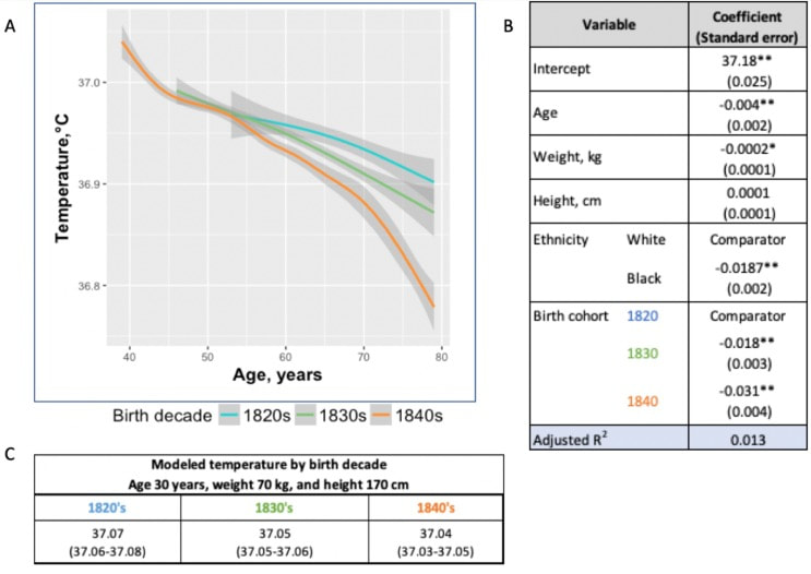
In recent years, researchers have uncovered a surprising trend in human physiology: a decline in body temperature over the past two centuries. Contrary to the long-standing belief that the normal body temperature is 37°C (98.6°F), studies spanning multiple cohorts and time periods have revealed a consistent decrease in average body temperature, suggesting a real physiological change rather than a mere artifact of measurement bias.

A groundbreaking study analyzed data from three cohorts spanning 157 years (over 600,000 data inputs), including Union Army Veterans of the Civil War, the National Health and Nutrition Examination Survey I, and the Stanford Translational Research Integrated Database Environment. The findings revealed a monotonic decrease in body temperature, with men born in the early 19th century having temperatures 0.59°C higher than men today. A similar decline was observed in women, indicating a significant shift in human physiology over time.
​
While some may attribute these findings to changes in measurement methods or biases, the study's authors argue that the observed drop in temperature reflects real physiological differences. Human body temperature is a crude surrogate for basal metabolic rate. These findings of a decrease in body temperature indicate a decrease in metabolic rate, which is supported in the literature when comparing modern experimental data to those from 1919. 

Resting metabolic rate, a key component of daily energy expenditure, has been linked to body temperature and longevity. The observed decrease in body temperature suggests a corresponding decline in metabolic rate, independent of changes in body size. This likely has implications for human health and longevity, as metabolic health underlies all vital organ functions. 
Picture
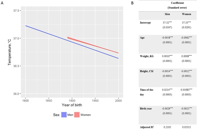
Additionally, changes in ambient temperature and the widespread adoption of heating and cooling systems in modern times may have influenced body temperature trends. Increased time spent in thermoneutral zones, where minimal energy is expended to maintain body temperature, could contribute to the observed decline in resting metabolic rate and body temperature.
​
In conclusion, body temperature has declined, implying lower metabolic rates (since heat is generated as a byproduct of energy production). This declining trend in human body temperature over the past two centuries offers valuable insights into the complex interplay between physiology, environment, and health. As researchers continue to unravel the mysteries of human biology, these findings pave the way for a deeper understanding of our species' evolution and resilience in the face of changing environments and lifestyles.

references

Protsiv, Myroslava, et al. “Decreasing Human Body Temperature in the United States since the Industrial Revolution.” ELife, vol. 9, 7 Jan. 2020, p. e49555, elifesciences.org/articles/49555, https://doi.org/10.7554/eLife.49555.

Dai, Dao-Fu, et al. “Mitochondrial Oxidative Stress in Aging and Healthspan.” Longevity & Healthspan, vol. 3, no. 1, 2014, p. 6, https://doi.org/10.1186/2046-2395-3-6. 
0 Comments

Unveiling Obesogens: The Hidden Culprits in Weight Gain

3/31/2024

0 Comments

 
Picture
In our modern world, where convenience often comes at a cost, the prevalence of obesogens – chemicals that disrupt the body's normal metabolism and contribute to weight gain – has emerged as a growing concern. From everyday products to industrial pollutants, obesogens permeate our environment, exerting subtle yet profound effects on our health and well-being.

Commonly encountered obesogens

Picture
Among the many obesogens encountered in daily life, several stand out for their widespread use and potential health impacts:
  1. Bisphenol A (BPA): Found in plastics, food can linings, and thermal paper receipts, BPA is notorious for its endocrine-disrupting properties, which can interfere with hormone signaling and contribute to weight gain.
  2. Persistent Organic Pollutants (POPs): These industrial chemicals, including polychlorinated biphenyls (PCBs), can accumulate in the environment and the food chain, posing risks to human health and metabolism.
  3. Phthalates: Commonly used in plastics, personal care products, and food packaging, phthalates have been linked to disruptions in metabolic processes and adipose tissue function.
  4. Artificial Sweeteners: While marketed as calorie-free alternatives to sugar, artificial sweeteners like aspartame can disrupt gut microbiota and metabolic signaling pathways, potentially contributing to weight gain.
  5. Pesticides: Chemicals used in agriculture to control pests, such as glyphosate, can also disrupt endocrine function and metabolic regulation, posing risks to human health.
  6. Polychlorinated biphenyls (PCBs): PCBs are a group of synthetic organic chemicals formerly used in various industrial applications (electrical equipment, hydraulic systems, and heat transfer fluids, and various consumer products such as paints, sealants, and plastics), known for their persistence in the environment (soil, water, air) and their potential to cause adverse health effects in humans and wildlife.
  7. Processed foods: Processed foods often contain obesogens like Bisphenol A (BPA) and phthalates, which can leach into the food from packaging materials or be introduced during processing. These chemicals can disrupt metabolic function and contribute to weight gain, emphasizing the importance of choosing whole, unprocessed foods whenever possible to minimize exposure.
Bisphenol F
Bisphenol P
Bisphenol S
Antibiotics
Aspartame
Soy
Fructose
Cow Milk (Pasteurized) 
Thimerosal
Monosodium Glutamate (MSG)
β-hexachlorocyclohexane (βHCH)
2,5-dichlorophenol (2,5-DCP)

Mechanisms of Action

Obesogens exert their effects through various mechanisms, including:
  • Endocrine Disruption: Many obesogens interfere with hormone signaling pathways, particularly those involved in metabolism and adipogenesis (the formation of fat cells).
  • Disruption of Gut Microbiota: Some obesogens alter the composition and function of gut bacteria, which play a crucial role in metabolic regulation and energy balance.
  • Epigenetic Modifications: Exposure to obesogens during critical periods of development, such as prenatal or early childhood, can lead to long-lasting changes in gene expression that predispose individuals to weight gain and metabolic disorders.
  • Inflammation and Oxidative Stress: Obesogens can trigger inflammatory responses and increase oxidative stress, both of which are implicated in the pathogenesis of metabolic and mitochondrial disorders.
Picture

Disruption of Metabolism via Mitochondria

Obesogens, through their pervasive presence in our environment, exert insidious effects on metabolic function, including the intricate workings of mitochondria – the cellular powerhouses responsible for energy production. By disrupting mitochondrial function, obesogens can contribute to metabolic dysregulation and, ultimately, weight gain.

Mitochondria play a central role in energy metabolism, converting nutrients into adenosine triphosphate (ATP), the primary source of cellular energy. However, exposure to obesogens can impair mitochondrial function through various mechanisms, including:
  • Oxidative Stress: Obesogens can promote the generation of reactive oxygen species (ROS) within mitochondria, leading to oxidative damage and dysfunction.
  • Mitochondrial Biogenesis: Some obesogens interfere with the process of mitochondrial biogenesis, the creation of new mitochondria, which is essential for maintaining optimal energy metabolism.
  • Respiratory Chain Dysfunction: Obesogens may disrupt the electron transport chain, a series of protein complexes within mitochondria that generate ATP, impairing energy production.
  • Mitochondrial Membrane Integrity: Obesogens can compromise the integrity of mitochondrial membranes, affecting the transport of ions and molecules critical for energy production.

​The disruption of mitochondrial function by obesogens can have profound implications for metabolic health and contribute to obesity through several pathways:
  1. Impaired Energy Expenditure: Dysfunctional mitochondria are less efficient at generating ATP, leading to reduced energy expenditure and a propensity for weight gain.
  2. Insulin Resistance: Mitochondrial dysfunction can impair insulin signaling pathways, contributing to insulin resistance, a hallmark of obesity and metabolic syndrome.
  3. Altered Lipid Metabolism: Mitochondria play a crucial role in lipid metabolism, including the breakdown of fatty acids for energy. Disrupted mitochondrial function can lead to aberrant lipid accumulation and adipogenesis, contributing to obesity.

causative relationship with health conditions

The impact of obesogens on human health extends beyond weight gain, with associations documented with various health conditions, including:
  • Obesity: Obesogens have been implicated in the global obesity epidemic, contributing to excess weight gain and adiposity. This includes being overweight, abdominal obesity (midsection fat), childhood and adult obesity.
  • Insulin Resistance and Diabetes: Disruption of metabolic pathways by obesogens can lead to insulin resistance, a precursor to type 2 diabetes.
  • Fatty Liver Disease: Chemical exposures, including those to bisphenols and phthalates, have been linked to the development of non-alcoholic fatty liver disease (NAFLD).

Additionally, obesogens are highly related to the following health conditions and physiologic imbalances:
Bisphenol Toxicity
Bisphenol-A Toxicity
​Chemically-Induced Liver Damage
​Fetal Origin of Adult Disease
​Fructose-Induced Toxicity
​Glyphosate Toxicity
​High Fat Diet
​Inflammation
Infant Chemical Exposures
Infant Nutrition
Insulin Resistance
​Oxidative Stress
​Phthalate Toxicity
​Prenatal Chemical Exposures
Triglycerides: Elevated

Unraveling the Role of Dysfunctional Adipose Tissue

Relatively little is known about the extent to which obesogen exposure programs dysfunctional adipose tissue that may store but not mobilize fat. However, emerging evidence suggests that obesogens may contribute to adipocyte dysfunction, leading to altered fat storage and metabolism. One potential underlying factor is suboptimal liver detoxification pathways due to inadequate micronutrient cofactors.
​
Inadequate levels of essential micronutrients, such as vitamins and minerals, can impair liver detoxification pathways responsible for metabolizing and eliminating obesogens from the body. As a result, obesogens may accumulate in adipose tissue, disrupting metabolic function and contributing to weight gain. Additionally, micronutrient deficiencies can compromise mitochondrial function, further exacerbating metabolic dysfunction and obesity risk.​

​​A Layman's Overview of Obesogens: Redefining the Weight Loss Paradigm

In the quest for weight loss, many of us often find ourselves fixating on calorie counting, fad diets, or intense workout regimens. However, what if I told you that the key to achieving a healthy weight isn't solely about shedding pounds but rather fixing your metabolism? Enter obesogens – a lesser-known yet influential factor in the obesity epidemic.

As mentioned, obesogens are chemicals found in our environment, ranging from pesticides and plastics to food additives and personal care products. These substances have the uncanny ability to disrupt our body's natural weight-regulating mechanisms, leading to weight gain and metabolic dysfunction. Instead of solely blaming calories in versus calories out, it's essential to recognize the role obesogens play in shaping our metabolism.

The Better Question: Fixing Metabolism

Rather than constantly asking ourselves, "How do I lose weight?" a more pertinent question would be:
"How do I fix my metabolism?"
Fixing metabolism involves addressing the root cause of weight gain – obesogen exposure and metabolic disruption. By eliminating or reducing our exposure to obesogens and ensuring our bodies receive essential micronutrients, we can optimize metabolic function and promote overall health.

The Two-Fold Solution

To achieve optimal health and maintain a healthy weight, a two-fold approach is necessary:
​

​1. Reduce Toxin Exposure: Minimize exposure to obesogens by making conscious choices in our daily lives. This includes opting for organic produce, using natural cleaning and personal care products, and avoiding plastic containers and food packaging whenever possible. By participating in a structured evidenced-based detoxification program, we in turn lower our toxic burden, and we can mitigate the adverse effects of obesogens on our metabolism.
Learn more about detoxification
2. Consume Micronutrients: Vital micronutrients, such as vitamins and minerals, serve as essential cofactors in metabolic pathways. Ensuring adequate intake of these micronutrients through a balanced diet rich in fruits, vegetables, whole grains, and lean proteins can support optimal metabolic function. Additionally, supplementation may be necessary to address any deficiencies and promote metabolic health.
​
The conventional approach to weight loss often overlooks the critical role obesogens play in metabolic dysfunction. Instead of solely focusing on calorie restriction or intense exercise, shifting our focus to fixing metabolism through toxin reduction and micronutrient consumption offers a more holistic and sustainable solution to achieving optimal health. By addressing the underlying factors contributing to metabolic disruption, we can pave the way for lasting weight management and overall well-being.

the harm of environmental toxins

The disruption of metabolic and mitochondrial function by obesogens represents a significant public health concern, with implications for obesity and metabolic disease. By understanding the mechanisms through which obesogens impair mitochondrial function and contribute to weight gain, researchers can develop targeted interventions to mitigate their adverse effects on metabolic health. Moreover, addressing underlying factors such as suboptimal liver detoxification pathways and micronutrient deficiencies is essential in combating the detrimental impact of obesogens on metabolic function and obesity prevalence.

The pervasive presence of obesogens in our environment underscores the need for greater awareness and regulation of these harmful chemicals. By minimizing exposure to obesogens and advocating for safer alternatives, we can mitigate their adverse effects on human health and combat the rising tide of obesity and metabolic disease. As we navigate the complexities of modern living, vigilance and informed consumer choices are essential in safeguarding our health and well-being against the hidden threats of obesogens.

Taking Action: The Integral Wellness Program​

For those seeking tangible solutions to combat the effects of obesogens and improve their overall well-being, the Integral Wellness Program offers a comprehensive approach to optimizing health and vitality. This flagship service provides personalized guidance and support in key areas of movement, nutrition, and lifestyle to directly enhance quality of life.
Learn more about the integral wellness program
Online/In-Person Guidance
One of the standout features of the Integral Wellness Program is its flexibility, offering both online and in-person consultations tailored to individual preferences and needs. Whether you prefer the convenience of virtual sessions or the hands-on approach of in-person coaching, our team of experienced wellness professionals is dedicated to supporting you every step of the way.
​Movement, Nutrition, and Lifestyle
The Integral Wellness Program takes a holistic approach to health, addressing modifiable factors and behaviors in three core areas:
  1. Movement: Through customized movement plans and exercise routines, participants are empowered to enhance physical fitness, flexibility, and overall mobility. Whether you're a seasoned athlete or new to fitness, our expert coaches will guide you towards achieving your movement goals safely and effectively.
  2. Nutrition: Central to the Integral Wellness Program is the emphasis on nutrient-dense foods and supplements to fuel and energize your body optimally. Our nutrition experts will work with you to develop personalized meal plans and dietary strategies tailored to your unique needs and preferences. By eliminating obesogen exposure and prioritizing wholesome, nourishing foods, you can support metabolic health and achieve sustainable weight management.
  3. Lifestyle: Beyond movement and nutrition, the Integral Wellness Program addresses lifestyle factors that contribute to overall well-being. From stress management techniques to sleep hygiene practices, our holistic approach encompasses all aspects of lifestyle optimization to promote balance, resilience, and vitality.

​Augmenting the Health Process
By participating in the Integral Wellness Program, you'll not only gain valuable knowledge and skills to navigate the challenges of modern living but also receive ongoing support and accountability to stay on track towards your health goals. Through targeted interventions aimed at eliminating obesogen exposure and promoting healthy behaviors, you can unlock your body's full potential and thrive in all aspects of life.
Learn more about the integral wellness program
The Integral Wellness Program offers a transformative journey towards optimal health and vitality. By prioritizing movement, nutrition, and lifestyle modifications, participants can take proactive steps to combat the effects of obesogens and reclaim control over their well-being. With the guidance and support of our dedicated wellness professionals, you'll embark on a path of self-discovery, empowerment, and lasting transformation.

references


Read More
0 Comments

Dr. Thomas Cowan: What Causes Heart Attacks

3/29/2024

0 Comments

 
Picture
This article challenges the conventional understanding of heart disease, particularly the widely accepted theory that attributes its cause primarily to events occurring in the coronary arteries. Instead, a paradigm shift is proposed, contending that a deeper understanding of heart disease, encompassing angina, unstable angina, and myocardial infarction (heart attack), necessitates a focus on events within the myocardium, the muscular tissue of the heart. Over the past decades, the prevailing belief in the coronary artery theory has led to costly surgical interventions, widespread medication use with questionable benefits, and dietary recommendations that may exacerbate rather than alleviate the problem. By delving into the precise pathophysiological events that underlie heart attacks, we can uncover alternative approaches to prevention and treatment, such as adopting a "Nourishing Traditions"-style diet and utilizing safe and affordable medicines like g-strophanthin. Furthermore, this shift in perspective prompts us to confront broader issues, including the impact of modern lifestyles on human health, the need for a new medical paradigm, and the importance of ecological consciousness. Ultimately, reexamining the root causes of heart disease offers a pathway to addressing this pervasive health challenge and forging a healthier future for all.

The information is summarized based on the work of Dr. Thomas Cowan, 
vice president of the Physicians Association for Anthroposophical Medicine and is a founding board member of the Weston A. Price Foundation. During his career he has studied and written about many subjects in medicine. These include nutrition, homeopathy, anthroposophical medicine, and herbal medicine.​
read Dr Cowan's article

Challenging the Conventional model: Revisiting the Causes of Heart Attacks

The traditional understanding of heart attacks, largely centered on arterial blockage due to plaque buildup, has faced challenges in recent years. Initially, it was believed that blockages in the major coronary arteries led to oxygen deficiency in the heart, causing chest pain (angina) and eventually progressing to a heart attack. This simplistic view prompted invasive procedures like angioplasty, stents, and coronary bypass surgery as standard treatments. However, clinical observations and research findings have cast doubts on this approach.
​
Anecdotal evidence (admittedly low quality evidence) from a trial in rural Alabama revealed surprising outcomes among individuals with single artery blockages. Contrary to expectations, less than 10% of those who experienced heart attacks did so in the region of the heart supplied by the blocked artery.

Similarly, a comprehensive study conducted by the Mayo Clinic highlighted the limited efficacy of bypass surgery in preventing future heart attacks. While the procedure offered relief from chest pain, it did not significantly reduce the risk of subsequent heart events, except in high-risk patients.

Contrary to popular belief, blockages exceeding 90% are often compensated for by collateral blood vessels, which develop over time to ensure uninterrupted blood flow to the heart. This extensive network of collateral vessels serves as a natural bypass system, mitigating the impact of arterial blockages on blood circulation.

However, diagnostic procedures like coronary angiograms, which rely on injecting heavy dye into the arteries, often fail to accurately assess the extent of blockages and the true blood flow in the heart. As a result, many patients undergo invasive treatments such as bypass surgery, stents, or angioplasty based on misleading information about the severity of their arterial blockages.

Moreover, studies have shown that these procedures provide minimal benefit, if any, to patients, particularly those with minimally symptomatic blockages exceeding 90%. Despite the widespread use of these interventions, their efficacy in restoring blood flow and preventing heart attacks remains questionable.
​
These revelations underscore the need for a reevaluation of conventional treatment strategies and a deeper exploration of the underlying mechanisms behind heart attacks. Rather than focusing solely on arterial blockages, a more holistic approach that considers factors beyond plaque buildup may offer greater insights into the prevention and management of heart disease.

Beyond the Coronary Artery Theory

The prevailing focus in cardiology has long been on the stable, progressing plaque within the coronary arteries, deemed responsible for heart attacks. However, recent insights challenge this notion, redirecting attention to the unpredictable nature of unstable plaques. Unlike their calcified counterparts, unstable plaques are soft and prone to rapid evolution, abruptly occluding arteries and triggering downstream oxygen deficits, angina, and ischemia.
​
These vulnerable plaques are believed to be a blend of inflammatory buildup and low-density lipoprotein (LDL), the primary targets of statin drugs. Consequently, the widespread adoption of statin therapy is advocated as a preventive measure against heart attacks, fueled by angiogram studies purportedly showcasing the prevalence of unstable plaques as the leading cause of myocardial infarctions (MIs).
Learn more about cholesterol
Yet, autopsies and pathology studies present a different narrative. Thrombosis, deemed crucial in precipitating MIs, is found in only a fraction of cases upon meticulous examination. Furthermore, measurements of myocardial oxygen levels during MIs reveal no discernible deficit, challenging the conventional understanding of ischemia as the primary mechanism.

While thrombosis does occur in conjunction with MIs, its occurrence in less than half of cases underscores the inadequacy of attributing MIs solely to arterial blockages. The timing of thrombosis, often post-MI, begs the question: what precipitated the event in the first place? These inconsistencies underscore the limitations of existing theories surrounding coronary artery involvement in MIs.

As the spotlight shifts away from stable plaques, a pressing question emerges: What truly underlies the genesis of heart attacks?

Unveiling the Autonomic Symphony: The Heart's Harmonious Balance

An accurate understanding of myocardial ischemia necessitates consideration of the primary risk factors associated with heart disease, including gender, diabetes, smoking, and chronic psychological stress. Curiously, none of these risk factors directly implicate coronary artery pathology; instead, they impact capillary health or exert indirect effects.

Over the past five decades, key medications in cardiology, such as beta-blockers, nitrates, aspirin, and statins, have demonstrated some benefits for heart patients. However, their mechanisms of action must be scrutinized within a comprehensive theory of myocardial ischemia.

A groundbreaking revelation in heart disease prevention and treatment stems from the autonomic nervous system's role in ischemia genesis, as illuminated by heart-rate variability monitoring. The autonomic nervous system comprises two branches—the sympathetic and parasympathetic—responsible for regulating physiological responses. Imbalance between these branches emerges as a significant contributor to heart disease.

Studies reveal a notable reduction in parasympathetic activity among patients with ischemic heart disease, particularly preceding ischemic events triggered by physical or emotional stressors. Conversely, abrupt increases in sympathetic activity rarely culminate in ischemia without antecedent parasympathetic decline. Notably, women exhibit stronger vagal activity than men, potentially influencing sex-based disparities in MI incidence.

Multiple risk factors, including hypertension, smoking, diabetes, and stress, diminish parasympathetic activity, underscoring the pivotal role of the regenerative nervous system in heart health. Conversely, pharmacological interventions like nitrates, aspirin, and statins stimulate parasympathetic mediators, promoting ANS balance.
​
In essence, while traditional risk factors and interventions influence plaque and stenosis development, their paramount impact lies in restoring ANS equilibrium. Thus, understanding the sequence of events leading to myocardial infarction demands a deeper exploration of autonomic nervous system dynamics.

The Underlying pathophysiology of Myocardial Ischemia

In the vast majority of cases, the pathology leading to myocardial infarction (MI) begins with a decreased tonic activity of the parasympathetic nervous system (rest and digest), often exacerbated by physical or emotional stressors. This reduction prompts an increase in sympathetic nervous system activity, triggering heightened adrenaline production and directing myocardial cells to break down glucose via aerobic glycolysis, rather than their preferred fuel source of ketones and fatty acids (often explaining why patients report feeling tired before a MI). Remarkably, despite these metabolic shifts, no change in blood flow, as measured by the myocardial cell oxygen level (pO2), occurs.

The shift towards glycolysis results in a surge of lactic acid production within myocardial cells, a phenomenon observed in nearly all MIs. This surge, coupled with localized tissue acidosis, impedes calcium entry into cells, compromising their contractility. Consequently, localized edema ensues, leading to hypokinesis—the hallmark of ischemic disease—and eventual tissue necrosis characteristic of an MI.
​
Moreover, the ensuing tissue edema alters arterial hemodynamics, escalating sheer pressure and exacerbating plaque instability. This process elucidates the rupture of unstable plaques and their role in exacerbating arterial blockage during critical, acute scenarios. This explanation accounts for all the observable phenomena associated with heart disease.

Understanding the etiology of heart disease holds profound implications beyond academic curiosity. It informs therapeutic strategies aimed at preserving parasympathetic activity, fostering holistic approaches to heart health, and challenging prevailing "civilized" industrial lifestyles. Central to this paradigm shift is the recognition of the vital role played by g-strophanthin—a hormone derived from the strophanthus plant. G-strophanthin is an endogenous hormone made in the adrenal cortex from cholesterol, whose production is inhibited by statin drugs, that does two things that are crucial for heart health and are done by no other medicine. G-strophanthin uniquely stimulates the production of acetylcholine, the primary neurotransmitter of the parasympathetic nervous system, while also converting lactic acid—the metabolic poison implicated in ischemic processes—into pyruvate, a preferred myocardial cell fuel. Perhaps this “magic” is why Chinese medicine practitioners say that the kidneys (i.e., adrenals, where ouabain is made) nourish the heart.

Embracing this understanding not only guides therapeutic interventions but also underscores the imperative of dietary modifications. A diet abundant in healthful fats and fat-soluble nutrients, while low in processed carbohydrates and sugars, emerges as a cornerstone of heart health—a departure from the industrialized diets synonymous with modern civilization.

In essence, unraveling the metabolic symphony orchestrating myocardial ischemia offers a transformative lens through which to perceive heart disease, fostering a holistic approach that transcends conventional paradigms and embraces the profound interconnectedness of mind, body, and environment.

references

Giorgio Baroldi. The Etiopathogenesis of Coronary Heart Disease. CRC Press EBooks, Informa, 20 Jan. 2004. Accessed 29 Mar. 2024.

Sroka K. On the genesis of myocardial ischemia. Z Kardiol. 2004 Oct;93(10):768-83. doi: 10.1007/s00392-004-0137-6. PMID: 15492892.

Helfant, R. H., et al. “Coronary Heart Disease. Differential Hemodynamic, Metabolic, and Electrocardiographic Effects in Subjects with and without Angina Pectoris during Atrial Pacing.” Circulation, vol. 42, no. 4, 1 Oct. 1970, pp. 601–610, www.ncbi.nlm.nih.gov/pubmed/11993303., https://doi.org/10.1161/01.cir.42.4.601. 

Takase, B., Kurita, A., Noritake, M., Uehata, A., Maruyama, T., Nagayoshi, H., ... & Nakamura, H. (1992). Heart rate variability in patients with diabetes mellitus, ischemic heart disease, and congestive heart failure. Journal of electrocardiology, 25(2), 79-88.

Sroka, K., Peimann, C. J., & Seevers, H. (1997). Heart rate variability in myocardial ischemia during daily life. 
Journal of electrocardiology, 30(1), 45-56.

Scheuer, J., & Brachfeld, N. (1966). Coronary insufficiency: relations between hemodynamic, electrical, and biochemical parameters. 
Circulation Research, 18(2), 178-189.

Schmid, P. G., Greif, B. J., Lund, D. D., & Roskoski Jr, R. O. B. E. R. T. (1978). Regional choline acetyltransferase activity in the guinea pig heart. 
Circulation Research, 42(5), 657-660.

​Katz, A. M. (1971). Effects of ischemia on the cardiac contractile proteins. 
Cardiology, 56(1-6), 276-283.

Manunta, Paolo, et al. “Endogenous Ouabain in Cardiovascular Function and Disease.” Journal of Hypertension, vol. 27, no. 1, 1 Jan. 2009, pp. 9–18, journals.lww.com/jhypertension/Abstract/2009/01000/Endogenous_ouabain_in_cardiovascular_function_and.3.aspx, https://doi.org/10.1097/HJH.0b013e32831cf2c6.

Doepp, Manfred. “May Strophanthin Be a Valuable Cardiac Drug ? .” American Journal of Medical and Clinical Research & Reviews, vol. 2, no. 9, 15 Sept. 2023, pp. 1–6, ajmcrr.com/index.php/pub/article/view/75/74, https://doi.org/10.58372/2835-6276.1069. Accessed 29 Mar. 2024.

​Thayer, J. F., Yamamoto, S. S., & Brosschot, J. F. (2010). The relationship of autonomic imbalance, heart rate variability and cardiovascular disease risk factors. International journal of cardiology, 141(2), 122-131.
0 Comments

Unlocking the Secrets of Longevity: How Your Microbiome Holds the Key

3/10/2024

0 Comments

 
Picture
Recent breakthrough studies have shone a light on the intriguing link between our microbiome – the diverse community of microorganisms residing in our gut and mouth – and the secret to a longer, healthier life. Scientists have long suspected that our genes, environment, and internal factors like the microbiome play a role in determining how long we live, but the specifics remained elusive. Now, thanks to cutting-edge research, we're getting closer to unraveling the mysteries of longevity.
​
In this groundbreaking exploration, scientists employed a sophisticated approach called Mendelian randomization (MR) to delve into the intricate relationships between the human microbiome and longevity. By analyzing genetic data from large cohorts, they uncovered some compelling associations that shed light on the microbial players in the quest for a longer life.
read the study

The Gut Chronicles: Microbial Superstars and Culprits

The gut microbiome, a bustling metropolis of bacteria, has been a focal point in the quest for longevity. The study identified certain gut microbes as potential champions in the battle against aging. Microbial heroes like Coriobacteriaceae, Oxalobacter, and the probiotic Lactobacillus amylovorus were found to be positively linked to increased odds of longevity.
​

On the flip side, a few gut microbes emerged as potential antagonists, with names like Fusobacterium nucleatum, Coprococcus, Streptococcus, Lactobacillus, and Neisseria negatively associated with longevity. These microbial foes might have a role in determining how gracefully we age.

Oral Health: More Than Just a Pretty Smile

The study didn't stop at the gut; it extended its gaze to the oral microbiome, a less-explored but equally important realm. The findings suggested a fascinating connection between the oral microbiome and longevity. Specific oral bacteria were identified as potential influencers in the longevity game.
​

Interestingly, the research hinted at a lower gut microbial diversity among centenarians (diversity appears to lower with age), but no significant difference in their oral microbiota. This finding underscores the importance of tracking the movements of these beneficial microbes across different parts of the body for a longer and healthier life.

Decoding the Genetic Blueprint for Longevity

The study leveraged Mendelian randomization to unravel the causality between the microbiome and longevity. This approach, using genetic variants as tools, allowed scientists to explore the potential causal links between specific microbial features and the length of our lives.
​

The bidirectional analyses provided a wealth of information, not only pinpointing specific microbes associated with longevity but also revealing the microbial preferences of genetically longevous individuals. For instance, genetic predisposition to longevity correlated with a higher abundance of Prevotella and a lower abundance of Bacteroides, suggesting a potential link between dietary choices and a longer life.

Microbes and Diseases: Unraveling the We

The study didn't just stop at longevity; it ventured into the realm of diseases. Certain microbes associated with longevity were found to have correlations with specific diseases. For example, Coriobacteriaceae, linked to longevity, was significantly reduced in patients with heart failure, suggesting a potential protective role against cardiovascular diseases.
​

This "microbiota—disease—longevity" axis provides a nuanced understanding of how our microbial companions might influence not only our lifespan but also our susceptibility to various health conditions.

What's Next in the Quest for a Longer Life

While the study opens exciting new avenues, there are some limitations to consider. The identified causalities didn't all reach statistical significance due to the vast number of microbial features tested. However, the robustness of the findings was supported by the replication of several identified causal links in independent datasets.

Moving forward, researchers aim to collect more comprehensive individual-level data, including microbiome profiles, genetics, socio-economic factors, behaviors, and environmental influences. This holistic approach will help tease apart the individual contributions of these factors to longevity.
​

In conclusion, this pioneering study, using Mendelian randomization, has provided us with a roadmap to explore the intricate connections between our microbiome and the quest for a longer, healthier life. As we unlock the secrets hidden in our genes and microbes, we inch closer to personalized approaches for healthy aging and interventions that could extend our time on this planet.

references

Liu, Xiaomin, et al. “Mendelian Randomization Analyses Reveal Causal Relationships between the Human Microbiome and Longevity.” Scientific Reports, vol. 13, no. 1, 29 Mar. 2023, p. 5127, www.nature.com/articles/s41598-023-31115-8, https://doi.org/10.1038/s41598-023-31115-8. 
0 Comments

Unpacking the Potential Harms of Wellbutrin: Neurotransmitter Modulation and Considerations for Special Populations

2/16/2024

0 Comments

 
Bupropion, originally named Amfebutamone, sold under the brand name Wellbutrin, is a medication commonly prescribed for the treatment of major depressive disorder (MDD), as is often used off-label for attention deficit hyperactivity disorder (ADHD), anxiety, obesity, and bipolar disorder. While it has demonstrated efficacy in addressing certain mental health issues, it is essential to examine the potential harms associated with its use, particularly considering its modulation of neurotransmitters like norepinephrine (NE) and dopamine. This article aims to shed light on the risks of Wellbutrin use, with a focus on its implications for pregnant or lactating women. Additionally, we'll explore the idea that the indications for Wellbutrin may stem from underlying nutrition and lifestyle factors rather than a deficiency of the medication.
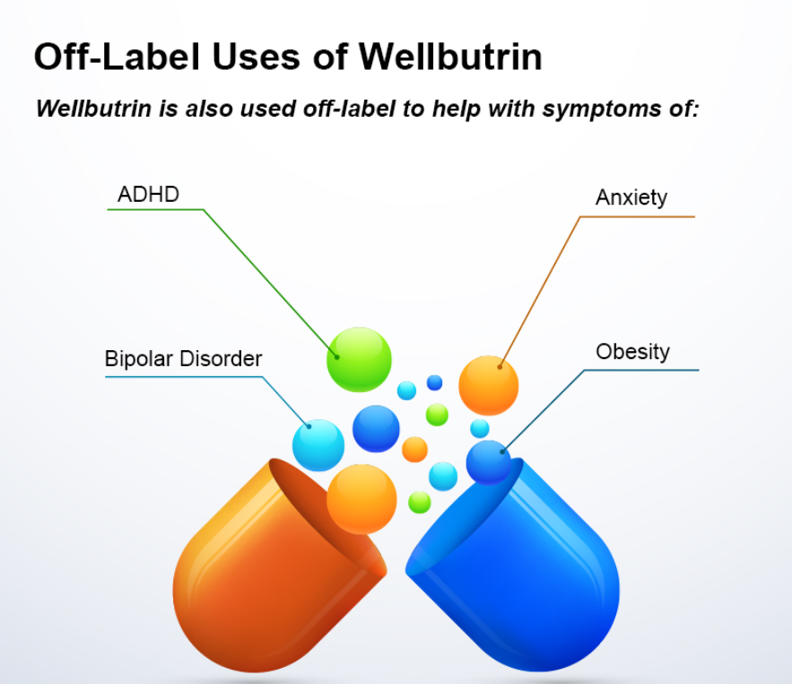
Picture

prescription trends

As of 2021, Bupropion, maintained its position as the 18th most prescribed drug in the United States. With an estimated 29,099,445 prescriptions filled, it remains a widely utilized medication in the realm of psychiatric pharmaceuticals. This notable figure underscores the prevalence of its use in addressing various conditions, including depression and smoking cessation.

The estimated number of patients in the United States receiving Bupropion in 2021 reached 6,412,363. This statistic reflects the significant impact and reach of Bupropion across diverse patient populations. Its popularity could be attributed to its purported effectiveness in managing depressive disorders, ADHD, anxiety, and aiding individuals in smoking cessation efforts.

description

Wellbutrin (bupropion hydrochloride), unlike any other antidepressant on the market, is chemically characterized as a monocyclic aminoketone, is chemically unrelated to tricyclic, tetracyclic, selective serotonin re‑uptake inhibitor, or other known antidepressant agents. Its structure closely resembles that of diethylpropion; it is related to phenylethylamines. It is designated as (±)-1-(3-chlorophenyl)-2-[(1,1-dimethylethyl)amino]-propanone hydrochloride. Bupropion hydrochloride powder is white, crystalline, and highly soluble in water. It has a bitter taste and produces the sensation of local anesthesia on the oral mucosa. There is documented interindividual variability - everyone responds differently.
Picture
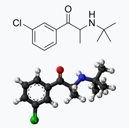
Molecular weight: 276.2 - Molecular formula: C13H18ClNO•HCl

Neurotransmitter Modulation: The Double-Edged Sword

Wellbutrin functions by influencing the levels of neurotransmitters in the brain, primarily inhibiting the breakdown of norepinephrine and dopamine. While this mechanism contributes to its antidepressant effects, it also raises concerns about potential side effects and risks. Altering neurotransmitter levels can lead to a range of adverse effects, with the most common including:
  • anxiety
  • dry mouth
  • hyperventilation
  • irregular heartbeats
  • irritability
  • restlessness
  • shaking
  • trouble sleeping

Here are some of the possible harms and common side effects associated with Wellbutrin:
  1. Insomnia: Difficulty sleeping is a frequently reported side effect.
  2. Dry Mouth: Many users experience a persistent dry mouth.
  3. Headache: Headaches are a common complaint.
  4. Nausea and Vomiting: Some people may feel nauseous or vomit.
  5. Constipation: Gastrointestinal issues, such as constipation, are possible.
  6. Dizziness: Users may experience dizziness or lightheadedness.
  7. Weight Loss: Appetite suppression can lead to weight loss.
  8. Increased Sweating: Excessive sweating is another potential side effect.
Serious Side Effects
  1. Seizures: Wellbutrin can increase the risk of seizures, especially at higher doses.
  2. Hypertension: Elevated blood pressure has been reported.
  3. Mania: In some cases, Wellbutrin can trigger manic episodes in individuals with bipolar disorder.
  4. Psychiatric Symptoms: Agitation, anxiety, and psychosis have been observed.
  5. Liver Damage: Although rare, Wellbutrin can cause liver injury.
Allergic Reactions
  1. Rash and Itching: Skin reactions such as rashes and itching can occur.
  2. Swelling: Swelling of the face, lips, tongue, or throat can indicate a severe allergic reaction.
  3. Difficulty Breathing: Respiratory issues are a severe sign of an allergic reaction.
Cardiovascular Effects
  1. Palpitations: Users may experience an irregular heartbeat or palpitations.
  2. Increased Heart Rate: Tachycardia, or an increased heart rate, can occur.
Psychiatric Risks
  1. Suicidal Thoughts and Behaviors: Like other antidepressants, Wellbutrin carries a risk of increased suicidal thoughts and behaviors, especially in young adults and adolescents.
Other Considerations
  1. Drug Interactions: Wellbutrin can interact with other medications, leading to adverse effects or reduced efficacy.
  2. Use in Pregnancy and Breastfeeding: See Below. The safety of Wellbutrin during pregnancy and breastfeeding is not well established and should be discussed with a healthcare provider.
Long-Term Risks
  1. Dependence and Withdrawal: Although less common, some users may develop dependence or experience withdrawal symptoms upon discontinuation.
View the full list of side effects
While the approach of inhibiting the reuptake of dopamine and norepinephrine aims to enhance the availability of these neurotransmitters in the brain, an imbalance can lead to adverse effects. Excessive levels or prolonged elevated concentrations of these neurotransmitters may contribute to overstimulation and disrupt normal neural signaling.

Norepinephrine is a neurotransmitter involved in the body's "fight or flight" response, influencing heart rate and blood pressure. Medications that impact norepinephrine reuptake can lead to cardiovascular side effects, including increased heart rate and elevated blood pressure. Individuals with pre-existing cardiovascular conditions may be at a higher risk for complications.

Altered levels of dopamine and norepinephrine can influence mood and behavior. In some cases, inhibiting reuptake may contribute to psychiatric symptoms such as anxiety, restlessness, or irritability. Balancing the desired therapeutic effects with potential adverse psychological consequences is a delicate consideration.

Abruptly discontinuing medications that inhibit dopamine and norepinephrine reuptake can lead to withdrawal symptoms. These symptoms may include mood swings, fatigue, and cognitive disturbances. Additionally, some individuals may develop a dependence on these medications, requiring careful management to taper off gradually.

Dopamine and norepinephrine play roles in regulating sleep and appetite. Disrupting these neurotransmitters can lead to sleep disturbances, insomnia, or changes in appetite. Monitoring and addressing these side effects are crucial for maintaining overall well-being during the course of treatment.

The response to medications that inhibit dopamine and norepinephrine reuptake can vary widely among individuals. Factors such as genetic predispositions, existing medical conditions, and concurrent medications may influence how the body reacts to these interventions. Individualized treatment plans and close monitoring are essential components of responsible prescribing.​

Impact on Pregnant or Lactating Women

Pregnancy and lactation introduce unique considerations when it comes to medication use. The use of Wellbutrin (bupropion) or any other medication during pregnancy requires careful consideration of potential risks and benefits. While studies on the safety of Wellbutrin during pregnancy are inconclusive, there is evidence suggesting a potential association with adverse outcomes, including preterm birth and low birth weight. Additionally, Wellbutrin and its metabolites are present in human breast milk excretions, raising concerns about its impact on nursing infants including alterations in neurotransmitters. Expectant or breastfeeding mothers should engage in thorough discussions with their healthcare providers to weigh the potential benefits against the risks and explore alternative treatment options.

Here are some considerations regarding the potential harms of taking Wellbutrin during pregnancy:

  1. Risk of Birth Defects: Researchers have observed ​a potential association between the use of certain antidepressants, including Wellbutrin, during the first trimester of pregnancy and a slightly increased risk of certain birth defects. However, the absolute risk is generally low. In rabbits, increased incidences of fetal malformations and skeletal variations were observed at the lowest dose tested (25 mg per kg per day, approximately equal to the maximum recommended human dose on a mg per m2 basis) and greater. 
  2. Neonatal Complications: Neonatal complications, such as withdrawal symptoms or adaptation issues, have been reported in infants born to mothers who took Wellbutrin during pregnancy. These symptoms are typically transient and managed by healthcare professionals.
  3. Preterm Birth and Low Birth Weight: Researchers have observed a possible association between antidepressant use during pregnancy and an increased risk of preterm birth or low birth weight. In rabbits, decreased fetal weights were observed at 50 mg per kg and greater. However, the overall impact is modest, and the reasons for these associations are complex.
  4. Persistent Pulmonary Hypertension of the Newborn (PPHN): There have been concerns about a potential link between maternal antidepressant use, including Wellbutrin, and an increased risk of PPHN. However, research results have been inconsistent, and the absolute risk remains low.

Wellbutrin should be used during pregnancy only if the potential benefit justifies the potential risk to the fetus. It's essential for individuals taking Wellbutrin and considering pregnancy, or those who become pregnant while on the medication, to discuss their situation with healthcare professionals. Abruptly stopping antidepressant medication can lead to a recurrence of depressive symptoms, which may have its own set of risks during pregnancy.

Healthcare providers will carefully weigh the potential risks and benefits based on the individual's mental health needs. In some cases, healthcare professionals may recommend continuing the medication, adjusting the dosage, or exploring alternative treatments.

Pregnant individuals should inform their healthcare providers about their medication use and work collaboratively to make informed decisions that prioritize both maternal mental health and the well-being of the developing fetus.

Inactive Ingredients

Wellbutrin is supplied for oral administration as 75‑mg (yellow‑gold) and 100‑mg (red) film‑coated tablets. Each tablet contains the labeled amount of bupropion hydrochloride and the inactive ingredients:
  • 75‑mg tablet – D&C Yellow No. 10 Lake, FD&C Yellow No. 6 Lake, hydroxypropyl cellulose, hypromellose, microcrystalline cellulose, polyethylene glycol, talc, and titanium dioxide
  • 100‑mg tablet – FD&C Red No. 40 Lake, FD&C Yellow No. 6 Lake, hydroxypropyl cellulose, hypromellose, microcrystalline cellulose, polyethylene glycol, talc, and titanium dioxide.
View the Package Insert
While the active ingredient in Wellbutrin plays a significant role in its pharmacological effects, it's important not to overlook the inactive ingredients in the formulation. While they make up a smaller portion of the overall product, their cumulative impact should not be underestimated, especially considering the frequent administration of the medication.

Though often considered inert, inactive ingredients can still interact with the body in various ways, potentially influencing drug absorption, metabolism, and overall therapeutic efficacy. Moreover, individual sensitivities or allergic reactions to certain inactive ingredients can occur, further emphasizing the importance of evaluating the entire list of components.
​
Even seemingly minor alterations in inactive ingredients can have profound implications, particularly when considering long-term usage. Over time, repeated exposure to these substances may contribute to cumulative effects or unexpected reactions. Therefore, comprehensive scrutiny of all ingredients, active and inactive alike, is essential for a thorough assessment of the safety profile of Wellbutrin and other pharmaceutical products.

Impact of Food coloring

The vibrant hues that adorn our favorite processed foods often come from artificial food colorings, but behind the visual appeal lies a potential risk, particularly for pregnant or lactating women. Artificial food color usually contains petroleum and is manufactured in a chemical process that includes formaldehyde, aniline, hydroxides, and sulfuric acids. Most impurities in the food color are in the form of salts or acids. Sometimes lead, arsenic, and mercury may be present as impurities. The U.S. FDA is yet to study the effects of synthetic dyes on behavior in children. 

To date, there are numerous scientific articles highlighting the relationship of consumption of food colorings to the following health conditions:
  • Attention Deficit Hyperactivity Disorder
  • Attention Deficit Disorder with Hyperactivity
  • Hyperkinetic Syndrome
  • Anaphylaxis
  • Chemically-Induced Liver Damage
  • Food Allergies
  • Leukocytoclastic Vasculitis
  • DNA damage
  • Oxidative Stress
  • Asthma
  • Allergies
  • Angioedema
  • Excitotoxicity
  • Neurodevelopmental Disorders
  • Urticaria

Underlying these conditions are the documented mechanics of action that cause physiologic imbalance, which include:
  • Cytotoxicity
  • Hepatotoxicity
  • Genotoxicity
  • Mutagenicity
  • Neurotoxicity

​Research has raised concerns about the consumption of food colorings and their potential adverse effects, including implications for conditions like ADHD, autism, and gastrointestinal dysfunction.

There is a substantial amount of data exploring the connection between artificial food colorings and ADHD. Researchers suggest that certain artificial colorings may exacerbate hyperactivity and inattention in children with ADHD. Pregnant women, mindful of their diet's impact on fetal development, may choose to limit the intake of foods containing these colorings.

The relationship between food colorings and autism spectrum disorders (and the underlying inflammation in the brain) is a topic of ongoing investigation. Meta-analysis have documented a link, emphasizing the need for caution, especially during pregnancy and lactation. As the developing brain is susceptible to external influences, limiting exposure to artificial additives becomes a consideration for expectant and nursing mothers.

Artificial food colorings have been associated with gastrointestinal disturbances, ranging from discomfort to more severe issues. Pregnant women, already navigating changes in digestion due to hormonal shifts, may choose to minimize exposure to food colorings to promote digestive well-being during this crucial period.

Potential Harms for Pregnant and Lactating Women:
  1. Fetal Development: The developing fetus is particularly vulnerable to external influences. Limited research suggests that certain food colorings may cross the placental barrier, potentially impacting fetal development.
  2. Breast Milk Composition: Lactating women should be mindful of their dietary choices, as food colorings have been detected in breast milk. While the significance of this transfer is still under investigation, cautious choices can contribute to ensuring the purity of breast milk.
  3. Gastrointestinal Sensitivity: Pregnancy often brings about changes in digestion, and artificial food colorings may exacerbate gastrointestinal discomfort. Sensitivity to these additives varies, and pregnant women may choose to adopt a more natural and minimally processed diet.

For pregnant or lactating women concerned about the potential harms of food colorings, opting for a diet rich in whole, unprocessed foods becomes a valuable strategy. Choosing fruits, vegetables, and other naturally colorful sources can provide vibrant flavors without the need for artificial additives.

While the connection between food colorings and adverse health outcomes is an area of ongoing research, pregnant and lactating women may choose to err on the side of caution. Prioritizing a diet that minimizes artificial additives and focuses on nutrient-dense, whole foods is a proactive step toward supporting both maternal and fetal health. Always consult with healthcare professionals for personalized guidance based on individual circumstances and health considerations.

Impact of PEG

​Polyethylene glycol (PEG) is considered "generally recognized as safe" (GRAS) when used in specific contexts, but some individuals may experience adverse effects. ​
Explore the concerns with GRAS
The concerns that arise with the safety of consuming PEG are multifold, including: 
  1. Pollution: The process to produce polyethylene glycol requires a chemical reaction known as ethoxylation and the use of compounds known as ethylene oxide and 1,4-dioxane – two chemicals that have well-documented toxic effects on humans.
  2. Contamination: PEGs are widely utilized for their ability to enhance penetration and absorption. But this also means that prolonged use or high doses of PEG can significantly enhance your body’s absorption of other toxins and harmful compounds that are found alongside PEGs or within the environment. 
  3. Lack of studies: Because polyethylene glycol has numerous derivatives and molecular weights, extensive studies have only been conducted on a handful of different PEG compounds. There is limited information on the real impact of PEGs as a whole and more PEG toxicity research is needed to truly understand this compound’s effects on the human body.

Depending on manufacturing processes, PEGs may be contaminated with measurable amounts of ethylene oxide and 1,4-dioxane. The International Agency for Research on Cancer classifies ethylene oxide as a known human carcinogen and 1,4-dioxane as a possible human carcinogen. Ethylene oxide can also harm the nervous system and the California Environmental Protection Agency has classified it as a developmental toxicant based on evidence that it may interfere with human development. 

1,4-dioxane is also persistent. In other words, it doesn’t easily degrade and can remain in the environment long after it is rinsed down the shower drain. 1,4-dioxane can be removed from cosmetics during the manufacturing process by vacuum stripping, but there is no easy way for consumers to know whether products containing PEGs have undergone this process.In a study of personal care products marketed as “natural” or “organic” (uncertified), U.S. researchers found 1,4-dioxane as a contaminant in 46 of 100 products analyzed. 

While carcinogenic contaminants are the primary concern, PEG compounds themselves show some evidence of genotoxicity and if used on broken skin can cause irritation and systemic toxicity. PEG itself is classified as expected to be toxic or harmful as mentioned on the Environment Canada Domestic Substance List. The industry panel that reviews the safety of cosmetics ingredients concluded that some PEG compounds are not safe for use on damaged skin (although the assessment generally approved of the use of these chemicals in cosmetics). Also, PEG functions as a “penetration enhancer,” increasing the permeability of the skin to allow greater absorption of the ingredients — including harmful ingredients.

Researchers have observed that a large percentage of parents, caregivers, and practitioners described an explosion of neurological side effects seemingly correlated to polyethylene glycol administration. Those side effects include:
  • Abnormal behavior
  • Anger
  • Anxiety 
  • Mood swings
  • Seizures
  • Sensory disturbances

Commonly found in various products, such as medications, laxatives, and skincare items, PEG may lead to the following potential harms:
  1. Gastrointestinal Issues: Oral ingestion of PEG, particularly in laxatives or medications, may cause gastrointestinal side effects such as bloating, cramps, gas, nausea, diarrhea, and vomiting.
  2. Dehydration: Excessive use of PEG laxatives without adequate fluid intake can lead to dehydration. It's important to follow the recommended dosages and stay hydrated while using PEG-containing products.
  3. Allergic Reactions: Although rare, some individuals may be allergic to PEG, leading to allergic reactions like rash, itching, swelling, or difficulty breathing. If any allergic symptoms occur, immediate medical attention is necessary.
  4. Renal Impairment: PEG has been associated with cases of renal injury, particularly in individuals with pre-existing kidney conditions. People with kidney issues should use PEG-containing products cautiously under medical supervision.
  5. Electrolyte Imbalance: Because of its ability to disrupt the flow of water, PEG laxatives can lead to electrolyte imbalances, which may result in abnormal levels of sodium, potassium, calcium, phosphate, and other electrolytes in the body. Severe electrolyte imbalances can have serious health consequences. Consumption of PEG has also been linked to an increased risk of metabolic acidosis, which is a build-up of acid and toxins in the body.
  6. Systemic Absorption: While the systemic absorption of PEG from the gastrointestinal tract is generally low, excessive use or prolonged exposure may increase the risk of systemic absorption, potentially leading to adverse effects.
  7. Hypersensitivity and Allergic Reactions: Individuals with a history of hypersensitivity to PEG or related compounds should avoid products containing PEG.  There are have been documented cases of an allergy to PEG. Some cases have even resulted in anaphylaxis – a severe and potentially life-threatening allergic reaction. Due to the risk of exposure to polyethylene glycol, the FDA has issued a warning to anyone with a known or suspected PEG allergy to communicate clearly with healthcare professionals as PEGs can be found lurking in medications, vaccines, contrast agents, and more. Researchers have estimated that approximately 72% of the US population has acquired anti PEG antibodies. The referenced study used blood samples taken from 1990-1999 and earlier, showing a steady increase over time in the percentage of those with antibodies to PEG, making it conservative to estimate, after two decades, that the incidence is closer to 80% today. 

People with pre-existing health conditions, especially those affecting the kidneys, should inform their healthcare providers before using PEG-containing products. As with any substance, individual responses to PEG can vary, and people experiencing adverse effects should seek medical attention promptly. It's important to weigh the risks and benefits of using PEG-based products based on individual health conditions and circumstances.

Impact of Talc

​Talc is a mineral commonly used in various products such as talcum powder, cosmetics, and personal care items. While talc is considered GRAS for external use, there have been concerns and controversies regarding potential health risks associated with its use, primarily when used in certain ways or in specific product formulations. Here are some of the concerns related to talc:
  1. Contamination Concerns: Talc products have been found to be contaminated with substances like asbestos, which is a known carcinogen. While regulations and testing standards are in place to monitor and limit asbestos contamination in talc products, Johnson & Johnson, the maker of Johnson’s Baby Powder, is facing more than 9,000 plaintiffs in cases involving body powders with talc contaminated with asbestos.
  2. Ovarian Cancer: There has been some controversy and litigation surrounding the potential link between talcum powder use in the genital area and an increased risk of ovarian cancer in women, due to contamination of asbestos. 
  3. Respiratory Issues: Inhalation of talc powder (likely not a concern with the consumption of Wellbutrin), particularly in occupational settings such as talc mining or during certain industrial processes, may pose respiratory risks. Talc is closely related to asbestos, and in some natural deposits, talc may be contaminated with asbestos fibers, which are known respiratory hazards.
  4. Pulmonary Effects in Infants: Although likely not a concern with the consumption of Wellbutrin, there have been reports of respiratory distress and other pulmonary issues in infants when talc-containing products, such as baby powders, are used excessively. Inhaling talc powder can be harmful to an infant's developing respiratory system.
  5. Skin Irritation: Although likely not a concern with the consumption of Wellbutrin, in some individuals, talc may cause skin irritation or allergies. Redness, itching, or rash may occur, especially in people with sensitive skin.

Individuals concerned about the potential risks of talc-containing products should consider alternatives. As with any substance, moderation and careful use are advisable, and individuals experiencing adverse effects should seek medical advice.

Impact of titanium dioxide

​Titanium dioxide is a widely used pigment and additive in various products, including cosmetics, sunscreens, paints, and food items. While it is generally recognized as safe when used in approved applications, there are concerns about potential health risks associated with certain forms and uses of titanium dioxide. Here are some considerations:
  1. Genotoxicity Concerns: Most relevant to the consumption of Wellbutrin, ome studies have suggested that certain forms of titanium dioxide nanoparticles may exhibit genotoxic effects, indicating potential damage to DNA. However, more research is needed to determine the relevance of these findings to human health and to establish safe exposure levels.
  2. Potential for Nanoparticle Absorption: Titanium dioxide nanoparticles have raised concerns regarding their potential to penetrate the skin. While research is ongoing, the skin barrier generally prevents the absorption of larger particles. However, nanoparticles may raise questions about their long-term safety, and more studies are needed to understand their effects thoroughly.
  3. Inhalation Risk (Unlikely a risk with Wellbutrin): Fine particles of titanium dioxide, particularly in the form of nanoparticles, can pose a respiratory risk when inhaled. Occupational exposure in industries such as manufacturing or handling titanium dioxide dust may be associated with respiratory irritation. However, consumer products like sunscreens or cosmetics typically use larger particles that are less likely to be inhaled.

It's important to note that regulatory agencies, such as the U.S. Food and Drug Administration (FDA) and the European Food Safety Authority (EFSA), have assessed the safety of titanium dioxide in approved uses. The permissible limits and specifications vary depending on the application.

Consumers concerned about titanium dioxide can choose products with alternatives or consult with healthcare professionals. As research continues, regulatory agencies may update guidelines to ensure the safe use of titanium dioxide in various products.

Root Causes vs. Medication

It's crucial to recognize that the conditions for which Wellbutrin is prescribed—ADHD, anxiety, obesity, and bipolar disorder—may not be caused by a deficiency of Wellbutrin itself. Rather, these conditions are complex and multifaceted, often influenced by underlying nutrition and lifestyle factors. Addressing these root causes is fundamental to comprehensive and sustainable mental health care.

​Nutrition and Lifestyle Factors: Unveiling the Roots of Depression

Depression, a complex and pervasive mental health condition, often finds its roots in a myriad of factors, extending beyond the realm of neurochemistry to include nutrition and lifestyle elements. Understanding and addressing these underlying factors can play a pivotal role in comprehensive depression management. Here's a closer look at the nutrition and lifestyle aspects that contribute to this intricate mental health landscape:
1. Dietary Choices:
  • Nutrient Deficiencies: Inadequate intake of essential nutrients, such as omega-3 fatty acids, vitamin D, B-vitamins, and minerals like zinc and magnesium, has been linked to an increased risk of depression. A diet rich in whole foods and diverse nutrients contributes to optimal brain function and mood regulation.
2. Gut-Brain Connection:
  • Microbiome Health: The gut-brain axis underscores the bidirectional communication between the gut and the brain. An imbalance in gut microbiota, often stemming from poor dietary habits and antibiotic use, may contribute to inflammation and impact mental health. Probiotics and a fiber-rich diet can foster a healthy gut environment.
3. Blood Sugar Regulation:
  • Stable Blood Sugar Levels: Fluctuations in blood sugar levels can affect mood and energy levels. Consuming a balanced diet with complex carbohydrates, fiber, and adequate protein helps regulate blood sugar and prevents energy crashes that may contribute to depressive symptoms.
4. Physical Activity:
  • Exercise Benefits: Regular physical activity has been consistently associated with improved mood and reduced depressive symptoms. Exercise promotes the release of endorphins, neurotransmitters that act as natural mood lifters. Incorporating movement into daily routines is a valuable component of depression management.
5. Sleep Hygiene:
  • Quality Sleep: Disrupted sleep patterns and insomnia are common features of depression. Prioritizing good sleep hygiene, including consistent sleep schedules, a comfortable sleep environment, and limiting screen time before bed, fosters better sleep quality and supports mental well-being.
6. Stress Management:
  • Mind-Body Practices: Chronic stress is a significant contributor to depression. Mindfulness techniques, meditation, yoga, and other stress-reduction practices play a crucial role in managing and preventing depressive symptoms by promoting relaxation and emotional resilience.
7. Social Connections:
  • Supportive Relationships: Social isolation and a lack of meaningful connections can contribute to depressive feelings. Nurturing positive relationships and maintaining a strong social support system are integral to emotional well-being.
Recognizing the impact of these nutrition and lifestyle factors on depression emphasizes the importance of a holistic approach to mental health. Integrating dietary changes, exercise, sleep hygiene, stress reduction, and social engagement into depression management plans provides individuals with a comprehensive toolkit for fostering mental well-being. As with any health concern, consultation with healthcare professionals is crucial to tailor interventions to individual needs and circumstances.

Nutrition and Lifestyle Factors: Off-Label Uses

  • ADHD and Anxiety: Many nutritional deficiencies, particularly in omega-3 fatty acids, and zinc, have been linked to ADHD and anxiety. Moreover, lifestyle factors, such as sleep patterns and physical activity, play a significant role in managing symptoms.
Learn More About ADHD
Learn More about Anxiety
  • Obesity: Obesity is often associated with dietary habits and sedentary lifestyles. Focusing on a balanced, nutrient-dense diet and regular physical activity can contribute to weight management without solely relying on medication.
  • Bipolar Disorder:​ Nutrition and lifestyle factors can influence the management of bipolar disorder. Adequate sleep, stress reduction, and maintaining stable blood sugar levels through a balanced diet are crucial components of a holistic approach.

Deprescription

As with many psychotropic medications, Wellbutrin (bupropion) requires careful consideration when discontinuing to minimize the risk of withdrawal symptoms. Abruptly stopping Wellbutrin, also known as going "cold turkey," can lead to uncomfortable and potentially distressing withdrawal symptoms. It is crucial to approach the discontinuation of Wellbutrin with a gradual tapering process, personalized to individual needs, to ensure a smoother transition.

Withdrawal symptoms, often associated with sudden cessation of psychotropic drugs, can manifest as a range of physical and psychological discomforts. These may include dizziness, headaches, irritability, mood swings, insomnia, and flu-like symptoms. The severity and duration of withdrawal symptoms can vary from person to person.

To mitigate the risk of withdrawal symptoms, the American Psychiatric Association recommends a tapering approach for all antidepressants, including Wellbutrin. Tapering involves gradually reducing the dosage over a specified period, allowing the body to adjust to the decreasing levels of the medication.

It is paramount not to discontinue Wellbutrin without consulting your healthcare provider. Your doctor can create a personalized taper schedule based on factors such as the duration of your medication use, your current dosage, and any specific symptoms you may be experiencing. Tapering is typically done over 6 to 8 weeks to provide a gradual adjustment.

Every individual responds differently to medication changes. Your doctor can tailor the taper schedule to your unique needs, ensuring a careful balance between minimizing withdrawal symptoms and maintaining mental health stability.

If you begin to experience withdrawal symptoms during the tapering process, it is crucial to communicate promptly with your healthcare provider. They may need to reassess your taper schedule or make adjustments based on your symptoms. Restarting Wellbutrin can often alleviate withdrawal symptoms within a few days.

The journey of tapering off Wellbutrin is a collaborative effort between you and your healthcare provider. Open communication about your experiences and any emerging symptoms allows for adjustments that prioritize your well-being throughout the process.

In the realm of psychotropic medications, a thoughtful and gradual approach to discontinuation is key. Tapering off Wellbutrin under the guidance of your healthcare provider not only minimizes the risk of withdrawal symptoms but also ensures a smoother transition, prioritizing your mental health. Remember, your doctor is your ally in this process, and together, you can navigate the complexities of tapering to promote your overall well-being.

Conclusion

While Wellbutrin has proven efficacy in certain contexts, it's imperative to approach its use with caution, especially considering its potential impacts on neurotransmitter modulation. Pregnant or lactating women should consult their healthcare providers to make informed decisions based on their unique circumstances. Moreover, recognizing that the indications for Wellbutrin may be rooted in broader lifestyle and nutritional factors encourages a more comprehensive approach to mental health care. Collaborative discussions between patients and healthcare professionals can pave the way for personalized and holistic treatment plans that address the underlying causes of mental health conditions.

references

“Bupropion - Drug Usage Statistics, ClinCalc DrugStats Database.” Clincalc.com, 2021, clincalc.com/DrugStats/Drugs/Bupropion. Medical Expenditure Panel Survey (MEPS) 2013-2021. Agency for Healthcare Research and Quality (AHRQ), Rockville, MD. ClinCalc DrugStats Database version 2024.01.

“Wellbutrin: Package Insert / Prescribing Information.” Drugs.com, www.drugs.com/pro/wellbutrin.html.

​Starr P, Klein-Schwartz W, Spiller H, Kern P, Ekleberry SE, Kunkel S. Incidence and onset of delayed seizures after overdoses of extended-release bupropion. Am J Emerg Med. 2009 Oct;27(8):911-5. doi: 10.1016/j.ajem.2008.07.004. PMID: 19857406.

​Spiller, Henry A., et al. “Bupropion Overdose: A 3-Year Multi-Center Retrospective Analysis.” The American Journal of Emergency Medicine, vol. 12, no. 1, Jan. 1994, pp. 43–45, https://doi.org/10.1016/0735-6757(94)90195-3. Accessed 28 Nov. 2021.

Bakthavachalu, Prabasheela, et al. “Food Color and Autism: A Meta-Analysis.” Advances in Neurobiology, vol. 24, 2020, pp. 481–504, pubmed.ncbi.nlm.nih.gov/32006369/#:~:text=Many%20families%20with%20autistic%20children, https://doi.org/10.1007/978-3-030-30402-7_15.

Hussain, Sunny Z., et al. “Probable Neuropsychiatric Toxicity of Polyethylene Glycol: Roles of Media, Internet and the Caregivers.” GastroHep, vol. 1, no. 3, May 2019, pp. 118–123, https://doi.org/10.1002/ygh2.336. 

Sellaturay, Priya, et al. “Polyethylene Glycol–Induced Systemic Allergic Reactions (Anaphylaxis).” The Journal of Allergy and Clinical Immunology: In Practice, Oct. 2020, https://doi.org/10.1016/j.jaip.2020.09.029.

“Drugs & Medications.” Webmd.com, 2019, www.webmd.com/drugs/2/drug-17118/polyethylene-glycol-3350-oral/details.

​Black RE, Hurley FJ, and Havery DC. “Occurrence of 1,4-dioxane in cosmetic raw materials and finished cosmetic products.” Int J PharJ AOAC Int. 84, 3 (May-Jun 2001):666-70.

Brashear, A. et al. “Ethylene oxide neurotoxicity: a cluster of 12 nurses with peripheral and central nervous system toxicity.” Neurology 46, 4 (Apr 1996):992-8.

California. EPA. Office of Environmental Health Hazard Assessment. Chemicals Known to the State to Cause Cancer or Reproductive Toxicity. February 5, 2010.https://www.oehha.org/prop65/prop65_list/files/P65single020510.pdf

Environmental Health Association of Nova Scotia. Guide to Less Toxic Products.Halifax: EHANS, 2004. https://www.lesstoxicguide.ca/index.asp?fetch=personal#commo.

OCA (Organic Consumer Association). 2008. Consumer alert. Cancer-causing 1,4-dioxane found in personal care products misleadingly branded as natural and organic. Available: https://www.organicconsumers.org/bodycare/DioxaneRelease08.cfm

Wangenheim J and Bolcsfoldi G. “Mouse lymphoma L5178Y thymidine kinase locus assay of 50 compounds.” Mutagenesis 3, 3 (May 1988):193-205.

Biondi O, Motta S, and Mosesso P. “Low molecular weight polyethylene glycol induces chromosome aberrations in Chinese hamster cells cultured in vitro.” _Mutagenesis_17, 3 (May 2002):261-4.

Lanigan, RS (CIR Expert Panel). “Final report on the safety assessment of PPG-11 and PPG-15 stearyl ethers.” Int J Toxicol.20 Suppl 4 (2001):13-26

Cosmetic Ingredient Review. Ingredient Reports — Quick Reference Table (summarizing publications through Dec 2009). https://www.cir-safety.org/staff_files/PublicationsListDec2009.pdf

Epstein, S with Fitzgerald, R. Toxic Beauty. Dallas: BenBella Books, 2009: 158-9.

Chen, Tao , et al. “Genotoxicity of Titanium Dioxide Nanoparticles.” Journal of Food and Drug Analysis, vol. 22, no. 1, 1 Mar. 2014, pp. 95–104, https://www.sciencedirect.com/science/article/pii/S102194981400009X, https://doi.org/10.1016/j.jfda.2014.01.008.
0 Comments

Resistance Training: A Key Player in Cardiovascular Health, Updated Insights from the American Heart Association

2/14/2024

0 Comments

 
Picture
In an update to its 2007 scientific statement, the American Heart Association (AHA) emphasizes the significant and multifaceted benefits of resistance training (RT) on cardiovascular health. Contrary to the misconception that RT solely enhances muscle mass and strength, the statement highlights the favorable physiological and clinical effects of this form of exercise on cardiovascular disease (CVD) and associated risk factors. The scientific statement aims to provide comprehensive insights into the impact of RT, either alone or in combination with aerobic training, on traditional and nontraditional CVD risk factors.

More is not always better

Epidemiological evidence suggests that RT is associated with a lower risk of all-cause mortality and CVD morbidity and mortality. Adults who participate in RT have ≈15% lower risk of all-cause mortality and 17% lower risk of CVD, compared with adults who report no RT. Approximately 30 to 60 minutes per week of RT is associated with the maximum risk reduction for all-cause mortality and incident CVD.
Picture
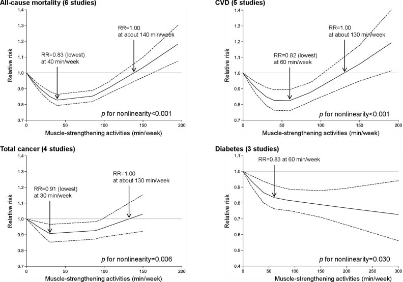
Notice this "U" shape in the curve when examining the relationship between RT and morbidity and mortality. This curve suggests that some RT is clearly beneficial, but has the volume of RT increases past a certain point the benefits drop and it becomes harmful. The concept of a "biphasic response" is fundamental to understanding hormesis. It describes the characteristic dose-response relationship observed in hormetic processes, where a substance or stressor elicits opposite effects at low and high doses. The response can be visualized as a U-shaped or J-shaped curve, illustrating the beneficial effects at low doses and potential harm at higher doses. ​
Picture
Learn more about hormesis

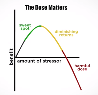
Benefits of RT on Traditional CVD Risk Factors

The AHA's scientific statement underscores the positive influence of RT on traditional CVD risk factors, including blood pressure (BP), glycemia, lipid profiles, and body composition. Numerous studies indicate that engaging in RT is associated with reduced resting BP, improved glycemic control, and favorable alterations in lipid profiles, contributing to a lower risk of all-cause mortality and CVD morbidity. Despite recommendations suggesting 2 days per week of RT, only 28% of U.S. adults adhere to this guideline, highlighting the need for increased awareness and promotion.

RT and resting blood pressure

RT has demonstrated the ability to reduce resting BP across diverse populations, with notable benefits observed in individuals with prehypertension and hypertension. The mechanisms behind these benefits include enhancements in endothelial function, vasodilatory capacity, and vascular conductance. The reductions in BP achieved through RT are comparable to those achieved with antihypertensive medications.

RT and Glycemia

RT shows promise in improving glycemia and insulin resistance, leading to a lower incidence of diabetes. The evidence suggests a nonlinear dose-response association, with up to 60 minutes per week of RT associated with the maximum risk reduction for diabetes.

RT and Lipid Profiles

While the effect on lipid profiles is modest, RT results in favorable changes in high-density lipoprotein cholesterol, total cholesterol, and triglycerides. These improvements are more pronounced in older adults and those with elevated cardiometabolic risk.

Rt, Body composition, and weight

RT positively influences body composition by increasing lean body mass and reducing body fat percentage. It is particularly effective in overweight or obese individuals, contributing to increased metabolic rate and mitigating weight gain over time.

Benefits of RT on Nontraditional CVD Risk Factors

In addition to traditional risk factors, the scientific statement highlights the potential mechanisms by which RT positively affects nontraditional CVD risk factors. These include increased cardiorespiratory fitness, improved endothelial function, and potential benefits for sleep quality, psychological health, and well-being.
Picture
Picture
Picture
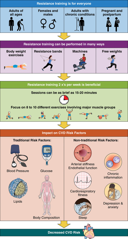
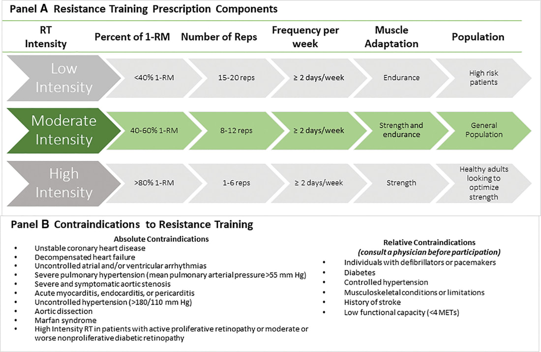
The AHA's updated scientific statement reinforces the pivotal role of resistance training in cardiovascular health, providing a comprehensive overview of its impact on both traditional and nontraditional risk factors. As the evidence supporting RT's benefits continues to grow, the statement serves as a valuable resource for clinicians and public health professionals, offering practical strategies for promoting and prescribing resistance training to enhance cardiovascular health in diverse populations.

References

Paluch, Amanda E, et al. “Resistance Exercise Training in Individuals with and without Cardiovascular Disease: 2023 Update: A Scientific Statement from the American Heart Association.” Circulation, 7 Dec. 2023, https://doi.org/10.1161/cir.0000000000001189. Accessed 11 Dec. 2023.

Momma H, Kawakami R, Honda T, Sawada SS. Muscle-strengthening activities are associated with lower risk and mortality in major non-communicable diseases: a systematic review and meta-analysis of cohort studies. Br J Sports Med. 2022 Jul;56(13):755-763. doi: 10.1136/bjsports-2021-105061. Epub 2022 Feb 28. PMID: 35228201; PMCID: PMC9209691.
0 Comments

Nasal Breathing: A Breath of Fresh Air for Cardiovascular Wellness – Insights from New Research

2/4/2024

0 Comments

 
Picture
Picture
The leading cause of death in the United States is cardiovascular disease, and the risk of cardiovascular issues can be predicted by factors such as blood pressure, heart rate variability, blood pressure variability, and cardiac vagal baroreflex sensitivity. The interplay between the cardiovascular and respiratory systems is highlighted, with a particular emphasis on how respiration affects key prognostic cardiovascular variables.

This study explores the impact of nasal breathing compared to oral breathing on cardiovascular health in young adults. Nasal breathing is associated with humidification, warming, and filtration of inhaled air, potentially leading to bronchodilation and improved breathing efficiency. While past research has shown nasal breathing to have positive effects on resting metabolic demands, its influence on cardiovascular markers is not well-understood.

The primary hypothesis is that nasal breathing, as opposed to oral breathing, will result in decreased blood pressure, improved heart rate variability, reduced blood pressure variability, and increased cardiac vagal baroreflex sensitivity at rest. The study aims to contribute to the understanding of how breathing patterns influence prognostic cardiovascular variables, aligning with the broader interest in the impact of breathing pace and training on cardiovascular health.

The secondary hypothesis focuses on the effects of nasal breathing during submaximal exercise. The expectation is that nasal breathing, by attenuating the ventilatory response and metabolic demands, will lead to reduced blood pressure responses, improved heart rate variability, and decreased blood pressure variability during exercise. This aspect is particularly relevant due to the association between elevated exercise blood pressure and the risk of developing hypertension and cardiovascular disease.

Findings

The study findings are summarized, focusing on the impact of nasal vs. oral breathing on physiological and subjective variables at rest and during exercise. At rest, nasal breathing is associated with lower mean and diastolic blood pressure, improved heart rate variability metrics, reduced LF/HF ratio, and lower ratings of perceived exertion (RPE) and breathlessness (RPB). However, it increased systolic blood pressure average real variability. During submaximal exercise, differences between nasal and oral breathing were observed for RPB, suggesting a modest effect on reducing breathlessness during acute exercise.

The discussion delves into the potential clinical significance of these findings, particularly the reduction in diastolic blood pressure during nasal breathing at rest. The study suggests a greater parasympathetic to sympathetic dominance during nasal breathing, indicated by changes in frequency-domain metrics of heart rate variability. Although nasal breathing did not significantly affect beat-to-beat blood pressure variability, there is speculation about potential connections between respiratory variables and blood pressure changes, emphasizing the need for further investigation.

The study notes that the impact of nasal breathing on cardiovascular variables may have implications for various populations and suggests avenues for future research, including examining nasal breathing's effects on blood pressure over longer durations, both at rest and during activities like exercise. The discussion also touches on the potential benefits of interventions like mouth-taping overnight, emphasizing the importance of considering nasal breathing in the context of broader health outcomes.
Picture
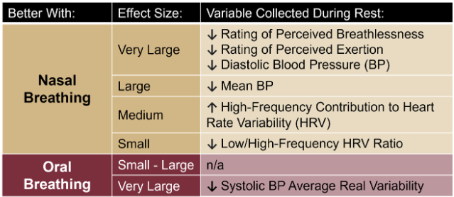
In summary, the study highlights the potential benefits of nasal breathing, with improvements in various cardiovascular and subjective measures at rest. While the effects during exercise are more modest, the findings contribute to understanding the nuanced relationship between respiratory patterns and cardiovascular health.

references

Watso, Joseph C., et al. “Acute Nasal Breathing Lowers Diastolic Blood Pressure and Increases Parasympathetic Contributions to Heart Rate Variability in Young Adults.” American Journal of Physiology. Regulatory, Integrative and Comparative Physiology, vol. 325, no. 6, 1 Dec. 2023, pp. R797–R808, pubmed.ncbi.nlm.nih.gov/37867476/, https://doi.org/10.1152/ajpregu.00148.2023.
0 Comments

Unveiling the Interplay Between Air Quality and Cardiometabolic Health: A Surprising Connection

1/29/2024

14 Comments

 
Picture
In a groundbreaking study, researchers have delved into the intricate relationship between air quality and cardiometabolic health, revealing startling findings that challenge conventional wisdom. Published in Environmental Research, this research sheds light on the impact of air pollutants, even at concentrations below the World Health Organization's (WHO) 2021 guidelines, on various aspects of cardiovascular and metabolic well-being.

Key Findings

The study, conducted over a period of 33 weeks with 82 participants grappling with obesity, examined the associations between air pollutants and cardiometabolic outcomes. Particulate matter emerged as a significant player, demonstrating a strong connection with blood lipids, hormones, and glucose regulation – key markers of cardiometabolic health.
​

Surprisingly, the research also uncovered a potential mitigating factor: diet. The participants' adherence to a Healthy Nordic diet, as measured by the Baltic Sea Diet score, showcased a remarkable ability to modify the impact of air pollution on certain cardiometabolic parameters.

Details of the Study

The study utilized linear mixed-effects models to analyze data gathered during a weight loss and weight loss maintenance intervention. The results revealed 17 significant associations between various air pollutants and 10 distinct cardiometabolic outcomes. The focus was primarily on blood lipids, hormones, and glucose regulation, providing a comprehensive understanding of the multifaceted effects of air pollution.
​
Interestingly, the Baltic Sea Diet score did not appear to mediate the association between air pollution and cardiometabolic outcomes. However, the diet quality factor emerged as a key player in modifying the impact of particulate matter (PM2.5) on total cholesterol. Furthermore, it influenced the associations of nitrogen dioxide (NO) and ozone (O3) with ghrelin, a hormone associated with appetite regulation.

References

Healy, Darren R., et al. “Associations of Low Levels of Air Pollution with Cardiometabolic Outcomes and the Role of Diet Quality in Individuals with Obesity.” Environmental Research, vol. 242, 1 Feb. 2024, p. 117637, www.sciencedirect.com/science/article/pii/S0013935123024416, https://doi.org/10.1016/j.envres.2023.117637.
14 Comments

Navigating Hormesis: Embracing the Balance Between Stress and Strength for Optimal Health

12/21/2023

8 Comments

 
In the intricate dance of biological responses to stress, hormesis emerges as a captivating phenomenon, challenging conventional notions of dose-response relationships. This blog post delves into the fascinating world of hormesis, where the subtle interplay between stressors and adaptive responses shapes our understanding of health and resilience.
Learn more about Hormesis

The Hormetic Curve: Unveiling the U-Shaped Story

Explore the dynamics of hormesis through the lens of a U-shaped or J-shaped curve, where low doses of stressors trigger beneficial responses while high doses lead to toxicity. Understand how this nonlinear relationship challenges traditional toxicological paradigms.
Picture

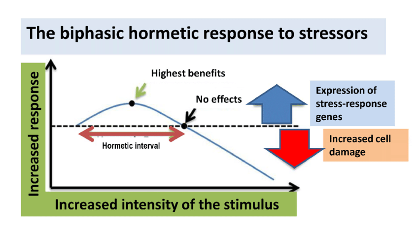
Small Doses, Big Impact: Hormetic Responses Unveiled

Delve into real-world examples of hormetic responses, from the beneficial effects of low-dose radiation to the adaptive mechanisms activated by moderate exercise. Learn how these responses stimulate resilience, adaptation, and overall well-being.

Hormesis in Toxicology: Redefining Risk Assessment

Unravel the complexities of hormesis in toxicology, where the concept challenges established risk assessment practices. Discover how hormesis introduces a nuanced understanding of dose-dependent effects, prompting a reevaluation of regulatory frameworks. ​

Hormesis and Adaptive Learning: "What Doesn't Kill You Makes You Stronger"

Connect the dots between hormesis and the age-old adage, "What doesn't kill you makes you stronger." Explore how the hormetic principle aligns with the idea that controlled exposure to stressors can lead to adaptive learning, fostering strength and resilience.
Learn more about Hormesis

Balancing Act: The Importance of Moderation

Emphasize the crucial role of balance and moderation in hormesis. Uncover the delicate equilibrium required for stressors to act as catalysts for positive adaptation without tipping into harmful territory.

From Sirtuins to Telomeres: Hormesis at the Cellular Level

Journey into the cellular realm and discover how hormesis influences sirtuins, telomeres, and other cellular processes. Explore the implications for cellular repair, mitochondrial function, and the potential impact on longevity.
Picture

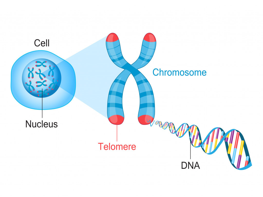
Practical Insights: Applying Hormesis in Everyday Life

Gain practical insights into how hormesis can be applied in daily life. Explore lifestyle choices, dietary considerations, and stress management techniques that harness the power of hormetic responses for enhanced well-being.
Picture
​Embark on a journey of discovery as we unravel the layers of hormesis, revealing its impact on biology, health, and our quest for a balanced and resilient life. Embrace the science behind stress and strength, and learn how hormesis invites us to rethink our approach to well-being.

Learn more about Hormesis
8 Comments

The War on Ivermectin

12/20/2023

0 Comments

 
The documentary "War on Ivermectin" explores the contentious landscape surrounding the drug Ivermectin during the COVID-19 pandemic. It delves into the controversy surrounding the use of Ivermectin as a potential treatment for COVID-19 and the challenges it has faced from regulatory bodies and mainstream medical establishments.

The documentary presents perspectives from advocates of Ivermectin, who argue for its efficacy and safety in treating COVID-19. It may shed light on the resistance faced by proponents of Ivermectin, exploring factors such as media portrayal, regulatory decisions, and the broader implications for the treatment landscape.
​
Additionally, the documentary might feature interviews with medical professionals, researchers, and individuals who have been affected by the debate over Ivermectin. It aims to provide a comprehensive overview of the complex and polarized discussions surrounding the drug in the context of the global response to the pandemic.
0 Comments

Documentary: The Business of Being Born

9/13/2023

0 Comments

 
​"The Business of Being Born" is a documentary that explores the maternity care system in the United States, shedding light on the medicalization of childbirth and advocating for a more holistic and woman-centered approach. Produced by Ricki Lake and Abby Epstein, the film challenges conventional hospital birthing practices and highlights the benefits of midwifery and home births.

The documentary features interviews with midwives, doctors, and mothers who share their experiences and perspectives on childbirth. It addresses concerns about the rising rates of cesarean sections, interventions, and the impact of hospital protocols on the birthing process. The film advocates for informed decision-making, empowering women to make choices that align with their preferences and needs during childbirth.

Through personal stories and expert opinions, "The Business of Being Born" encourages viewers to rethink the way society approaches and views childbirth, urging a return to more personalized, woman-centered, and natural birthing experiences. The documentary contributes to the ongoing conversation about maternity care practices and promotes awareness of alternative options for expectant mothers.
0 Comments

Andropause Explained: A Deep Dive of the Effects, Causes & Solutions of Low Testosterone

8/5/2023

0 Comments

 
Picture
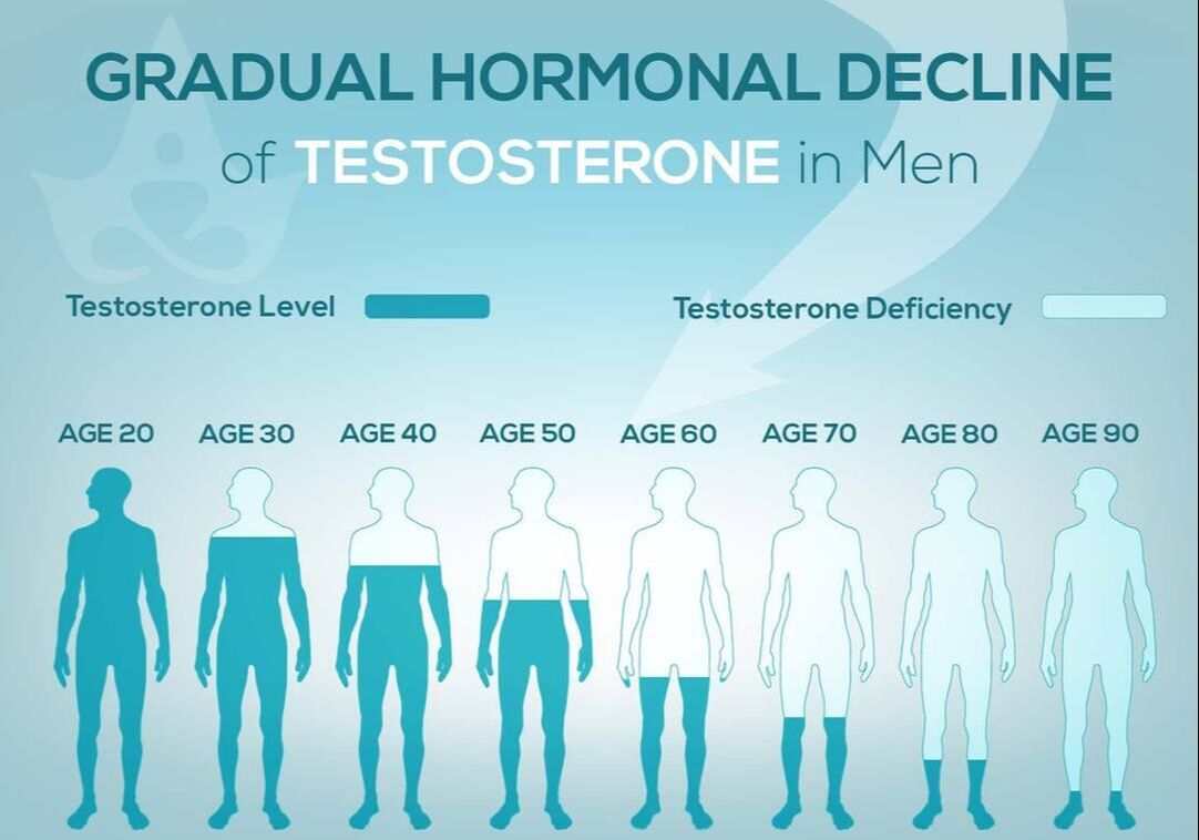
Women are not the only ones who experience hormonal changes in midlife. Men also undergo a transition similar to menopause called andropause, also known as late-onset hypogonadism (LOH), during which vitality hormones, particularly testosterone (T), but also human growth hormone (HGH), are produced in lower quantities. As men age, not only does the body start making less testosterone, but also the levels of another hormone called sex hormone binding globulin (SHBG), which pulls usable testosterone from the blood, begins to increase. 

Andropause is a natural phase of aging, but is accentuated by many factors including, but not limited to age-associated comorbid illnesses, medications, and malnutrition.
 The age at which symptoms of andropause may manifest can vary, but it typically occurs in middle-aged and older men, beginning in the late 40s to early 50s, with the most frequent age occurring between 51-60 years, with patients reporting symptoms such as impotence, weakness, and memory loss. Other age-related alterations due to andropause include body composition, mood, cognitive function, sleep, and erythropoiesis.

​The pharmaceutical industry has capitalized heavily on this 'change of life' phase, with Viagra, among other pharmaceuticals, but these pharmaceuticals have severe, if not sometimes deadly side effects. All the more reason why modifiable factors and natural alternatives are in great need today.


While the process of andropause is considered inevitable, understanding the causes and adopting proactive nutrition and lifestyle strategies can prevent an early onset of this condition, and significantly alleviate symptoms thereby enhancing overall quality of life.

normalization of Low T

Testosterone is a crucial hormone for both men and women, contributing to muscle bulk, strength, fat-burning, and overall vitality. Adequate testosterone levels provide a chiseled look, high energy, and strength in men, and definition, muscle, and energy in women. However, declining testosterone levels can lead to increased fatigue, difficulty building muscle, and higher fat accumulation, posing a risk to overall health.

Contrary to the common belief that decreasing testosterone is solely an aging-related issue, it has been observed that testosterone levels, even in young men, have been declining for decades. Factors contributing to this decline include the Standard American Diet, characterized by high sugar intake, imbalanced omega-6 to omega-3 ratios, processed foods lacking essential nutrients, and the attack on cholesterol. Additionally, environmental toxins play a role in lowering testosterone levels.

Notably, the Standard American Diet, abundant in sugars, particularly processed sugars and carbohydrates, elevates cortisol levels, which inversely affects testosterone. The imbalance of omega-6 to omega-3 ratios, primarily due to the prevalence of corn and soy in processed foods, further contributes to cortisol elevation, adversely impacting testosterone levels. Processed foods with low nutritional value, combined with the demonization of cholesterol, essential for testosterone production, also play a role in this decline.
​
Addressing these issues through dietary adjustments, such as choosing organic and grass-fed options, avoiding processed foods and sugars, ensuring adequate protein intake, and engaging in regular exercise, can positively impact testosterone levels and overall health. Understanding and addressing these lifestyle factors is essential for maintaining optimal hormonal balance and supporting longevity.
Picture
Standard American Diet (SAD)

PSYCHONEUROIMMUNOLOGY:
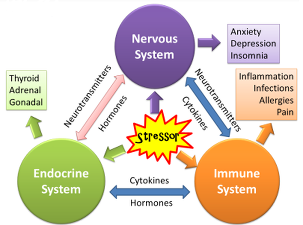
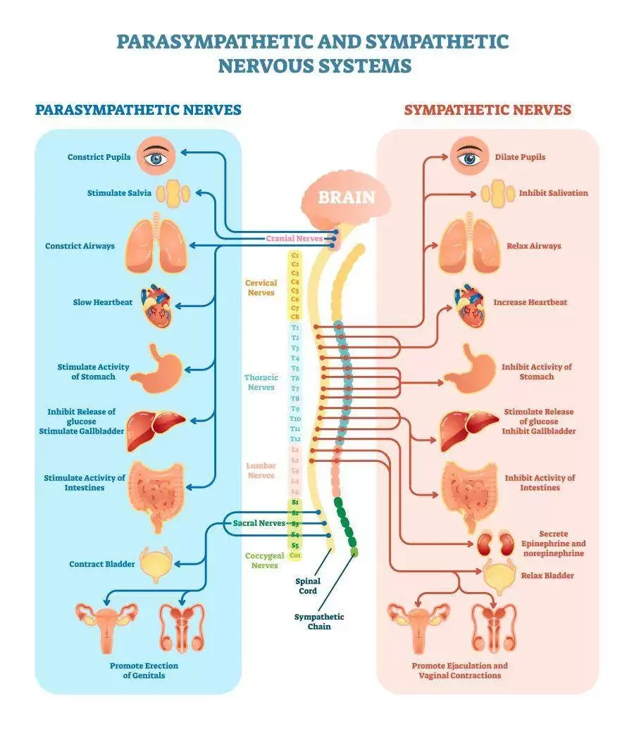
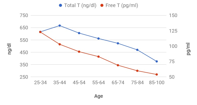
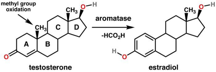
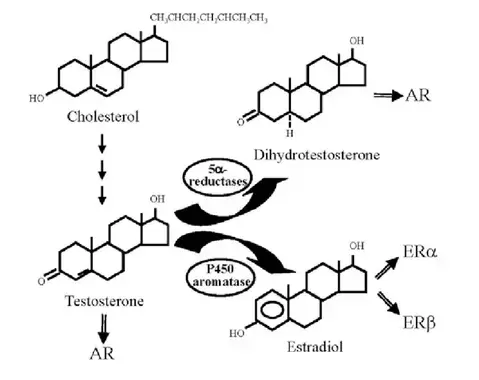
​The interconnectedness of the endorcrine system

Psychoneuroimmunology (PNI) explores the dynamic interconnections between psychological processes, the nervous system, the immune system, and the endocrine system. When delving into the context of andropause, PNI provides insights into how psychological factors can influence the hormonal shifts associated with this transitional phase in life. PNI acknowledges the body's ability to adapt hormonally to various stressors, including the changes associated with andropause. Understanding how the mind and body interact during this phase can inform strategies to support hormonal adaptation and mitigate associated symptoms.
Picture
Here are some Key Aspects of PNI as it relates to Andropause:
  1. Psychological Impact on Hormones: Psychological factors, such as perceived and sub-conscious stress, anxiety, emotions, personality traits, mental states, and mood disorders, can impact the endocrine system, potentially exacerbating hormonal imbalances. The endocrine system, which includes glands that produce hormones, plays a crucial role in regulating various physiological processes, such as metabolism, growth, and stress responses. Acute and chronic psychological stress activates the sympathetic or "fight or flight" response, leading to the release of stress hormones such as cortisol and adrenaline from the adrenal glands. Prolonged or chronic stress can dysregulate the endocrine system, impacting hormonal balance, including testosterone. Additionally, mood disorders like depression can be associated with alterations in the levels of neurotransmitters and hormones.  
  2. Neuroendocrine Connections: PNI investigates the intricate bidirectional communication between the nervous and endocrine systems. Neurotransmitters, neuropeptides, and hormones act as messengers, transmitting signals between these systems. The brain's responses to stress or emotional stimuli can influence hormonal and immune responses. Stress, for example, triggers the release of cortisol, a stress hormone, which can further influence the production and regulation of testosterone in men.
  3. Immune System Modulation: Considering psychological states can modulate the nervous system via changes communication, this in turn can disrupt immune function, affecting the body's ability to defend against pathogens or regulate inflammation. Conversely, immune responses can signal the brain and influence mood and behavior via changes in the endocrine system.

Psychological well-being plays a significant role in navigating andropause. PNI explores how mood changes, such as irritability or depressive symptoms, may be linked to hormonal fluctuations. Conversely, positive psychological states can contribute to overall well-being during this transitional period.

PNI also explores the placebo and nocebo effects, where psychological factors, the presence of absence of support and/or a proactive mindset can influence  how individuals experience and cope with the body's physiologic hormonal responses. Positive expectations (placebo) or negative beliefs (nocebo) can impact hormonal and immune functions.

Understanding the complex interplay between psychological states, the nervous system, the immune system, and the endocrine system during andropause is crucial for developing holistic approaches to support men's health and overall quality of life. PNI provides a framework for exploring the mind-body interactions that shape the experience of andropause, offering insights into potential interventions to promote parasympathetic nervous system activation thereby enhancing psychological and hormonal well-being, which includes nutrition and lifestyle interventions, such as stress management techniques.
Picture

Systemic effects of Andropause

The concept of late-onset hypogonadism (LOH) in aging men has specific characteristics with respect to the symptoms and clinical features associated with LOH. Low testosterone levels can have diverse effects on various aspects of the body (men and women included), influencing hormones, neurotransmitters, energy levels, and potentially contributing to the development of chronic diseases, and had been observed to lead to various symptoms including, but not limited to, sexual dysfunction, reduced vitality, and mood disturbances. Additionally,  comprehensive meta-analyses have revealed low testosterone in males is associated with increased all-cause mortality.

Hormonal Imbalance

Low testosterone levels can impact various physiological processes, decreasing quality of life, via the disrupted balance of other hormones, including but not limited to:
  1. Effects on Estrogen Levels: Testosterone is converted to estrogen through the aromatization process (explained further below). Low testosterone levels may lead to a reduction in estrogen levels, potentially impacting various estrogen-dependent functions. Prolonged activation of estrogen has been observed to cause a variety of abnormal health effects including various comorbidities, from heart attacks, to neurodegenerative diseases, including Alzheimer's, Parkinson's, stroke, as well as all types of cancer. In fact, in 2001 the National Institutes of Health declared estrogen as a known human carcinogen. Estrogen from a biochemical view is known to heal wounds. Estrogen is the de-differentiating factor to replace damaged tissue, essentially reverting the healing tissue back to a stem cell state, with the expectation of a pro-differentiating factor to come in an stop the effects of estrogen. That pro-differentiating factor in healthy individuals is typically progesterone in women, and androgens in men. And as it turns out, these pro-differentiating factors decline with age whereas the de-differentiating factors (estrogen) does not.
  2. Impact on Insulin Sensitivity: Testosterone plays a role in maintaining insulin sensitivity. Low testosterone levels are associated with insulin resistance, which can contribute to metabolic disorders and disrupt the hormonal balance.
  3. Influence on Cortisol Levels: Testosterone has been suggested to have a regulatory effect on cortisol levels. Low testosterone may contribute to altered cortisol regulation, impacting the body's stress response. The combination of low T and high cortisol has been observed to drive mental health conditions, such as PTSD.
  4. Association with Thyroid Hormones: Some studies indicate a potential association between testosterone levels and thyroid hormones. Disruptions in testosterone levels may contribute to alterations in thyroid and metabolic function, thereby resulting in weight gain among many other downstream effects.
  5. Interplay with Growth Hormone: Testosterone and growth hormone exhibit intricate interactions. Low testosterone levels may influence the secretion and effectiveness of growth hormone, impacting growth and metabolism.
Understanding the interconnectedness of hormones is crucial for comprehending the broader implications of low testosterone levels. Researchers collectively suggest that disruptions in testosterone levels can reverberate through the endocrine system, potentially contributing to various health issues. However, it's essential to note that the hormonal system is complex, and the relationships between hormones are multifaceted, requiring further research for a comprehensive understanding.

Neurotransmitter Regulation

Testosterone plays a role in neurotransmitter regulation, and low levels may contribute to changes in mood, cognition, and overall mental well-being.

Testosterone, primarily recognized for its influence on reproductive tissues, extends its impact to the central nervous system, playing a crucial role in neurotransmitter regulation. Here's an exploration of how testosterone affects neurotransmitters with specific examples:
  1. Serotonin Modulation: Testosterone has been associated with the regulation of serotonin, a neurotransmitter crucial for mood stability. It is important to maintain a balance of serotonin, as high levels of serotonin have been linked to accelerated aging. A study using PET imaging found that the dorsal raphe nucleus (DRN), a region in the brain responsible for serotonin production, showed increased serotonin synthesis capacity in older adults compared to younger individuals. This heightened serotonin activity was identified as a potential marker for unfavorable aging outcomes. Additionally, researchers have observed that individuals with atherosclerosis alongside chronic lung issues also exhibited imbalances in hormones like corticosteroids and estrogen, lower testosterone levels, and increased production of serotonin and noradrenaline.
  2. Dopamine Influence: Testosterone contributes to the modulation of influencing the synthesis, release, and reuptake of dopamine levels, thereby impacting reward and pleasure pathways in the brain. With low testosterone, this orchestration may falter, contributing to a muted motivational crescendo and potentially influencing a decline in overall energy levels. This interplay between testosterone and dopamine interplay contributes to the maintenance of optimal energy levels, and disruptions in this delicate dance may manifest as fatigue and lethargy. Beyond the realms of physiology, the testosterone-dopamine connection extends its influence to mental health. Research suggests that alterations in this hormonal duet may be associated with mood disturbances, further emphasizing the importance of hormonal balance for a holistic sense of vitality.
  3. Gamma-Aminobutyric Acid (GABA) Interaction: Testosterone has been linked to the regulation of GABAergic transmission, affecting inhibitory processes in the brain. Literature suggests that low testosterone is associated with alterations in GABA receptor expression and function, potentially disrupting the inhibitory balance maintained by GABA in the central nervous system. GABA, as a primary inhibitory neurotransmitter, plays a crucial role in regulating excitatory signals in the brain. Studies propose that disruptions in the testosterone-GABA axis may contribute to mood disorders, anxiety, and other mental health challenges, emphasizing the intricate interplay between hormonal status and mental well-being. The influence of testosterone on GABA extends beyond the realms of the central nervous system. Researchers have observed that alterations in GABAergic function may have systemic effects, impacting various physiological processes, including immune function and cardiovascular health. ​
  4. Glutamate Regulation: Testosterone may influence glutamate levels, impacting excitatory neurotransmission in various brain regions. Glutamate is the primary excitatory neurotransmitter in the central nervous system. It is involved in synaptic plasticity, learning, and memory. Changes in testosterone levels may influence the release, reuptake, or sensitivity of glutamate receptors, thereby modulating excitatory signaling. Some studies suggest that testosterone may have neuroprotective effects, including protecting against excitotoxicity—a process where excessive glutamate leads to neuronal damage or cell death. Testosterone's potential neuroprotective role might involve interactions with glutamate pathways. Testosterone has been associated with cognitive functions, and alterations in glutamate levels or receptor function may contribute to cognitive changes observed in conditions associated with low testosterone.
These findings collectively emphasize the intricate role of testosterone in shaping neurotransmitter activity, contributing to the understanding of its impact beyond reproductive functions, including overall quality of life.

Low Energy and Fatigue

Low testosterone levels can be linked to reduced energy levels and increased fatigue, impacting overall vitality and motivation.

Mitochondria, revered as cellular powerhouses, dance to the tune of testosterone's regulatory prowess. Scholarly investigations affirm that testosterone safeguards mitochondrial integrity, enhancing their efficacy in the production of adenosine triphosphate (ATP), the cellular coinage of energy. The intricate orchestration of oxidative phosphorylation, orchestrated by mitochondria, falters in the face of low testosterone, culminating in decreased ATP production and a consequential dip in overall energy levels.

In the grand theater of bodily performance, testosterone takes center stage in preserving muscle mass and strength. Skeletal muscles, integral to the poetry of physical activity, face discord when testosterone levels wane, potentially leading to muscle wasting, compromised energy utilization, and heightened fatigue.

As highlighted above, testosterone extends its influence to the cerebral realm, engaging in a nuanced dance with neurotransmitters, including dopamine. This dance, when disrupted by low testosterone, may contribute to a reduction in motivation and a perception of fatigue.

Testosterone's anti-inflammatory cadence resonates through the body. Its scarcity tilts the balance towards inflammation, a known harbinger of fatigue. By modulating inflammatory pathways, testosterone establishes an environment conducive to sustained vitality

Lastly, testosterone's temporal ballet adheres to a circadian rhythm, with crescendos during specific periods, notably during sleep. Perturbations in these hormonal rhythms may contribute to sleep disturbances, amplifying fatigue and affecting overall well-being

Association with Chronic Diseases

Due to the aforementioned effects of low testosterone levels, andropause has been associated with an increased likelihood of developing chronic conditions, including metabolic syndrome and cardiovascular disease. There are various downstream effects that occur as a result of metabolic syndrome. Metabolic syndrome is a cluster of conditions that include central obesity, insulin resistance, type-2 diabetes, high blood pressure, dyslipidemia, increased inflammation (which can lead to a whole host of other chronic diseases), and reduced muscle mass and strength. Researchers have even found a relationship between metabolic syndrome and a dysregulated immune system, which has been observed to lead to various types of cancer. Additionally, low testosterone has been observed to drive kidney and liver disease (non-alcoholic fatty liver). Mental health disorders, including depression and anxiety have also been observed, likely due to changes in neurotransmitters.

It is clear that optimal levels of testosterone, or a lack of thereof, has vast implications on systemic health. It's important to note that while associations between low testosterone and metabolic syndrome exist, their interconnectedness is complex, and individual responses may vary. Lifestyle factors, and other health conditions can also contribute to the development of metabolic syndrome and related diseases.

Association with Increased mortality

As testosterone levels decline there often is an accompanied increased in luteinizing hormone (LH) levels, indicating impaired testicular function. Factors such as obesity and other medical conditions can further reduce testosterone by affecting the hypothalamic-pituitary-testicular (HPT) axis. Given testosterone's role in libido, muscle mass, and fat distribution, understanding its health impacts is crucial.

Testosterone in circulation is bound to sex hormone-binding globulin (SHBG), which modulates its availability. Low testosterone with high LH suggests primary testicular issues, while low testosterone with low or normal LH indicates central causes or HPT axis suppression due to conditions like obesity. Testosterone converts to dihydrotestosterone (DHT) and estradiol, influencing various tissues.

Men with low testosterone, high LH, or very low estradiol concentrations had increased all-cause mortality. In men aged 40-69, lower testosterone correlates with higher all-cause mortality but not necessarily cardiovascular disease (CVD) deaths. Studies using mass spectrometry, a more accurate measurement method, found higher testosterone linked to lower CVD risks, especially stroke. 

A meta-analysis of cohort studies using mass spectrometry aimed to clarify these findings. Men with low testosterone (<7.4 nmol/L) had higher all-cause mortality, independent of LH levels, indicating the risk is associated with low testosterone itself. Very low testosterone (<5.3 nmol/L) was linked to increased CVD mortality. Low testosterone might lead to poorer outcomes due to its association with lower muscle mass, greater adiposity, and other cardiovascular risk factors.

Additionally, low testosterone combined with high SHBG posed a higher mortality risk, suggesting the combination's significance. Studies also indicated a U-shaped relationship between DHT and mortality, with risks at both low and high DHT levels.
​
In summary, this analysis highlights the importance of maintaining balanced testosterone levels for health and longevity in men. Low testosterone is consistently linked to higher mortality risks, emphasizing the need for further research into testosterone's role in aging and health.

Causes of andropause

Attention to appropriate exercise and nutrition, and evaluation and treatment of other etiological factors that may contribute to clinical manifestations are essential for optimal management of age-related functional decline in older men.

Because age-related alterations in physiological function are usually a result of multiple etiologies, it is important to evaluate and treat other factors (e.g., inadequate nutritional intake, confounding illness and medication, inactivity or poor conditioning, excessive alcohol, and smoking) in addition to low T levels that may contribute to the clinical syndrome.

Hormone decline

The primary factor in andropause is a gradual decline in testosterone levels, impacting various bodily functions. ​It's important to note that individual responses to low testosterone can vary, and the impact on overall health is influenced by various factors, including age, lifestyle, and underlying health conditions. Regular medical monitoring and consultation with healthcare professionals are essential for a comprehensive understanding of one's hormonal health.
Picture
Average T Levels by Decade: 720 ng/dl for a man in his 30’s; 667 ng/dl for a man in his 40’s; 606 ng/dl for a man in his 50’s; 562 ng/dl for a man in his 60’s; 523 ng/dl for a man in his 70’s.

Aromatization

Aromatization is a biological process where testosterone, the primary male sex hormone, is converted into estrogen, a key female sex hormone. This conversion is facilitated by the enzyme aromatase, which is expressed by all cells in the human body (for both men and women), including the gonads, brain, adipose tissue (fat cells), and skin. Aromatase is a member of the cytochrome P450 superfamily, and is responsible for catalyzing the conversion of androgens (such as testosterone) into estrogens (such as estradiol), a process known as aromatization. While aromatization is a natural and necessary process in both men and women, an imbalance or excessive aromatase activity can lead to various health issues.
Picture
Before assuming that estrogen is useless, it is important to realize that estrogen serves many roles. Consider estrogen as a useful tool in times where physiologic growth is needed - however growth at all costs is harmful. Estrogen is essential for both men and women for various physiological functions, including carbohydrate and lipid metabolism, bone health, cardiovascular health, and reproductive function. In men, a certain level of estrogen is necessary for the regulation of libido, sperm production, and overall well-being.

So indeed, there is a function for estrogen, as there is a function for aromatase, but when unchecked can certainly cause harm. While there are likely various function for the aromatase enzyme, including the synthesis of estrogen, some researchers speculate that aromatization is required to inhibit the secretion of luteinizing hormone (LH) in men. 
Picture
Luteinizing hormone (LH)
LH can have various effects on the reproductive system. LH is a hormone produced by the pituitary gland, and its primary role in men is to stimulate the Leydig cells in the testes to produce testosterone. However, an excess of LH can disrupt the normal functioning of the male reproductive system in several ways:
  1. Negative Feedback Mechanism: The male reproductive system has a negative feedback mechanism to regulate hormone levels. Elevated testosterone levels can signal the hypothalamus and pituitary gland to reduce the production of gonadotropin-releasing hormone (GnRH) and LH. This is a natural regulatory mechanism to prevent excessively high testosterone levels.
  2. Desensitization of Leydig Cells: Luteinizing hormone (LH) from the pituitary gland stimulates the Leydig cells in the testes to produce testosterone, which in turn supports sperm production and other reproductive functions. Prolonged exposure to high levels of LH might lead to desensitization of Leydig cells in the testes. Over time, the cells may become less responsive to LH, potentially reducing testosterone production. The conversion of cholesterol to testosterone occurs in the interstitial Leydig cells. Leydig cells will release testosterone, and respond to testosterone via receptors. 
  3. Impacts on Spermatogenesis: Excessive LH levels may also affect spermatogenesis, the process of sperm production. Changes in hormone balance can influence the quantity and quality of sperm produced. Semen and testosterone are closely related in several ways, as testosterone plays a crucial role in male reproductive health and function. Testosterone is essential for spermatogenesis, the process of sperm production in the testes. It stimulates the Sertoli cells in the seminiferous tubules of the testes to support the development and maturation of sperm cells. Testosterone influences the production of seminal fluid, which is the liquid part of semen. This fluid is produced by the seminal vesicles, prostate gland, and bulbourethral glands and provides a medium for sperm to travel and survive. Adequate levels of testosterone are necessary for maintaining libido (sexual desire) and sexual function, which are important for ejaculation and the release of semen during sexual activity.
    Testosterone helps maintain the health and function of the testes, where sperm are produced. Low testosterone levels can lead to testicular atrophy and reduced sperm production. Testosterone levels are regulated by the hypothalamic-pituitary-gonadal (HPG) axis. 
  4. Potential Health Issues: Chronic elevations in LH, especially if associated with imbalances in other hormones, may contribute to various health issues, including reproductive disorders and hormonal imbalances.

Dihydrotestosterone (DHT)
Another pathway for testosterone metabolism involves its conversion to DHT. This conversion is facilitated by the enzyme 5-alpha-reductase. DHT is a more potent androgen than testosterone and plays a crucial role in the development of male reproductive tissues, including the prostate and external genitalia. It is particularly important during fetal development and puberty.
​
While DHT is essential for certain physiological processes, excess levels of DHT can lead to various issues, including:
  • Prostate Enlargement (BPH): Elevated DHT levels have been associated with benign prostatic hyperplasia (BPH), a condition characterized by an enlarged prostate. This can lead to urinary symptoms such as difficulty in urination.
  • Male Pattern Baldness: Excessive DHT is linked to the miniaturization of hair follicles, contributing to male pattern baldness (androgenetic alopecia).
  • Acne and Oily Skin: Increased DHT levels can stimulate the sebaceous glands, leading to excess oil production and contributing to acne.
  • Aggravation of Prostate Cancer: While the relationship is complex, some studies suggest that high DHT levels may contribute to the growth of prostate cancer cells.

There are various causes of aromatase upregulation, including but not limited to adipose tissue, inflammation and age. Fat cells, particularly in abdominal adipose tissue, can produce increased amounts of aromatase. This is one reason why obesity is associated with higher estrogen levels in both men and women. Additionally, inflammatory signals can stimulate the expression of aromatase, leading to increased conversion of testosterone to estrogen. Aging is associated with changes in hormone levels, and older individuals may experience increased aromatase activity.

As noted earlier, in response to aromatase imbalances, disruptions to the delicate balance of hormones occurs, including but not limited to estrogen dominance, where estrogen levels outweigh testosterone levels. States of estrogen dominance have intriguing presentations, often causing males to become more feminine in phenotype (e.g., gynecomastia, excess adipose deposition), and vice versa for females (females become more masculine). Additionally, aromatase upregulation can disrupt contribute to symptoms such as fatigue, reduced libido, and mood changes.

Apnea

The potential connection between apnea (specifically sleep apnea) and hormonal changes, particularly focusing on estrogen and testosterone levels, is well cited in the scientific literature.  Apnea, characterized by under-breathing and a buildup of carbon dioxide as in the case during sleep, might play a role in exacerbating symptoms of hormonal decline, likely due to alterations in metabolic system.
​
There exists a complexity in the relationship, noting that it's unclear whether reductions in estrogen or testosterone cause apnea or if apnea contributes to hormonal changes. There's evidence indicating estrogen receptors on lung neurons, suggesting a possible influence of estrogen on breathing sensitivity. Similarly, testosterone reductions are associated with apnea, given the presence of testosterone receptors in visceral cells, including the lungs.
​
The aspect that breathing patterns during sleep, especially in cases of apnea, could actively modulate hormones is intriguing none the less. The significance of optimizing both estrogen and testosterone levels, among generalized hormonal regulation, regardless of gender, through actionable changes in breathing patterns is a worthwhile modifiable risk factor that appears to carry no risk. 
Picture

Aging & lifestyle

Aging itself contributes to hormonal changes. However it is worth noting that aging is accelerated by many lifestyle choices and behaviors. Therefore, unhealthy lifestyle habits such as poor nutrition, lack of exercise, and chronic stress can exacerbate andropause symptoms. 

Researchers have investigated how stress contributes to age-related changes in the male reproductive system. Stress is a multifaceted physiological response, and can occur in response to various factors including the following stimuli: mental, physical, emotional, chemical, thermal, electromagnetic, etc. Examined areas of interest include hormonal regulation, testicular function, and sperm quality, aiming to elucidate the mechanisms through which stress may influence reproductive aging.
​
Key findings highlight the intricate connections between stress and age-related alterations, suggesting that chronic stress can accelerate certain aspects of the aging process in the male reproductive system. There are various involved pathways, such as the involvement of stress hormones, oxidative stress, and inflammation, in mediating the effects on reproductive tissues.

The need for a comprehensive understanding of the complex interactions between stress and aging is especially concerning with respect to the reproductive health in males in our modern world. 

Endocrine disruptors

Endocrine disruptors are substances that can interfere with the normal functioning of the endocrine system, which is responsible for regulating hormones in the body. These disruptors, including certain chemicals, pollutants, and synthetic compounds, have been associated with adverse effects on testosterone levels and overall quality of life. 

Endocrine disruptors can mimic or block the action of natural hormones, leading to hormonal imbalances. This interference can affect the delicate equilibrium between testosterone and other hormones, such as estrogen. Hormonal imbalance, particularly a decrease in testosterone levels, can have various consequences on physical and mental well-being.

Some endocrine disruptors may interfere with the production of testosterone. For example, they might affect the function of Leydig cells in the testes, which are responsible for testosterone synthesis. Lower testosterone levels can impact muscle mass, bone density, energy levels, and sexual function.

Endocrine disruptors can interfere with the signaling pathways that regulate hormone production and release. This disruption can lead to impaired communication between the endocrine glands, affecting the feedback loops that maintain hormonal balance.

Several chemicals and substances have been identified as known or suspected endocrine disruptors that may influence testosterone levels. It's important to note that research in this area is ongoing, and the effects of these substances can vary depending on factors such as exposure levels, duration, and individual susceptibility. Here are some known or suspected endocrine disruptors associated with potential impacts on testosterone levels, supported by evidence:
  1. Bisphenol A (BPA): Found in plastics, epoxy resins, and some food containers.
  2. Phthalates: Commonly found in plastics, personal care products, and certain medications.
  3. Organophosphate Pesticides (including glyphosate): Used in agriculture and can be found in residues on fruits and vegetables.
  4. Polychlorinated Biphenyls (PCBs): Industrial chemicals that were once used in electrical equipment, but are now banned.
  5. Perfluorinated Compounds (PFCs): Used in the production of non-stick cookware, waterproofing agents, and flame-retardant materials.
  6. Heavy Metals (Lead, Mercury, Cadmium): Found in contaminated water, certain fish, and some industrial processes.
  7. Triclosan: Commonly used in antibacterial soaps, toothpaste, and other personal care products.
  8. Parabens: Preservatives used in cosmetics, pharmaceuticals, and personal care products.
  9. Phenols: Found in some detergents, disinfectants, and personal care products.​
  10. Dioxins: Environmental pollutants formed during the combustion of certain chemicals.
  11. Atrazine: A widely used herbicide in agriculture.
​Acid Blockers
ADHD drugs
​Adjuvant
Adderall
Adrenaline
Aluminum
Antidepressants
Antihypertensive drugs
Antipsychotic drugs
Antiretroviral drugs
Atorvastatin
​Benzophenones
Bile Acid Sequestrants (+ binding resins)​
Bisphenols (BPA, BPF, BPS)
Cell Phone Exposure
​Cesium-137
Cholesterol Lowering Drugs
Concerta
Corticosteroid
Dexamethasone
​Electronic Cigarettes
Ethinyl Estradiol (plus Lynestrenol)
Ethylene Glycol
Fluoride
Fructose
​Gluten 
(and Exorphins)
Hexachlorocyclohexane
Histamine Receptor Antagonists
Ibuprofen
Infant Formula
Lead
Levonorgestrel/ethinyl estradiol
Lovastatin
Mercury
​​Monosodium Glutamate (MSG)
Mycoestrogens
Nanoparticles
Nonylphenol [and ​Ethoxylate (NPE)]
Oral Contraceptives
Organochlorine
Pesticides &
Compounds
Organophosphate Pesticides
Persistent Organic Pollutants (POPs)
Pesticides
Phenothrin
Polybrominated Diphenylethers (PBDEs)
Polyoxyethylene Amine
Prednisone
Pravastatin
Progestins
Rosuvastatin
Simvastatin
Sodium Fluoride
Soy
Statin Drugs
Sugar Sweetened Beverages
​​Tamoxifen
Thimerosal
Thiazide Diuretics
Tin
Titanium Nanoparticles (including Dioxide)
Tween 80
(Polysorbate 80)
Vinclozolin
Vitamin A Palmitate
Zearalenone (ZEA)
 
It is important to note that the chart above is composed of substances that have evidence supporting endocrine disrupting abilities, but that does not inherently mean that testosterone will lower. The endocrine system is complex, and all hormones are connected in some fashion.

It's important to be aware of potential exposure to these substances and take steps to minimize risks. This includes choosing products that are labeled as BPA-free, using natural and organic personal care products, and being mindful of pesticide residues on food. Additionally, maintaining a healthy lifestyle, including a balanced diet and regular exercise, can help support overall endocrine health.

Bisphenol-A (BPA)

BPA is a known endocrine disruptor, meaning it can interfere with the normal functioning of the endocrine system, including the production, release, transport, metabolism, and elimination of hormones. BPA is a synthetic compound used in the production of plastics, and it can be found in various everyday items, including receipt paper, food containers, water bottles, and the lining of food cans.

The mechanism by which BPA may lead to lower testosterone levels involves its ability to mimic or interfere with the action of hormones. BPA is known to have estrogenic properties, meaning it can bind to estrogen receptors in the body, thereby mimicking the effects of estrogen. This can disrupt the delicate hormonal balance, leading to alterations in the normal regulatory processes of the endocrine system.

Excessive estrogenic activity, whether from BPA or other sources, can negatively impact the production of testosterone. Estrogen and testosterone are usually balanced in the body, and disruptions in this balance can lead to a decrease in testosterone levels. BPA may interfere with the function of Leydig cells in the testes, which are responsible for producing testosterone. This interference can result in reduced testosterone synthesis.

BPA may interfere with the signaling pathways involved in hormone production and regulation. This disruption can lead to a cascade of effects, including reduced stimulation of testosterone production by luteinizing hormone (LH) from the pituitary gland.

BPA exposure has been associated with testicular abnormalities, including changes in testicular morphology and function. These changes can contribute to lower testosterone levels.

It's important to note that the impact of BPA on testosterone levels can be influenced by factors such as the duration and level of exposure, individual sensitivity, and overall health. Chronic exposure to BPA, particularly during critical developmental periods, may have more pronounced effects. Reducing exposure to BPA by using BPA-free products, choosing fresh foods over canned goods, and being mindful of plastic usage may help mitigate potential risks.
Picture
Picture

Smoking

Smoking, and even many vaporizers, have been associated with lower testosterone levels, and the mechanism of action involves several factors related to the endocrine system.

Leydig cells in the testes are responsible for producing testosterone. Smoking exposes the body to various harmful chemicals, including those in cigarette smoke, and glycerin. These toxic substances can directly affect Leydig cells, leading to dysfunction and a decrease in testosterone production.

Smoking generates oxidative stress in the body due to the production of free radicals and reactive oxygen species. Oxidative stress has been linked to damage to testicular cells, including Leydig cells. This damage can interfere with the normal process of testosterone synthesis.

Smoking is known to constrict blood vessels and impair blood flow. This vasoconstriction can affect blood supply to the testes, compromising their function. Inadequate blood flow to the testes may contribute to decreased testosterone production.

Smoking can disrupt the delicate balance of hormones involved in reproductive health. For example, it may lead to an increase in cortisol, a stress hormone, which can negatively influence testosterone levels. Hormonal imbalances, particularly elevated stress hormones, can interfere with the normal regulatory mechanisms of testosterone production.

Smoking has been associated with increased aromatase activity. As mentioned, higher aromatase activity can lead to a greater conversion of testosterone to estrogen, resulting in lower testosterone levels.

Smoking has been linked to structural damage in the testes. This damage can impact the overall health of testicular tissue and contribute to lower testosterone levels.

Luteinizing hormone (LH) stimulates the production of testosterone by the Leydig cells. Smoking has been associated with decreased levels of LH. Reduced LH levels can result in diminished stimulation of testosterone production.
​
It's important to note that the impact of smoking on testosterone levels can vary among individuals, and factors such as the duration and intensity of smoking, overall health, and genetic predisposition may influence the extent of the effect. Quitting smoking is a crucial step in promoting overall health, including reproductive health, and may contribute to the restoration of normal testosterone levels over time.
Picture

Alcohol

Alcohol consumption has been associated with a potential decrease in testosterone levels, and several mechanisms may contribute to this effect. 

Chronic alcohol consumption has been linked to testicular atrophy, which is a reduction in the size and function of the testes. Testicular atrophy may result in a decreased ability of Leydig cells (which produce and respond to testosterone) to function optimally, leading to lower testosterone levels.

Alcohol can disrupt the normal hormonal regulation of the endocrine system. Chronic alcohol use may alter the balance of hormones involved in reproductive health. Alcohol consumption has been associated with increased cortisol levels (a stress hormone), which can have inhibitory effects on testosterone production.

Alcohol can suppress the release of GnRH, a hormone that stimulates the production of luteinizing hormone (LH) and follicle-stimulating hormone (FSH) from the pituitary gland. Reduced GnRH levels may lead to lower LH levels, which, in turn, can diminish the stimulation of testosterone production by Leydig cells.

Chronic alcohol consumption has been associated with increased aromatase activity. Elevated aromatase activity can lead to a greater conversion of testosterone to estrogen, resulting in lower testosterone levels.

The liver is involved in the metabolism of hormones, including testosterone. Chronic alcohol use can lead to liver damage and impaired liver function. Liver dysfunction may impact the normal clearance and metabolism of hormones, potentially contributing to hormonal imbalances, including lower testosterone levels.

Alcohol interferes with the absorption and utilization of certain nutrients, including zinc. Zinc is an essential mineral for testosterone production. Nutrient deficiencies, particularly zinc deficiency, may contribute to decreased testosterone synthesis.

Excessive alcohol consumption can disrupt sleep patterns. Sleep is crucial for the natural production of testosterone during the night. Poor sleep quality or insufficient sleep may negatively impact testosterone levels.
​It's important to note that the impact of alcohol on testosterone levels can vary among individuals, and factors such as the amount and duration of alcohol consumption, overall health, and genetic predisposition may influence the extent of the effect. Moderation in alcohol consumption and maintaining a healthy lifestyle are important considerations for overall well-being, including reproductive health.
Picture

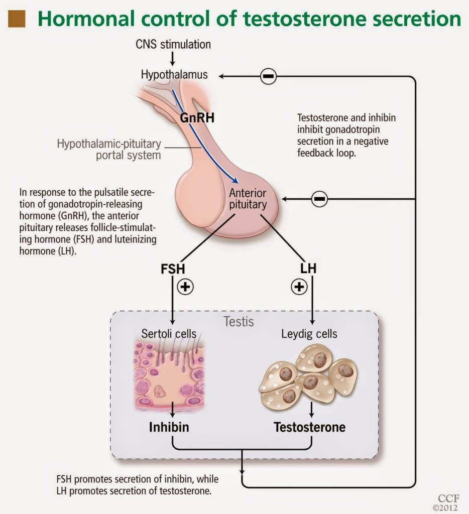
Excess Environmental Heat (sauna)

Deliberate heat exposure is associated with various health benefits, including indirect mechanisms through which sauna sessions may influence hormonal balance and overall quality of life. The proposed mechanisms by which sauna exerts positive health effects, potentially indirectly benefiting testosterone, includes the following:
  1. Improved Circulation and Blood Flow: Improved blood flow may enhance nutrient and oxygen delivery to tissues, including the testes, potentially supporting optimal Leydig cell function responsible for testosterone production.
  2. Heat Stress and Hormetic Response: Hormetic stressors, including heat stress from sauna use, may stimulate the release of certain hormones, potentially influencing the endocrine system, including testosterone regulation.
  3. Stress Reduction and Cortisol Modulation: Chronic stress and elevated cortisol levels have been linked to disruptions in testosterone balance. Sauna-induced relaxation may help modulate cortisol levels and support hormonal balance.
  4. Detoxification and Elimination of Toxins: Some environmental toxins may interfere with hormone balance, and the elimination of these toxins through the skin may indirectly support hormonal health, including testosterone levels.
  5. Enhanced Recovery and Exercise Benefits: Regular exercise is associated with improved testosterone levels. Sauna acts as a non-impact cardio sessions, and especially when performed post-exercise may enhance recovery, potentially supporting the overall positive effects of exercise on hormonal health.
  6. Improved Sleep Quality: Sauna use, particularly in the evening, has been reported to promote relaxation and improve sleep quality. Quality sleep is crucial for the natural regulation of hormones, including testosterone. Improved sleep may indirectly contribute to hormonal balance.
  7. Cardiovascular Health Benefits: Sauna use has been associated with cardiovascular benefits, including improved endothelial function and reduced blood pressure. Cardiovascular health is linked to overall well-being, and maintaining a healthy cardiovascular system may positively influence hormonal balance.

Given the notable benefits of sauna, it has been demonstrated that prolonged exposure to excessive heat, such as in hot environments or the use of hot baths and saunas, has been associated with potential impairment of testosterone levels. Several mechanisms may contribute to the negative impact of heat on testosterone.

The testes are located outside the body in the scrotum, a sac of skin. This positioning is crucial for maintaining a lower temperature than the core body temperature, which is necessary for optimal sperm and testosterone production. Prolonged exposure to heat, especially if the testes are subjected to elevated temperatures, can disrupt the normal temperature regulation and impair the function of Leydig cells, which produce testosterone.

Elevated testicular temperatures resulting from prolonged exposure to heat have been linked to a decrease in sperm production (spermatogenesis). The same conditions that inhibit spermatogenesis may also impact Leydig cell function, leading to a reduction in testosterone synthesis.

GnRH stimulates the release of luteinizing hormone LH from the pituitary gland. LH, in turn, stimulates the Leydig cells to produce testosterone. Prolonged heat exposure has been associated with a decrease in GnRH and LH levels, potentially leading to reduced stimulation of testosterone production.

The body perceives excessive heat as a stressor, leading to the activation of the stress response, including the release of cortisol. Elevated cortisol levels can negatively impact the balance of sex hormones, potentially suppressing testosterone synthesis. While sauna has been observed to lower cortisol, perception and experience are important to note. In other words, if an individual does not have much experience with deliberate heat exposure and decides to enter extreme heat for long durations, their body likely cannot compensate to the stressor. Gradual progressions in intensity and durations for sauna use are recommended.

High temperatures can affect the quality of sperm, leading to decreased motility and fertility. The relationship between impaired sperm quality and testosterone levels suggests that the detrimental effects of heat may extend to Leydig cell function and testosterone synthesis.
​
It's important to note that the impact of heat on testosterone levels can vary among individuals, and the body's ability to regulate temperature may differ. Additionally, the body has mechanisms to cope with short-term variations in temperature. However, chronic or extreme heat exposure may pose risks to reproductive health.

Non-native EMFs

With the increasing use of mobile phones, laptops, and wireless technologies like Wi-Fi and 5G globally, there is growing concern about the non-native electromagnetic frequencies (EMFs) , also known as, electromagnetic radiation (EMR) these devices emit. EMFs are known to interact with the male reproductive system through thermal and nonthermal mechanisms, negatively affecting testicular functions essential for testosterone and sperm production.

A comprehensive review of studies from 2003 to 2020 highlighted several key findings:
  1. Sperm Quality: Both human and animal studies indicate that exposure to EMR from mobile phones leads to reduced sperm motility, structural anomalies, and increased oxidative stress due to overproduction of reactive oxygen species (ROS).
  2. Semen Analysis: Human semen samples exposed to mobile phone EMR showed significant reductions in motility and viability, and increased ROS levels. Laboratory-controlled exposure of semen samples to mobile phone radiation resulted in decreased sperm concentration and quality.
  3. Epidemiological Studies: Research involving large cohorts of men demonstrated a correlation between mobile phone usage and lower semen volume, sperm concentration, and total sperm count. Constant use of mobile internet services was particularly linked to poorer sperm quality.
  4. Survey Results: Surveys of men referred for semen analysis revealed that prolonged phone usage (over an hour per day) and usage while charging were associated with higher percentages of abnormal sperm concentrations.
  5. Experimental Findings: Studies on male rats exposed to mobile phone radiation showed slight decreases in serum testosterone levels and testicular weight. Other experiments demonstrated that EMR exposure led to genotoxic effects on spermatozoa and altered pituitary function, affecting the Leydig and Sertoli cells critical for male fertility.
  6. Laptop Exposure: Sperm samples exposed to Wi-Fi radiation from laptops for extended periods showed reduced motility and increased DNA fragmentation.
  7. Military Study: An increased rate of childlessness was observed among military men exposed to RF electromagnetic fields (EMF), further supporting the link between EMR exposure and male infertility.​

Here are some proposed mechanisms through which exposure to non-native EMFs, such as Wi-Fi, Bluetooth, cellphones, and sources of "dirty electricity" including electric-generated heaters, negatively influence testosterone levels:
  1. Scrotal Hyperthermia and Oxidative Stress: These were identified as primary mechanisms through which EMR affects male fertility. Long-term and frequent use of mobile phones exacerbates these effects.
  2. Increased Oxidative Stress: Exposure to EMFs has been associated with increased oxidative stress in some studies. Oxidative stress refers to an imbalance between free radicals and antioxidants in the body. Elevated oxidative stress may have the potential to disrupt the endocrine system, including the regulation of testosterone.
  3. Disruption of Melatonin Production: EMF exposure, especially from devices used at night like cellphones, may interfere with melatonin production. Melatonin is a hormone that regulates sleep-wake cycles. Disruption of melatonin levels can impact the circadian rhythm and potentially influence testosterone production, as testosterone follows a circadian pattern with higher levels during sleep.
  4. Alteration of Calcium Ion Movement: Researchers have observed that non-native EMFs increase the movement of calcium ions in cells. Calcium ions play a role in various cellular processes, including hormone production. Disruption of calcium ion movement influences signaling pathways involved in testosterone synthesis, among other harmful effects.
  5. Impact on Leydig Cells: As mentioned, Leydig cells in the testes are responsible for producing testosterone. Researchers have observed exposure to EMFs affects Leydig cell function, thereby altering testosterone synthesis.
  6. Heat Generation: Certain devices emitting EMFs, especially those with high power, may generate heat. Prolonged exposure to localized heat, particularly in the groin area where the testes are located, could potentially impact sperm quality and testosterone production
​Individual responses to EMFs can vary, and the potential effects may depend on factors such as the duration and intensity of exposure, individual health status, and the specific frequencies involved. 

Environmental Toxins

Environmental toxins can interfere with testosterone levels through various mechanisms, disrupting the normal functioning of the endocrine system. Here are several ways in which environmental toxins may lower testosterone levels:
  1. Endocrine Disruption: Many environmental toxins are classified as endocrine-disrupting chemicals (EDCs). These substances can mimic or interfere with the actions of hormones, including testosterone. EDCs may bind to hormone receptors, blocking or activating them inappropriately. This interference can lead to imbalances in hormonal signaling, including the regulation of testosterone production.
  2. Aromatase Activity and Estrogen Dominance
  3. Disruption of Leydig Cell Function
  4. Inhibition of Gonadotropins: Some environmental toxins can interfere with the secretion of gonadotropins, such as LH and FSH, which regulate testosterone production. Inhibition of gonadotropin release may lead to diminished stimulation of Leydig cells and, consequently, lower testosterone levels.
  5. Testicular Toxicity: Certain environmental toxins may exhibit testicular toxicity, causing damage to the testes and impairing their function. Testicular damage can impact Leydig cell activity and overall testosterone synthesis.
  6. Oxidative Stress: Some environmental toxins can induce oxidative stress, resulting in an imbalance between free radicals and antioxidants in the body. Oxidative stress has been associated with testicular damage and impaired testosterone production.
  7. Impaired Sperm Quality: Environmental toxins may affect sperm quality, including motility and morphology. Sperm abnormalities can be indicative of disruptions in the testicular microenvironment, potentially influencing testosterone levels.
  8. Epigenetic Changes: Exposure to environmental toxins may lead to epigenetic changes, alterations in gene expression without changes to the underlying DNA sequence. Epigenetic modifications in genes related to testosterone synthesis and regulation can impact hormonal balance.

Below is a list of known compounds, chemicals, and environmental toxins that reduce testosterone levels, as supported by scientific literature:
Anti-Androgens
​Atorvastatin
Bisphenol A
​Ethinyl Estradiol (plus Lynestrenol)
Glyphosate (Roundup
​Ibuprofen
Levonorgestrel
​/ ethinyl estradiol
​Lovastatin
Organophosphate pesticides
Parabens
Pesticides
Phthalates
Simvastatin
Statin Drugs
Sugar Sweetened Beverages
Titanium Dioxide
Titanium Nanoparticles
Vitamin A Palmitate
 
 
Reducing exposure to environmental toxins by choosing products with fewer harmful chemicals, adopting a healthy lifestyle, and avoiding environmental pollution can contribute to overall well-being and support hormonal health.

Solutions to andropause

Improving testosterone levels through lifestyle interventions involves adopting positive habits that contribute to overall health and hormonal balance. Several lifestyle interventions have been observed to enhance testosterone levels, including changes in nutrition, exercise, cold water immersion, and avoiding harmful environmental stressors.

It's important to note that individual responses to lifestyle interventions can vary, and results may take time.
Picture

Nutritional Strategies

​In an effort to enhance testosterone levels it is crucial to consume a well-balanced diet rich in essential micronutrients (vitamins and minerals) and antioxidants, and void of harmful toxins, such as pesticides and plastics, to the best of one's ability. Adequate nutrition is crucial for overall health and hormone production. Include sources of healthy fats in your diet, such as omega-3 fatty acids found in fish, flaxseeds, and walnuts. Healthy fats, including those sourced from animals, such as pasture-raised beef, are important for hormone synthesis, including testosterone.

Here is an evidence-based list of foods, compounds, and substances known to enhance testosterone levels:
​Astragalus
Astaxanthin
Bitter Melon
Biochanin A
Caffeine
Calcium*
Coconut (+ Oil)
Coleus Forskohlii
Curcumin
Daidzein
Dogwood
Fermented Foods and Beverages
Fenugreek
Formononetin
Genistein
Ginseng (Korean)
Ginsenosides
Ginkgo biloba
Isoflavones
Linoleic acid^
​(Conjugated) ​
Maca
 Magnesium 
Molybdenum
Mulberry
 NAC (N-acetyl-L-cysteine) 
Olive
Onion
Pantothenic Acid (Vitamin B-5)
Phosphatidylserine
Phytoestrogens (+/-)
Raspberry​
Resveratrol
Saffron
Saw Palmetto
Selenium
Shilajit
Squalene
Suma (Pfaffia Paniculata)
Taro
Tauroursodeoxycholic acid
Tongkat Ali
Tribulus
Vitamin A (Retinol)
Vitamin E
Zinc
Enhancing testosterone levels involves a multifaceted approach that goes beyond simply incorporating specific compounds into one's routine. While various compounds, herbs, and nutrients are touted for their potential to support testosterone production, it's crucial to approach supplementation with caution and a well-informed mindset.

Before embarking on a supplement regimen, prioritize the quality of ingredients. Opt for reputable brands that use high-quality, pure ingredients. The effectiveness and safety of a supplement are inherently linked to the quality of the components it contains.

Understanding the appropriate dosage for each compound is paramount. Dosage recommendations can vary based on factors such as age, health status, and individual response. Always follow recommended dosages and, if uncertain, consult with a healthcare professional for personalized advice.

Enhancing testosterone is not just about isolated compounds; it's a holistic journey. Lifestyle factors, including nutrition, exercise, sleep, and stress management, play pivotal roles in hormonal balance. Consider adopting a well-rounded approach that encompasses these lifestyle elements.

Before introducing additional compounds, it's wise to address and minimize harmful stressors in your life. Chronic stress, inadequate sleep, and poor dietary choices can negatively impact hormone levels. Individuals may find more significant benefits by first focusing on stress reduction and overall well-being.

It's essential to recognize that responses to supplements can vary widely among individuals. What works for one person may not yield the same results for another. Pay attention to how your body responds, and be patient; changes may take time.

*^The hormonal system is complex and nuanced. Just because researchers have observed the calcium and linoleic acid (LA) can increase testosterone, more is not better. Excess LA (an essential fatty acid - the body cannot make it, and must consume it in the diet) is well established to disrupt metabolic function, which can thereby lead to lower testosterone. Innumerable amounts of food contain LA, therefore to call it "essential" can be deceiving. It is important to note that both calcium and LA should be consumed in whole food sources. 

In conclusion, while compounds like zinc, magnesium, and various herbs have been associated with potential testosterone support, a thoughtful and informed approach is crucial. Prioritize high-quality ingredients, determine suitable dosages, and consider the broader lifestyle factors influencing hormonal health. Taking proactive steps to reduce harmful stressors can set a solid foundation for any testosterone-enhancing efforts. Before making significant changes to your supplement routine, it's advisable to consult with healthcare professionals for personalized guidance tailored to your unique needs. Remember, optimizing testosterone is a holistic endeavor that encompasses both supplementation and a balanced, healthy lifestyle.
Picture

Aromatase inhibitors

Aromatase inhibitors are compounds that have the function to block or inhibit the activity of the aromatase enzyme. They are commonly used in the treatment of conditions where reducing estrogen levels is beneficial, such as in certain types of breast cancer. In the context of testosterone replacement therapy (TRT) or hormonal imbalances in men, aromatase inhibitors may be prescribed to prevent excessive conversion of testosterone to estrogen.

Aromatase inhibitors are often utilized in medical scenarios where reducing estrogen levels is crucial, such as in the treatment of hormone-sensitive breast cancer in postmenopausal women. In men undergoing TRT, aromatase inhibitors may be used to manage or prevent symptoms of estrogen dominance that can occur with exogenous testosterone administration. Aromatase inhibitors should be used judiciously, as completely suppressing estrogen levels in men can have adverse effects on bone health, libido, and overall well-being. Additionally, as paradoxical as it might sound,  some aromatase inhibitors are actually estrogenic (e.g. anastrozole). The use of aromatase inhibitors should be tailored to individual needs, and regular monitoring of hormone levels is essential to ensure a balanced hormonal profile.

There are many natural aromatase inhibitors including progesterone, Maca, Grape seed extract, Nettle, Saw Palmetto, and more. ​The following foods contain compounds that have been shown to inhibit aromatase activity, thereby suppressing estrogen biosynthesis:

In summary, aromatization is a natural and complex process with essential physiological functions. However, imbalances in aromatase activity can have implications for hormonal health. Aromatase inhibitors, when used under medical supervision, can help manage hormonal imbalances and associated symptoms. It's crucial to approach hormone management with a comprehensive understanding of individual health needs and regular monitoring.
 ​  
Artichokes
Arugula
Black Tea
Blueberries
Broccoli
Brussel Sprouts
Cabbage
Cauliflower
Celery
Cherries
Chives
Cilantro
Collard Greens
Corn
Cranberries
Ligonberries
Currants
Bilberries
Grapes
Green Onions
Green Tea
Honey (Raw)
Horseradish
Peppers
Lemons & Limes
Mexican Oregano
Mushrooms
Mustard
Mustard Greens
Oats
Oranges
Parsley
Passion Fruit
Pomegranates
Radishes
Saffron
Turnips
Turnip Greens
Walnuts
Watercress

Exercise

Regular exercise stands as a formidable influence on testosterone levels, a key factor in maintaining overall well-being. Achieving hormonal balance requires a thoughtful blend of cardiovascular and resistance training exercises. Let's explore how different facets of exercise harmonize to foster optimal testosterone levels:

1. Resistance Training: Regular engagement in resistance or strength training exercises holds a pivotal role in sustaining hormonal equilibrium. Compound movements like squats, deadlifts, and weightlifting emerge as catalysts for increased testosterone production. By activating large muscle groups, such as incorporating lower-body exercises like squats and lunges alongside upper-body workouts, trigger a substantial hormonal response, elevating testosterone levels. 

Heavy lifting, especially with full-body exercises like squats, deadlifts, and bench presses, is crucial for boosting testosterone. Use weights at 85-95% of your one-rep max (1RM) and aim for 2-3 full-body workouts per week. Beginners can start with weight machines before transitioning to free weights.

Longer rest periods (around 120 seconds) between sets are better for testosterone production. To make the most of your time, alternate between exercises that don't stress the same muscles. For example, pair bench presses with squats, taking shorter breaks between each.

Forced reps involve performing as many reps as possible, then having a partner assist with a few additional reps. This method has been shown to increase testosterone more effectively than solo reps. Incorporate forced reps into the last set of your exercises.

2. High-Intensity Interval Training (HIIT): HIIT workouts introduce brief, intense bursts of exercise followed by rest or lower-intensity periods. Integrating HIIT into your routine exhibits positive effects on testosterone levels. The dynamic nature of HIIT prompts the body's adaptive response, nurturing hormonal balance. Studies show that short, intense sprints can significantly boost testosterone levels. For optimal results, perform 5-10 sprints lasting no more than 15-30 seconds each, with full recovery between sprints (typically 3-4 times the sprint duration). Aim to do sprint workouts 2-3 times a week.

3. Cardiovascular Exercise: While resistance training takes center stage, cardiovascular exercise contributes significantly to overall health. Moderate-intensity cardio activities like jogging, cycling, jumping rope, rebounding, or sauna enhance cardiovascular well-being, complementing the body's holistic fitness.

4. Avoid Overtraining: Guarding against overtraining, characterized by excessive exercise without adequate recovery, is crucial for hormonal health. Prolonged intense workouts may elevate cortisol levels, a stress hormone with adverse effects on testosterone production. Adequate recovery time is essential to prevent the pitfalls of overtraining. Checking biometrics such as HRV is a great evidence-based indicator to quantify stress in the system.

A sample full-body workout three times a week might include:
  • Warm-up
  • 4 sets of 8 reps of bench press and squats
  • 4 sets of 8 reps of deadlifts and pull-ups
  • 6 sets of 10-second sprints
  • Cool-down

A well-rounded exercise routine embraces resistance training (consisting of novel exercises to address specific musculoskeletal, biomechanical imbalances, and breathing mechanics), HIIT, and moderate-intensity cardio. Each type of exercise brings unique contributions to hormonal balance, offering comprehensive benefits for overall health. The body intricately adapts hormonally to the demands imposed during exercise. Thoughtfully designed, regular exercise routines can instigate positive hormonal adaptations, fostering improved testosterone regulation. 

Acknowledging the diversity of individual responses to exercise is paramount. Tailoring routines to personal preferences and fitness levels ensures sustainable and enjoyable exercise habits, promoting long-term commitment. Consistency emerges as the linchpin for reaping long-term hormonal benefits from exercise. Establishing a regular routine that integrates various exercise types contributes not only to hormonal well-being but also to overall health.

In conclusion, the synergy of cardiovascular and resistance training exercises presents a potent strategy for optimizing testosterone levels. Resistance training, with a focus on compound movements and weightlifting, sparks testosterone production, while the inclusion of HIIT and cardiovascular exercise contributes holistically to health. Vigilance against overtraining and allowing sufficient recovery time are pivotal in maintaining hormonal balance. By adopting a balanced and personalized exercise routine, individuals can actively support hormonal health, enhancing their overall well-being. 
Picture

Stress Management

Chronic stress can have profound effects on hormonal health, including the disruption of cortisol and testosterone levels. Implementing stress management techniques, from meditation, yoga,  mindfulness, even listening to music, dancing, walking in nature, breathwork, and laughter - anything that helps you relax - plays a crucial role in mitigating the impact of chronic stress and fostering hormonal balance, particularly with respect to testosterone levels. These practices activate the body's relaxation response, reducing cortisol levels, thereby optimizing hormonal balance. 

Techniques that induce relaxation activate the parasympathetic nervous system (PNS), which counters the fight-or-flight response associated with chronic stress. By calming the SNS, they help restore hormonal balance, positively impacting testosterone levels. This increase in the PNS can directly improve sleep quality. Quality sleep is essential for optimal hormonal function, including testosterone production during the night.

As with many of the solutions addressed, there is trend in the underlying mechanisms that result in improved hormonal balance, including reduced inflammation, improved mood and mental health, which fosters a mind-body connection that allows individuals to better understand and manage stress triggers. By increasing self-awareness, individuals can make conscious choices that positively impact their hormonal responses, thereby facilitating changes in thought patterns and behaviors related to stress. A more adaptive response to stressors can reduce the physiological impact on hormones, including testosterone.

Stress management practices positively influence the communication between the brain and endocrine glands. Improved hormonal communication supports optimal functioning of the hypothalamus, pituitary gland, and testes, essential for testosterone regulation.

As with most practices that induce positive health effects, regular practice offers cumulative benefits over time. Consistency in these techniques contributes to sustained stress resilience and supports ongoing hormonal health. Anyone interested in optimizing hormone levels is encouraged to adopt a holistic perspective of well-being by addressing physical, mental, and emotional aspects of health. A balanced and integrated approach to well-being positively influences hormonal health, including testosterone levels.

As mentioned, incorporating meditation, yoga, or mindfulness, music, dancing, humor and laughter - whatever helps you relax - into one's routine can be a powerful strategy for managing chronic stress and supporting hormonal health. It's essential to choose techniques that resonate with individual preferences and consistently practice them to reap the long-term benefits.
Picture

Cold Water Immersion

Cold water immersion, often referred to as cold water therapy or cold exposure, such as cold showers or baths, has been studied for its potential impact on testosterone levels in men. ​Cold exposure may stimulate the production of certain hormones and support overall endocrine function, via various mechanisms of action. 

Cold exposure activates the hypothalamus, a region of the brain that plays a central role in the regulation of hormonal balance. The hypothalamus controls the release of gonadotropin-releasing hormone (GnRH), which, in turn, stimulates the pituitary gland to release luteinizing hormone (LH). LH acts on the testes, promoting the synthesis and release of testosterone.

From a vascular perspective, cold water immersion may lead to vasoconstriction (narrowing of blood vessels) followed by vasodilation (widening of blood vessels) in response to rewarming. This cycle of vasoconstriction and vasodilation can enhance blood flow, potentially increasing perfusion to the testes. Improved blood flow to the testes may support optimal Leydig cell function, which is crucial for testosterone production.

Metabolically, exposure to cold activates brown adipose tissue (BAT), a type of fat tissue that generates heat. BAT activation is associated with increased energy expenditure and metabolic activity. Some studies suggest that BAT activation may positively influence hormonal regulation, including testosterone production.

Cold water immersion has also been demonstrated to have anti-inflammatory effects. Chronic inflammation is associated with disruptions in not only energy production, but hormonal balance as well, including reduced testosterone levels. By reducing inflammation, cold water immersion may support a more favorable hormonal environment.

Additionally, some individuals report improved sleep quality following cold water immersion, likely due to enhanced thermoregulation. Quality sleep is crucial for overall health, including hormonal regulation. Improved sleep may indirectly contribute to optimal testosterone levels.

Cold exposure is considered a form of hormetic stress, stimulating cold shock proteins a mild stressor that, when applied in moderation, may lead to adaptive responses. Hormetic stressors, such as cold exposure, have been proposed to stimulate the body's adaptive mechanisms, including the endocrine system, potentially leading to increased testosterone production.
learn more about hormesis
An important variable to consider with any hormetic stressor is the biphasic response - some is good, too much can be harmful. While there is evidence supporting the potential positive effects of cold water immersion on testosterone, more research is needed to fully understand the mechanisms and establish clear guidelines. Additionally, individual responses to cold exposure can vary, and caution should be exercised, especially for individuals with existing health conditions.

From a neurobiological perspective, cold water immersion has been associated with an increase in neurotransmitters, such as dopamine and norepinephrine levels, which can beneficially impact mood and behavior. However, what is not often described by proponents of cold water immersion​ is that exposure to extreme cold (determined by the individual's physiology, experience and perception) can trigger a cascade of physiological responses leading to elevated adrenaline (epinephrine) levels, thereby causing excess stress, and moving the needle in the opposite direction, away from optimal hormonal balance. Cold water immersion can certainly activate the sympathetic nervous system, otherwise known as the body's "fight or flight" response, which is why it is best to perform cold water immersion sometime in the morning or mid-day.

Dopamine and norepinephrine are neurotransmitters that play key roles in the regulation of mood, attention, and arousal. Cold water immersion has been shown to stimulate the release of both dopamine and norepinephrine in response to the stress of cold exposure. Dopamine has been suggested to have a regulatory role in the release of adrenaline. Studies indicate that dopamine, acting through specific receptors, may influence the release of adrenaline from the adrenal medulla. The overall response to cold stress involves the activation of the hypothalamus-pituitary-adrenal (HPA) axis, leading to the release of stress hormones, including cortisol and adrenaline.

It is important to realize that just because some is good, more is not always better. Deliberate cold exposure can absolutely improve quality of life, via hormonal mechanisms. However, if performed improperly, cold water immersion can disrupt hormonal effects. It is ideal to start low and slow. In other words, use a low dose and progressively increase as tolerance improves. As always, listen to your body. Shivering is a sign that the body is too cold. 

Picture

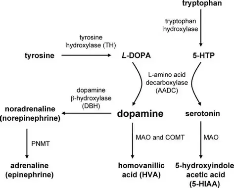
Regulating Ejaculation Frequency

According to Taoist philosophy, regulating ejaculation frequency and retaining semen can lead to increased strength, mental clarity, and sustained high levels of testosterone, sperm, and semen. This practice is believed to enhance sexual appetite by allowing the body to rebuild sexual energy between ejaculations.

Recommended Ejaculation Frequency by Age:
  • 20s: As desired
  • 30s: 3-4 times per week
  • 40s: 2-3 times per week
  • 50s: 1-2 times per week
  • 60+: Once a week, depending on health

A popular method among Tao practitioners is to ejaculate only two or three times out of every ten sexual encounters. Sun Simiao, a notable Tao theorist, advises men over 50 to ejaculate no more than once every 20 days, and men over 60 no more than once every 100 days.
​
To practice "injaculation" rather than ejaculation, men can squeeze the muscles used to stop urine flow while "breathing the energy" up the spine. If ejaculation seems imminent, applying pressure to the perineum (the area between the scrotum and anus) can help delay it. Elaboration of this technique can be found in the book, The Multiorgasmic Man. 
​
This technique is cost-free but may require some patience to avoid becoming moody or aggressive due to sexual frustration. On the other hand, researchers at Boston University School of Public Health found that ejaculating at least 21 times a month may reduce the risk of prostate cancer. 

Other Lifestyle Strategies

​If you have made it this far in the article, you may have gathered a trend by now: the things that optimize hormones are the things that help establish balance in overall health. Eliminate the bad, and integrate the good. Here are some other helpful strategies that would beneficial to incorporate:
  1. Adequate Sleep: Ensure sufficient and quality sleep. Sleep plays a crucial role in hormonal regulation, including testosterone production. Aim for 7-9 hours of sleep per night. This also include mitigating non-native EMF exposure via blue lights in the environment. Blue light exposure is well documented to disrupt circadian rhythms, thereby impairing recovery mechanisms, and disrupting the delicate balance of hormones. 
  2. Maintain a Healthy Weight: Achieve and maintain a healthy weight. Obesity is associated with lower testosterone levels, and losing excess weight can positively impact hormonal balance. A general rule of thumb is a healthy BMI, although for individuals who have large amounts of muscle, the reliability of BMI falls short. 
  3. Vitamin D: Ensure adequate vitamin D levels. Vitamin D deficiency has been linked to lower testosterone levels, among many other comorbidities and increased all-cause mortality. It is ideal to spend time in sunlight at solar noon, and avoid vitamin D supplements. Our ancient ancestors spent nearly all of their time outside, and did not have access to synthetic products.
  4. Limit Alcohol Consumption: Moderate alcohol intake, as excessive alcohol consumption has been associated with lower testosterone levels. To date, there are zero benefits of alcohol consumption, perhaps (although loosely) with exception to social connection - of course, there are limits to the risk:benefit ratio with respect to the amount of alcohol consumed. Alcohol is a known toxin directly connected to a variety of comorbidities.

Navigating andropause involves a multifaceted approach that addresses both hormonal changes and lifestyle factors. By incorporating these science-backed nutrition and lifestyle strategies, men can optimize their well-being during this natural phase of life. Always consult with a healthcare professional for personalized advice based on individual health needs.

Optimizing Your Testosterone: A Day in the Life

Morning:
Start your day with getting sun into your eyes. Afterwards, consume a breakfast rich in healthy fats like avocado, eggs, grass-fed butter and cheese, oyster mushrooms, sauerkraut, and coconut milk. Along with breakfast, take the following supplements:
  • 5g creatine
  • 50mg supplemental DHEA
  • 10mg boron
  • 30g cocoa powder
  • 2g maca root extract
  • 250 mg shilajit
For added benefits, consider these optional supplements:
  • 500mg fenugreek extract
  • 200mg Pycnogenol
  • 200-300mg Eurycoma longifolia
  • 300mg Tribulus

Throughout the Day:
Be sure to get sun on your skin at solar noon in an effort to create vitamin D. Implement EMF mitigation strategies by keeping your phone in airplane mode when not in use, avoiding using your laptop on your lap (or using an anti-radiation devices, such as Aires Tech), and turning off Wi-Fi on devices when using ethernet. Detox your home and consider auditing your workspace for EMF with an acoustimeter or by hiring a building biologist. Manage stress to maintain a high testosterone ratio. Practice relaxation techniques like deep nasal and belly breathing, laugh, smile, get outdoors, and be mindful of stress mitigation strategies. Spend time with people, especially women, as their presence can boost testosterone levels. Avoid pornography as it can negatively impact hormonal balance.

Afternoon Workout:
For an effective testosterone-boosting workout, do the following exercises with heavy weights, ensuring good form. Perform 5 sets of 5 reps each:
  • Bench Press
  • Deadlift
  • Front Squat
  • Shoulder Press
  • Clean
During the 90-second to two-minute rest periods between sets, do light mobility or core exercises like opposite-arm and opposite-leg extensions, planks with shoulder taps, side lunges, or jump rope. Repeat this workout twice a week, increasing the weight with each set, and use a partner for assistance on the final set if needed.

Evening:
Enhance carbon dioxide levels to augment the efficiency of oxygen transport and energy production.
Learn more about Carbon dioxide
Increase nitric oxide levels by including foods like arugula, spinach, beets, carrots, red onions, walnuts, pumpkin seeds, extra virgin olive oil, pomegranate, cubed watermelon, dark chocolate, and red wine with your dinner. Set aside time a couple of times a week for sex, making it meaningful by practicing techniques from "The Multi-Orgasmic Man" such as reverse orgasm, tantric practices, or reducing ejaculation frequency. Before bed, take 400–500mg of magnesium.

Integral Wellness Program: All in one Approach

For individuals on a quest to elevate their testosterone levels through a hands-on, step-by-step approach, the Integral Wellness Program offers a comprehensive guide. This program, accessible at Mindful Wellness, integrates a holistic perspective on well-being (Movement, Nutrition and Lifestyle), aiming to address various facets of life to enhance hormonal health. Here are some of the key elements:
1. Evidence-Based & Holistic Approach:
  • Rooted in evidence-based practices, the Integral Wellness Program prioritizes approaches backed by scientific research.
  • This credible and reliable program adopts a holistic model, recognizing the interconnectedness of physical, mental, and emotional well-being.
  • By addressing multiple dimensions of health, it aims to create a synergistic effect, optimizing the conditions for hormonal balance.
2. Step-by-Step Protocols & Customized Guidance:
  • The program unfolds in a step-by-step manner, offering clear protocols for implementation.
  • This structured approach simplifies the journey, making it accessible for individuals seeking a systematic and manageable process.
  • Recognizing that each individual is unique, the program provides personalized guidance tailored to specific needs and goals.
  • Customization ensures that the approach resonates with individual preferences and aligns with their health objectives.
3. Movement, Nutritional Lifestyle Optimization:
  • Integral to the program are nutritional strategies designed to support hormone optimization.
  • These strategies likely include guidance on nutrient-dense foods, dietary patterns, and specific nutrients beneficial for hormonal health.
  • Beyond nutrition, the program delves into lifestyle optimization.
  • Factors such as sleep, stress management, and physical activity are likely addressed, recognizing their significant impact on hormonal balance.
  • This holistic approach acknowledges the influence of mental well-being on hormonal health.
4. Educational Resources:
  • The program likely provides educational resources, empowering individuals with knowledge about testosterone, hormonal health, and the impact of lifestyle choices.
  • Informed decisions are pivotal to sustained well-being.
5. Accessible Platform:
  • The program is available through an accessible online platform, enabling participants to engage at their own pace and convenience.
  • Flexibility in participation facilitates seamless integration into daily life.

In essence, the Integral Wellness Program serves as a comprehensive guide for those embarking on a journey to optimize testosterone levels. Through its holistic and step-by-step approach, individuals can navigate the intricacies of well-being, unlocking the potential for sustained hormonal health.

Movement

nutrition

lifestyle

Learn More about the integral wellness program

References


Read More
0 Comments
<<Previous
    The Awareness domain contains research, news, information, observations, and ideas at the level of self in an effort to intellectualize health concepts.
    Mind
    Body
    Spirit
    The Lifestyle domain builds off intellectual concepts and offers practical applications.
    Lifestyle
    Taking care of yourself is at the core of the other domains because the others depend on your health and wellness.
    Community
    World
    Cosmos

    Archives

    March 2025
    December 2024
    October 2024
    September 2024
    August 2024
    July 2024
    May 2024
    April 2024
    March 2024
    February 2024
    January 2024
    December 2023
    September 2023
    August 2023
    June 2023
    November 2022
    October 2022
    July 2022
    February 2022
    January 2022
    December 2021
    November 2021
    October 2021
    August 2021
    July 2021
    June 2021
    March 2021
    February 2021
    January 2021
    December 2020
    November 2020
    September 2020
    August 2020
    March 2020
    December 2019
    November 2019
    October 2019
    September 2019
    August 2019
    July 2019
    May 2019
    April 2019
    March 2019
    February 2019
    January 2019
    December 2018
    November 2018
    October 2018
    September 2018
    August 2018
    July 2018
    June 2018
    April 2018
    March 2018
    February 2018
    January 2018
    December 2017
    November 2017
    October 2017
    September 2017
    August 2017
    July 2017
    June 2017
    May 2017
    April 2017
    March 2017
    February 2017
    January 2017

    Categories

    All
    5G
    Adductors
    Anxiety
    Autism
    Ayurveda
    Big Pharma
    Body
    Breathing
    Cancer
    Cannabis
    Carbohydrates
    Cardiovascular Disease
    Cell Phones
    Children
    Chronic Disease
    Cognition
    Consciousness
    Coronavirus
    Covid
    Cryotherapy
    Depression
    Deuterium
    Diet
    Dietary Guidelines
    EMFs
    Emotions
    Endocrine Disruptors
    Environment
    Exercise
    Farming
    Fasting
    Fats
    Fluoride
    Food
    Food Like Product
    Food-like Product
    Forest
    Gardening
    Genetics
    Glyphosate
    GMOs
    Hamstrings
    Healing
    Health
    Herbalism
    Hormones
    HRV
    Immunity
    Infertility
    Laughter
    Light
    Lockdowns
    Loneliness
    Longevity
    Masks
    Meditation
    Metabolism
    Microbiome
    Microwave Radiation
    Mind
    Mortality
    Musculoskeletal System
    Nature
    Neuroplasticity
    Nutrition
    Obesity
    Omega 3
    Omega-3
    Organic
    Pelvis/Thigh
    Pesticides
    Physical Therapy
    Placebo
    Plastic
    Pollution
    Positive
    Pregnancy
    Prevention
    Processed Foods
    Protein
    Psi
    Quadriceps
    Research
    Retirement
    Salt
    Sleep
    Spine/Thorax
    Spirit
    Stress
    Sugar
    Technology
    Touch Screens
    Toxicity
    Vaccines
    Vegetable Oils
    Well Being
    Well-Being

    RSS Feed

Picture
Picture
Picture
Picture
Picture
Picture
Picture
  • Home
    • Start Here >
      • Look Around
      • Find Your Why >
        • Affirm Your Values
      • Identify Imbalances >
        • Shift Your Paradigm
  • Learn
    • Pages >
      • Inner World >
        • Awareness >
          • The Human >
            • Awareness Systems >
              • Mind >
                • Psychology >
                  • Biological >
                    • Sensation & Perception >
                      • Stress >
                        • Hormesis
                      • Visualization
                  • Behavioral >
                    • Emotions
                  • Cognitive >
                    • Conscious Mind
                    • Subconscious Mind
                  • Social
                  • Personality >
                    • Ego >
                      • Cognitive Dissonance >
                        • Anxiety
              • Body >
                • Anatomy >
                  • Organ Systems >
                    • Endocrine System >
                      • Pineal Gland
                    • Fascia
                • Physiology >
                  • Bioenergetics >
                    • Mitochondria
                  • Hormones >
                    • Testosterone
                    • Thyroid
                    • Pregnenalone
                    • Progesterone
                  • Bone Biomechanics
                  • Inflammation
              • Spirit >
                • Altered States >
                  • Entheogens
                • Intention >
                  • Creating Reality
                • Parapsychology
            • Core Pillars >
              • Happiness
              • Nutrition >
                • Nutrients >
                  • Macronutrients >
                    • Carbohydrates >
                      • Inositol
                    • Fat >
                      • Linoleic Acid
                    • Protein >
                      • Amino Acids >
                        • L-Theanine
                      • Collagen
                    • Water
                  • Micronutrients >
                    • Vitamins >
                      • Vitamin A
                      • Vitamin C
                      • B Vitamins
                      • Vitamin D
                    • Minerals >
                      • Copper
                      • Electrolytes
                      • Magnesium
                      • Boron
                  • Antioxidants >
                    • Glutathione
                  • Flavonoids >
                    • Greens
                    • Diosmin / Hesperidin
                  • Antinutrients >
                    • Histamine
                    • Mycotoxins
                  • Phytonutrients
                  • Therapeutics >
                    • Shilajit
                    • GABA
                    • Probiotics
                    • Enzymes
                    • Hydrochloric Acid
                    • Methylene Blue
                    • Adrenal Cocktail
                    • Diatomaceous Earth
                    • Mushrooms
                  • Energy Metabolism >
                    • Alcohol
                    • Caffeine
                    • Sugar-Sweetened Beverages
                  • Nutrient Timing >
                    • Intermittent Fasting >
                      • Calorie Restriction
                • Steps to an Optimal Diet >
                  • Optimal Diet Foundation >
                    • Digestion
                  • Create Your Own Food >
                    • Impact of Supplements >
                      • BCAAs
                      • Creatine Monohydrate
                      • Diatomaceous Earth
                      • Omega-3 Fatty Acids
                      • Probiotics
                      • Spirulina
                  • Whole Foods >
                    • Alkaline vs. Acidic >
                      • Acidic Foods
                      • Alkaline foods
                  • Plant-Based >
                    • Vegetarianism
                  • Animal Products >
                    • Dairy
                  • Organic
                  • Local & Seasonal
                  • Biochemical Individuality
                • Sample Meal Plans
                • Obesity >
                  • Cholesterol
                  • Weight Management
              • Fitness >
                • Active Warmup
                • Concurrent Training >
                  • Aerobic Exercise >
                    • HIIT
                  • Resistance Training >
                    • Push
                    • Pull
                    • Legs
                    • Abdominals
                    • Accessory Muscles
                  • Sports-Specific Training
                • Stretching
                • Exercise Adherence
              • Silence >
                • Sleep
          • Remedies & Therapies >
            • Ayurveda >
              • Detoxification >
                • Methylation
                • Basti
              • Doshas
              • Chakras
            • Functional Medicine >
              • FODMAP
              • Testing
              • Parasites
            • Physical Therapy >
              • Lumbar Spine
            • Contrast Therapy >
              • Deliberate Heat Exposure
              • Deliberate Cold Exposure
            • Herbalism >
              • Milk Thistle
              • Cannabis
              • Ginkgo biloba
              • Ginseng
            • Subtle Energy >
              • Earthing
          • Risk Factors >
            • Chemicals
            • Cigarettes
        • Lifestyle Applications >
          • Conscious Creations >
            • The Basics >
              • How to Shop
              • Kitchen Essentials
            • Food >
              • Sprouting
              • Vegetables >
                • Cruciferous Vegetables >
                  • Maca
              • Fiber
              • Fruits
              • Whole Grains >
                • Gluten
            • Recipes >
              • Appetizers >
                • Appetizers
              • Breakfast
              • Entrées >
                • Entrées
              • Sweets
              • Beverages >
                • Beverages
              • Dressings
          • Mindful Movement >
            • Breathing
            • Mobilization
            • Stretches
            • Stabilization
            • Exercises
            • Program Design
          • Personal Growth >
            • Communication
            • Effectiveness >
              • Goal Setting
            • Meditation >
              • Mindfulness
            • Motivation
            • Human Relations
            • Simple Living >
              • Self-sufficiency
              • Minimalism >
                • Quality Over Quantity
              • Basic Needs
      • Outer World >
        • Relationship Concepts
        • Community Concepts >
          • Development >
            • Decentralization
            • Infrastructure
            • Sustainability >
              • Permaculture
          • Values >
            • Do No Harm
            • Cooperation
            • Sense of Community
            • Service
          • Culture
        • World concepts >
          • Geopolitics >
            • Agriculture
            • Government >
              • Politics >
                • Systems >
                  • Agorism
                  • Democracy >
                    • Voting
              • Military Industrial Complex >
                • Declassified Projects >
                  • Bluebeam
                  • Highjump
                  • Manhattan
                  • MKUltra
                  • Mockingbird
                  • Northwoods
                  • Paperclip
                  • Popeye
                  • Stormfury
              • Healthcare >
                • Pharmaceuticals
                • Vaccines
                • Water Fluoridation
            • Infrastructure >
              • Energy >
                • Hydrocarbons
            • Economy >
              • Banks >
                • Federal Reserve >
                  • Money
              • Corporatocracy >
                • Alphabet
                • Facebook
          • Technology >
            • Biotechnology >
              • Genetic Engineering
            • EMFs >
              • Cell Phones
              • Microwaves
              • Smart Meters
            • Geoengineering
            • Internet >
              • Smart Grid >
                • Artificial Intelligence
                • Biometrics
              • Social Media
            • Zero-Point
          • Media >
            • Context >
              • Mainstream Media
            • Advertising >
              • Television
            • Propaganda >
              • Education >
                • Conditioning
                • History of Humanity
              • Psychological Operations >
                • False Flag Attacks >
                  • 9/11
              • Symbols >
                • Language
        • Cosmological Concepts >
          • Philosophy >
            • Hermetic Philosophy >
              • Mentalism >
                • Law of One
              • Correspondence
              • Vibration
              • Polarity
              • Gender
              • Rhythm
              • Cause & Effect
            • Metaphysics >
              • Cosmology
              • Ontology >
                • Hidden Messages >
                  • Numeric >
                    • Numerology
                  • Visual >
                    • Sacred Geometry >
                      • Fibonacci Sequence
            • Logic
            • Knowledge
          • Science >
            • Life >
              • Biology
            • Physical >
              • Physics >
                • Astronomy >
                  • Contents >
                    • Solar System >
                      • Earth
                      • Sun
                • General >
                  • Entropy
                  • Time
                  • Frequencies >
                    • Dimensions
                • Theorectical >
                  • Quantum Mechanics
                  • Multiverse
                  • Holographic Universe
          • Phenomena >
            • Extraterrestrials
            • Interplanetary Climate Change
    • Domains >
      • I >
        • Awareness
        • Lifestyle
      • We >
        • Relationships
        • Community
      • All >
        • World
        • Cosmos
  • Offerings
    • All Products >
      • Body Care >
        • Bath Salts >
          • Relax
        • Soap Bars >
          • Gentle Lavender Soap
        • Herbal Oils >
          • Radiance Skin Serum
        • Salves/Lotions >
          • Relief Body Butter (CBD + Magnesium)
          • CBD Body Butter - Herbal Salve
          • Dirty Chai Body Butter
          • Sweet Rose Cream
          • Golden Body Butter
          • Boo-Boo Salve
          • Loving Sister Salve
          • Sacred Moon Body Butter
      • Extracts >
        • Tinctures >
          • Mycelium Mix
          • Balance Formula
          • Respire
          • Endurance
          • Vital Roots - Detox
          • Bio Shield - Microbe Detox
          • Progesterone Balance
          • Bone Strength
        • Tonics / Syrups >
          • Fire Cider
          • Nutri-Tonic
          • Sour Cider
          • Bitter Tonic
          • Elderberry Immunity
          • Resilience
      • Herbal Tea >
        • Afternoon Delight
        • Happy Tummy
        • Homestead Garden
        • Mystic Garden
        • Nutri Tea
        • Serenity
        • Sleepy Tea
        • 5th Chakra Evolved Chai
      • Supplements >
        • BPC-157
    • Wellness Programs >
      • Movement Program
      • Nutrition Program
      • Lifestyle Program
      • Integral Wellness Program
    • Leave A Review
  • About
    • Mindful Wellness
    • Subscribe
    • How to Use This Website >
      • Research
    • Resources >
      • Sponsors >
        • Products
      • Questionnaires >
        • Nutrition & Lifestyle Questionnaire
        • Toxicity Questionnaire
      • Free Guides >
        • Kickstart Your Path To Holistic Vitality
      • Workshops >
        • Low Back Pain Workshop
      • Film & Literature
      • Quote Archive
    • Disclaimer
    • Support