The COVID-19 pandemic led to a unforeseen advancement in medical product technology with the emergency use authorization of the first-ever mRNA gene technology ("COVID vaccines")—BNT162b2 (Pfizer-BioNTech) and mRNA-1273 (Moderna). These "vaccines" utilized synthetic mRNA molecules that encode the SARS-CoV-2 Spike protein, encapsulated in synthetic lipid nanoparticles (LNPs), allowing for the widespread delivery of mRNA into virtually all human cells. This technology is intended to mimic a natural infection, enabling the host cells to produce the viral spike protein (which happens to induce the well-known cytokine storm, and lasts for months), which then triggers a hyperactive immune response that can lead to severe inflammation and damage to tissues. One of the more concerning observations is that mRNA vaccines may stimulate the production of IgG4 antibodies, otherwise known as immunoglobulin G, subtype 4. More specifically, IgG4 antibodies induced by repeated vaccination appear to generate immune tolerance to the SARS-CoV-2 spike protein. Immune tolerance means the immune system is less likely to mount a strong inflammatory response to the antigen (in this case, the Spike protein of SARS-CoV-2), which can lead to a host of various immune problems, including but not limited to cancer. "Emerging evidence suggests that the reported increase in IgG4 levels detected after repeated vaccination with the mRNA vaccines may not be a protective mechanism; rather, it constitutes an immune tolerance mechanism to the spike protein that could promote unopposed SARS-CoV2 infection and replication by suppressing natural antiviral responses." What are immunoglobulins?Immunoglobulins (antibodies) play a vital role in the human immune system, acting as the primary defense against infections and foreign substances. Among the various immunoglobulin classes (IgA, IgE, IgM, and IgG), IgG is the most abundant and is subdivided into four subclasses: IgG1, IgG2, IgG3, and IgG4. These subclasses differ in their structure, physiological roles, and interaction with immune cells. While IgG1 is the most common, accounting for the majority of serum immunoglobulins (66%), IgG4 represents a smaller fraction (4%) but has gained significant interest due to its unique properties and effects on the immune system. Structure and Unusual Behavior of IgG4IgG4 differs from other subclasses due to its limited ability to activate the complement system, a key immune mechanism responsible for destroying infected cells. This characteristic has led to IgG4 being classified as an unusual antibody. One of its most intriguing features is Fab arm exchange, a process in which the two halves of an IgG4 antibody can dissociate and recombine with halves from other IgG4 antibodies. This creates bi-specific antibodies with two distinct Fab arms, reducing their ability to form immune complexes and stimulate immune responses. IgG4 antibodies have low affinity for C1q and Fc receptors, which limits their capacity to initiate effector responses. However, this bi-specific structure enables IgG4 to potentially block the inflammatory effects of other antibody classes, such as IgG1 or IgE, by displacing their antigen-binding capabilities. This property contributes to IgG4's role as a "blocking antibody", known for its anti-inflammatory effects. Think of C1q as a "signal booster" in the immune system. When an antibody (like other IgGs) sticks to a virus or bacteria, C1q can attach to the antibody and kickstart a bigger immune response. This response helps destroy the invader by activating a defense system called the complement system, which can puncture the bad cell or attract more immune cells to the area to help fight. However, IgG4 doesn’t do this as effectively as other types of IgG antibodies. It has a much weaker ability to call on C1q, meaning it doesn't start this complement attack as strongly. This makes IgG4 more of a "quiet observer" in some situations, which can be helpful in avoiding too much inflammation. Fc receptors are like "docking stations" found on the surface of immune cells. These docking stations grab onto the "tail" end of antibodies (this part of the antibody is called the Fc region) and help immune cells, like phagocytes (cells that eat invaders), detect and destroy harmful germs or infected cells. IgG4 doesn’t bind well to these Fc receptors, so it doesn’t trigger a strong immune attack like other antibodies (such as IgG1). In Other Words:
IgG4-Related Systemic DiseaseIgG4 is linked to a broad range of clinical conditions, collectively referred to as IgG4-related systemic disease. This condition involves multiple organs, characterized by significant fibrosis, infiltration of IgG4-positive plasma cells, and dispersed immune cell infiltrates. Although the disease can affect different organs, common histological (anatomical tissue) features are observed, such as tissue fibrosis and immune cell infiltration. IgG4: Friend or Foe?The role of IgG4 in human health is complex, with evidence pointing to both protective and pathogenic effects. In allergy immunotherapy, for example, IgG4 is often viewed as a protective antibody due to its ability to reduce inflammation and prevent IgE-mediated allergic reactions. Elevated levels of antigen-specific IgG4 are associated with successful desensitization to allergens, allowing for the development of immune tolerance. During prolonged exposure to allergens, IgG4 competes with IgE for antigen binding, effectively neutralizing allergic responses without activating immune effector cells. However, IgG4 can also be involved in pathogenic processes. In some autoimmune diseases, such as pemphigus vulgaris (autoimmune disease that causes blistering of the skin and mucous membranes, such as the mouth, throat, and genitals), IgG4 plays a role in the disease’s progression by contributing to tissue damage. Additionally, IgG4 has been linked to the suppression of immune responses in certain types of cancer, where its blocking ability may inhibit the immune system's capacity to fight tumor cells. Protective Role in Allergy ImmunotherapyIn the context of allergy immunotherapy, IgG4’s lack of effector function and its capacity for half-antibody exchange raise questions about its role in modulating immune responses. Several studies have demonstrated that high levels of antigen-specific IgG4 are associated with successful outcomes in allergen-specific immunotherapy. This is because IgG4 can inhibit the effects of IgE, the antibody responsible for allergic reactions. Through allergen-specific memory T- and B-cell responses, the immune system adapts to tolerate allergens, reducing chronic inflammation and allergic responses. This process is crucial for building a more robust and balanced immune system. IgG4-Related Disease and Its PathogenesisIgG4-related disease (IgG4-RD) is a condition that causes inflammation and tissue damage in various parts of the body. It is marked by high levels of a specific type of immune cell called IgG4 plasma cells, which are found in the affected tissues, and higher-than-normal levels of IgG4 antibodies in the blood. IgG4-RD includes a wide variety of diseases:
It also plays a significant role in the pathogenesis of at least 13 autoimmune disorders, including rheumatoid arthritis, and myasthenia gravis. The clinical manifestations of IgG4-RD are usually tumor-like masses or organ enlargement, which result from dense tissue infiltration by immune cells and expansion of the extra-cellular matrix. One or more organs are affected; the 11 organs considered typical of IgG4-RD including the:
In certain autoimmune diseases, IgG4 levels correlate with disease severity, and in experimental models, IgG4 has been shown to cause disease manifestations when injected into animals. This indicates the pathogenic role of IgG4 in these disorders. IgG4’s Role in CancerRecent studies suggest that IgG4 antibodies may play a role in immune evasion by cancer cells, contributing to cancer progression. Immune checkpoint inhibitors (ICBs) are commonly used in cancer immunotherapy to block proteins like PD-1 (programmed cell death protein 1) and allow the immune system to attack cancer cells. However, IgG4 antibodies, including PD-1 antibodies, have been linked to cases of rapid disease progression, known as hyper-progressive disease (HPD). IgG4 antibodies were found to interfere with anti-tumor immune responses, blocking the ability of other antibodies (such as IgG1) to target and destroy cancer cells. This was demonstrated in studies of cancers like malignant melanoma, where locally produced IgG4 antibodies hindered the immune system's response, allowing tumors to grow unchecked. Studies using animal models confirmed that IgG4 promotes tumor growth by blocking local immune responses. For example, in a breast cancer model, the local administration of IgG4 significantly accelerated tumor growth compared to controls. These findings suggest that IgG4 antibodies assist cancer cells in escaping immune detection, facilitating tumor progression. Although immune checkpoint inhibitors targeting PD-1 are effective in some cancers, their IgG4 subclass raises concerns about potential side effects, including autoimmune reactions and rapid tumor progression. IgG4's role in blocking immune responses may explain the occurrence of hyper-progressive disease in some patients undergoing cancer immunotherapy. Studies have shown that certain cancers, including malignant melanoma, extrahepatic cholangiocarcinoma, and pancreatic cancer, often have an abundance of IgG4-positive plasma cells in and around the tumor. A groundbreaking study by Karagiannis et al. revealed that cancer-specific IgG4 antibodies, unlike their IgG1 counterparts, do not activate the immune processes required to destroy cancer cells. Instead, IgG4 appears to interfere with IgG1's ability to promote tumor cell death, allowing the cancer to evade immune attack. This mechanism of immune escape enables the tumor to grow unchecked, making IgG4 a key player in cancer progression. IgG4's Impact on Tumor Immune Evasion Karagiannis and colleagues demonstrated that tumors producing IgG4 can actively inhibit the immune system’s ability to kill cancer cells. In their study, they found that IL-4 and IL-10, cytokines associated with immune regulation, were elevated in cancerous tissues, leading to increased IgG4 production. The research showed that IgG4 not only failed to fight tumors but also blocked other immune antibodies, like IgG1, from effectively doing so. This was further supported by experiments using immunocompetent mice models, where the introduction of IgG4 antibodies sped up tumor growth. These findings suggest that IgG4 plays a direct role in helping cancers evade the immune system. IgG4 and Cancer Immunotherapy IgG4’s interference with the immune system extends to cancer immunotherapy. Specifically, nivolumab, a PD-1 blocking antibody used in cancer treatment, belongs to the IgG4 class. While effective in some cases, its IgG4 nature may also contribute to the rapid progression of cancer in others. In mouse studies, treatment with nivolumab led to faster tumor growth when compared to controls, suggesting that IgG4’s role in cancer therapy may have unintended negative effects. The connection between IgG4 and cancer highlights the challenges of using ICIs. On the one hand, PD-1 inhibitors can stimulate the immune system to fight cancer, but on the other hand, they may inadvertently promote immune evasion and tumor growth when IgG4 is involved. Cancers Appearing in Ways Never Before Seen After COVID Vaccinations: Dr. Harvey RischDr. Harvey Risch, professor emeritus of epidemiology at the Yale School of Public Health and Yale School of Medicine, has voiced concerns about a potential rise in cancer cases following COVID-19 vaccinations. Dr. Risch, whose research focuses on cancer causes and prevention, recently shared his observations in an interview with EpochTV’s American Thought Leaders. According to him, oncology clinics are facing significant delays in appointment availability, especially in New York, where patients are now waiting months instead of weeks for cancer-related consultations. One of the key points Dr. Risch highlights is the appearance of unusual cancer cases, particularly in younger individuals. For instance, he cites cases of 25-year-olds developing colon cancer despite having no family history, a rare occurrence under normal circumstances. He believes that something must be triggering these early-onset cancers, which don't align with traditional understandings of cancer development, which can take years or even decades to manifest. Dr. Risch explained that a healthy immune system plays a crucial role in detecting and neutralizing cancerous cells before they can multiply. However, if the immune system is weakened or compromised, it may fail to perform this function effectively, allowing cancerous cells to grow unchecked. He believes that in some people, the COVID-19 vaccines have impaired their immune systems to varying degrees, potentially leading to an increased risk of developing cancer, recurring infections, or other serious health conditions. Dr. Risch used the term “turbo cancers” to describe aggressive forms of cancer that seem to develop and progress at an unusually fast rate. He mentioned cases where cancers, such as breast cancer, are reappearing in vaccinated women much sooner than expected. Typically, breast cancer that returns after surgery takes around two decades to reappear, but in some of these cases, it has resurfaced in much shorter timeframes. He also reported instances where tumors grew dramatically in the short period between initial diagnosis and follow-up appointments, surprising oncologists who are accustomed to slower cancer progression. In light of these findings, Dr. Risch encourages individuals to be particularly attentive to any new or unusual symptoms in their bodies. Being proactive and aware of potential warning signs could help detect issues earlier. Dr. Risch also discussed the way medical agencies track adverse events following vaccination. Officially, a person is not considered "vaccinated" until two weeks after their shot, meaning any negative reactions occurring before then are often counted as happening to unvaccinated individuals. However, Dr. Risch emphasized that a significant portion of adverse reactions, including serious health issues, can occur within the first few days of vaccination, yet they are being incorrectly attributed. Dr. Risch criticized how public health policies were handled during the pandemic, saying key principles of public health were abandoned early on. He cited the denial of early treatments for COVID-19 and what he believes were unnecessary vaccinations, calling the approach a “colossal failure.” In his view, a lot of the current fear surrounding new COVID variants is being fueled by propaganda designed to promote more vaccinations, rather than genuine concern for public health. While Dr. Risch acknowledges that the individual risk of a severe adverse reaction to the vaccine is relatively low, when millions of people are vaccinated, even small risks can translate to large numbers of individuals experiencing serious health consequences. According to him, these reactions can sometimes be worse than the virus itself, leaving hundreds of thousands of people with injuries or long-term health problems. Given the mild nature of current COVID-19 variants, Dr. Risch strongly advises against receiving more mRNA vaccines. He believes that most people already have some immunity from previous infections and that the new variants are not life-threatening. For Dr. Risch, the focus should be on managing these illnesses as we do with other common infections, like the flu, without resorting to unnecessary vaccinations. In summary, Dr. Risch’s concerns reflect growing skepticism over the long-term effects of COVID-19 vaccines, particularly their potential link to an increase in cancer cases. His call for awareness, both from individuals and the medical community, underscores the need for further research into the vaccines’ impact on the immune system and overall health. Hyper-Progressive Disease and Immune EscapeThe phenomenon of HPD—where patients experience accelerated cancer progression during treatment—may be partly explained by IgG4’s involvement. As tumors produce more IgG4 antibodies, they hinder immune responses and facilitate tumor survival and growth. This could explain why a subset of patients receiving PD-1 inhibitors experience HPD rather than remission. IgG4 and AutoimmunityInterestingly, while IgG4 can contribute to immune suppression in cancer, it may also lead to autoimmune reactions. In some cases, the use of PD-1 inhibitors has been associated with the development of acute myocarditis, a severe inflammation of the heart muscle. This potentially life-threatening condition highlights the delicate balance ICIs strike between immune stimulation and suppression. antibody class switchingAntibody class switching is like your immune system changing the type of weapon it uses to fight an infection. When you get vaccinated, your immune system makes antibodies—proteins that help protect you by recognizing and neutralizing harmful invaders like viruses. At first, your body might make certain types of antibodies, like IgG1 and IgG3, which are really good at attacking and destroying a virus. But after repeated exposure to the same vaccine or virus (like with the repeated doses of the COVID-19 mRNA vaccines), your body may shift gears and start producing a different type of antibody, called IgG4. IgG4 antibodies act more like peacekeepers than attackers. Instead of creating a strong inflammatory response to destroy invaders, IgG4 helps calm the immune system down. This happens after the immune system has been repeatedly exposed to the same thing over time, which is why it can occur after multiple mRNA vaccine doses. While this shift to IgG4 antibodies can help prevent excessive inflammation, it also means the immune response might be less aggressive, particularly in detecting and neutralizing immune threats. In the case of the COVID-19 mRNA vaccines, scientists have noticed that repeated doses can lead to higher levels of IgG4, and it appears to leading to higher rates of cancer and other IgG4-related diseases. The body might be learning to tolerate the spike protein (the part of the virus the vaccine targets), but the effects of this shift on long-term immunity are arguably worse than the disease itself. The Impact of Antigen Dose and Repeated Vaccination on IgG4 Antibody ProductionVaccines have long been claimed to be a cornerstone in disease prevention, but recent research has shown that certain vaccines can induce the production of IgG4 antibodies. While the mRNA COVID-19 "vaccines" have brought this response into focus, it's not unique to them—vaccines for diseases like HIV, malaria, pertussis, tetanus toxoid (TT) vaccine and the respiratory syncytial virus (RSV) have also been associated with IgG4 production. This antibody class switch is influenced by three key factors: antigen concentration, repeated vaccination, and the type of vaccine used.
In contrast, adenovirus-based vaccines like AstraZeneca's did not elicit such a long-lasting IgG4 response (despite them being suspended in 18 countries for adverse events). The link between higher antigen doses and immune tolerance is well-documented: too much antigen can lead to T-cell exhaustion and immune tolerance, weakening the immune system’s ability to fight infections. While the traditional view has supported the idea that “more is better” when it comes to antigen doses for vaccines, especially in cases like HIV or tuberculosis where there are no clear immune predictors of protection, this approach is not without drawbacks. The following key concerns arise from excessive antigen dosing:
Studies have also shown that in some cases, lower vaccine doses can result in better T-cell responses. This has led experts to reconsider vaccine dosing strategies, suggesting that smaller doses may sometimes be more effective, especially in boosting immunity. 2. Repeated Vaccination Repeated exposure to vaccines, especially mRNA-based vaccines, can significantly influence the type of antibodies produced. After the initial two doses of COVID-19 mRNA vaccines, most individuals develop IgG1 and IgG3 antibodies, which are typically pro-inflammatory and play a key role in fighting infections. However, as more doses are administered, a shift occurs, with IgG4 levels increasing significantly, particularly after a third dose or subsequent infection with a SARS-CoV-2 variant. A group of 29 people got three doses of the Pfizer mRNA vaccine, Comirnaty. Blood samples were taken from them at different points: after each dose, and later during two follow-up visits, one about 7 months after the second shot and the other about 6 months after the third. During this time, 10 people got infected despite being vaccinated. Researchers measured specific immune responses (different types of antibodies) in their blood using special tests. Results below a certain threshold were marked as very low. The graphs show individual results and averages for the group. Only certain comparisons between the time points are shown in the data. This IgG4 response was not observed in those who received adenovirus-based vaccines. In one study, only recipients of the Pfizer mRNA vaccine exhibited this significant increase in IgG4 levels, particularly 5–6 months after the second vaccination. In contrast, other vaccine schedules, such as mixing Pfizer with AstraZeneca, did not show a similar IgG4 rise, emphasizing the unique nature of the mRNA vaccines in inducing this response. 3. The Consequences of Over-Vaccination Recent studies have raised concerns about the potential negative effects of over-vaccination with mRNA boosters (as of September 2024, the CDC has recommended the American public to administer nine doses of COVID "vaccines" since the onset of the pandemic). In mouse models, extended booster vaccination schedules diminished the effectiveness of the immune system against new infections, particularly for Delta and Omicron variants. The findings showed that excessive boosting resulted in:
This suggests that repeated vaccination may diminish the immune system's ability to respond to new infections or reinfections, potentially leading to more severe disease outcomes for those who become infected again after multiple booster doses. Interestingly, the increase in IgG4 antibodies after mRNA COVID-19 vaccinations does not appear to be caused by genetic predisposition. Around 50% of individuals showed a significant increase in IgG4 after their second mRNA vaccination, and this was consistent across different populations, indicating that repeated exposure to the antigen was the primary cause. This finding contradicts the traditional paradigm of vaccinology, where low antigen doses are generally recommended for booster shots. Both Pfizer and Moderna vaccines used the same antigen doses for primary and booster shots, leading to elevated IgG4 levels. The production of IgG4 antibodies following vaccination is influenced by several factors, including antigen dose, repeated exposure, and the type of vaccine. The unique ability of the mRNA vaccines to induce IgG4 antibody production—especially after multiple doses—raises important questions about long-term immunity and potential immune tolerance. As research continues, striking a balance between sufficient immune response and avoiding immune exhaustion will be crucial in optimizing vaccination strategies for future diseases. REferencesUversky, Vladimir N, et al. “IgG4 Antibodies Induced by Repeated Vaccination May Generate Immune Tolerance to the SARS-CoV-2 Spike Protein.” Vaccines, vol. 11, no. 5, 17 May 2023, pp. 991–991, www.ncbi.nlm.nih.gov/pmc/articles/PMC10222767/, https://doi.org/10.3390/vaccines11050991.
Koneczny, Inga. “Update on IgG4-Mediated Autoimmune Diseases: New Insights and New Family Members.” Autoimmunity Reviews, vol. 19, no. 10, Oct. 2020, p. 102646, https://doi.org/10.1016/j.autrev.2020.102646. Boretti, Alberto. “MRNA Vaccine Boosters and Impaired Immune System Response in Immune Compromised Individuals: A Narrative Review.” Clinical and Experimental Medicine, vol. 24, no. 1, 27 Jan. 2024, www.ncbi.nlm.nih.gov/pmc/articles/PMC10821957/#:~:text=Immunocompromised%20individuals%20may%20not%20mount, https://doi.org/10.1007/s10238-023-01264-1. Perugino, Cory. “IgG4-Related Disease - Musculoskeletal and Connective Tissue Disorders.” Merck Manuals Professional Edition, Aug. 2023, www.merckmanuals.com/professional/musculoskeletal-and-connective-tissue-disorders/igg4-related-disease/igg4-related-disease. Rispens, Theo, and Maartje G Huijbers. “The Unique Properties of IgG4 and Its Roles in Health and Disease.” Nature Reviews Immunology, 24 Apr. 2023, https://doi.org/10.1038/s41577-023-00871-z. Brogna, Carlo, et al. “Detection of Recombinant Spike Protein in the Blood of Individuals Vaccinated against SARS‐CoV‐2: Possible Molecular Mechanisms.” PROTEOMICS - Clinical Applications, 31 Aug. 2023, https://doi.org/10.1002/prca.202300048. Irrgang, Pascal, et al. “Class Switch toward Noninflammatory, Spike-Specific IgG4 Antibodies after Repeated SARS-CoV-2 MRNA Vaccination.” Science Immunology, vol. 8, no. 79, 27 Jan. 2023, https://doi.org/10.1126/sciimmunol.ade2798. Efthymis Oraiopoulos, and Jan Jekielek. “Cancers Appearing in Ways Never before Seen after COVID Vaccinations: Dr. Harvey Risch.” The Epoch Times, 20 Sept. 2023, web.archive.org/web/20230923113807/www.theepochtimes.com/health/cancers-appearing-in-ways-never-before-seen-after-covid-vaccinations-dr-harvey-risch-5495364. Accessed 4 Oct. 2024. Goldman, Serge, et al. “Rapid Progression of Angioimmunoblastic T Cell Lymphoma Following BNT162b2 MRNA Vaccine Booster Shot: A Case Report.” Frontiers in Medicine, vol. 8, 25 Nov. 2021, www.ncbi.nlm.nih.gov/pmc/articles/PMC8656165/, https://doi.org/10.3389/fmed.2021.798095. Accessed 15 May 2024.
0 Comments
For years, drinking from water bottles and using plastic containers seemed like a health-conscious choice. Plastic was seen as convenient, durable, and safe. However, recent research has begun to challenge this perception, especially when it comes to microplastics, or even worse nanoplastics—tiny particles that can enter our bodies through ingestion, inhalation, and even skin contact. Difference Between Microplastics and NanoplasticsMicroplastics and nanoplastics are small plastic particles that have significant environmental and health impacts, but they differ in size and behavior.
Both microplastics and nanoplastics can carry harmful chemicals and disrupt biological processes, but their difference in size affects how they interact with the environment and the body. Microplastics tend to accumulate in larger quantities in the digestive systems, while nanoplastics can penetrate tissues and organs more deeply. Sources of Microplastics and Nanoplastics: Primary vs. SecondaryMicroplastics and nanoplastics are pervasive in the environment, originating from a variety of sources that are broadly categorized as primary or secondary.
Both primary and secondary sources contribute significantly to the environmental burden of microplastics and nanoplastics, with secondary sources often accounting for the majority due to the widespread use and disposal of plastic products. The journey of discarded plasticThe journey of microplastics from production to human consumption is complex and concerning. Over 80% of microplastics originate on land, with less than 20% coming from marine sources. Due to their light and durable nature, these particles can travel vast distances across the globe, contributing to widespread environmental contamination. Processes such as thermal degradation, photodegradation, and hydrolysis ensure that microplastics persist in the environment, breaking down into even smaller nanoplastics. A single microplastic particle can fragment into billions of nanoplastic particles, suggesting a ubiquitous presence of nanoplastic pollution worldwide. It is estimated that unless practices change, the amount of plastic entering the ocean by 2025 could be as high as 26 million metric tons per year. According to environmental advocacy group Ocean Conservancy, some plastics resist degradation so long they may be in a recognizable shape for up to 400 years. GREAT PACIFIC GARBAGE PATCHHeavily polluted areas of the ocean are referred to as garbage patches, and now cover nearly 40% of the world's ocean surfaces. The Great Pacific Garbage Patch (GPGP) stands as a stark testament to the widespread pollution caused by discarded plastic, both large and microscopic. This massive accumulation of plastic debris is located in the North Pacific Subtropical Gyre, an area often described as "a gyre within a gyre," where ocean currents converge, trapping floating debris. The GPGP has grown to an estimated size of approximately 1.6 million square kilometers—about twice the size of Texas—and is so vast that it is now visible from space. Plastic waste, which comprises over 60% of less dense material than seawater, floats on the ocean's surface, driven by currents and winds. As these plastics travel across the globe, they encounter various environmental factors such as sunlight, temperature fluctuations, waves, and marine life, which gradually degrade them into smaller pieces known as microplastics. These microplastics are then transported offshore and become trapped within the circulating currents of oceanic gyres, particularly in the North Pacific. The Great Pacific Garbage Patch has formed through the convergence of these buoyant plastics, accumulating in a vast area within the North Pacific Subtropical Gyre. This region, with its circular ocean currents, acts as a sink for plastic debris, drawing in and concentrating floating plastic waste. The result is a massive, swirling mass of plastic pollution that not only threatens marine ecosystems but also infiltrates our food supply. As plastics degrade in the ocean, they break down into microplastics, which are then mistaken for food by marine life. Fish and other sea creatures ingest these tiny particles, which then enter the food chain. When we consume seafood, we are also ingesting these microplastics, which can accumulate in our bodies over time. The presence of microplastics in the food we eat is a direct consequence of the plastic pollution in our oceans, particularly in regions like the Great Pacific Garbage Patch. The Environmental and Health ImpactThe Great Pacific Garbage Patch is not just an environmental disaster; it is a growing health concern. As plastics degrade and release toxic chemicals, they pose a threat to marine life and humans alike. These microplastics and nanoplastics do not simply pass through our bodies; they can accumulate in our organs, leading to long-term health effects. The vastness of the GPGP, combined with its persistent growth, highlights the urgent need to address plastic pollution on a global scale. Microplastics and nanoplastics are emerging as significant environmental contaminants with profound ecotoxicological effects on aquatic wildlife. These tiny plastic particles, which can result from the breakdown of larger plastic debris or be intentionally manufactured, have been shown to cause harm to marine organisms through a variety of mechanisms. The impacts are far-reaching, affecting everything from individual cellular functions to entire ecosystems. Studies have demonstrated that plastics are so ingrained in the ocean food chain they have contaminated the bodies of living creatures from zooplankton to lobster, crab and fish — all creatures eaten by other animals further up the food chain. Mechanisms of Harm to Aquatic Wildlife
The ecotoxicological effects of microplastics and nanoplastics on aquatic wildlife are not limited to individual organisms. The accumulation of these particles in marine environments can lead to broader ecological disruptions. For example, reduced feeding activity and growth delays in key species can affect the entire food web, leading to declines in predator populations and altering ecosystem dynamics. Moreover, the persistence of microplastics in the environment means that these impacts can accumulate and intensify over time, potentially leading to long-term declines in biodiversity and the health of marine ecosystems. The Great Pacific Garbage Patch is a glaring example of how our discarded plastic waste has come to dominate the world's oceans, creating a cycle of pollution that impacts both the environment and human health. As this floating mass of debris continues to grow, so does the urgency to find solutions to the plastic pollution crisis. Microplastics and nanoplastics are not just passive pollutants; they actively harm aquatic wildlife through a variety of mechanisms, including genotoxicity, cytotoxicity, oxidative damage, and neurotoxicity. These impacts, coupled with the physical presence of microplastics in the digestive systems of marine animals, can lead to significant ecological and biological disruptions, underscoring the urgent need for action to reduce plastic pollution in our oceans. Microplastics in sea saltIn recent years, microplastics have become an increasingly concerning contaminant, even infiltrating the very salt we consume. A 2015 study published in Environmental Science and Technology revealed alarming findings: salt sold and consumed in China contained microsized particles of plastics derived from disposable bottles, polyethylene, cellophane, and other materials. Notably, the highest concentrations of these plastic particles were found in salt harvested from seawater. To put this into perspective, the study identified over 250 particles of plastic in just one pound of sea salt. Sherri Mason, Ph.D., a professor of chemistry at State University New York Fredonia, highlighted the ubiquity of plastic contamination, suggesting that it doesn’t matter whether you purchase sea salt from Chinese or American supermarkets—the issue persists globally. In fact, Mason went on to lead another study in 2017 that demonstrated Americans could be ingesting up to 660 microparticles of plastic annually if they adhere to the recommended daily intake of 2.3 grams of salt. Given that nearly 90% of Americans consume more salt than this, the actual intake of microplastics is likely higher. Mason's research, conducted in collaboration with the University of Minnesota, analyzed plastics found in various consumer products including beer, tap water, and salt. They discovered that sea salt is particularly susceptible to plastic contamination due to its production process, which involves evaporating saltwater and leaving behind the solid salt—along with any microplastics present in the water. Mason emphasized that this contamination is not unique to any one region, stating, "It's not that sea salt in China is worse than sea salt in America, it's that all sea salt—because it's coming from the same origins—is going to have a consistent problem." She urged consumers to reconsider their plastic usage and its pervasive role in our society, suggesting that addressing the flow of plastic into the environment is essential to curbing this widespread contamination. As consumers become more aware of the hidden dangers in everyday products, the need for alternative materials and reduced plastic consumption becomes increasingly critical for both environmental and public health. A Word on the Benefits of Consuming Salt Contrary to popular belief, consuming high amounts of salt does not necessarily lead to increased thirst or elevated blood pressure. In fact, studies have consistently failed to support these common assumptions. Instead, elevated insulin levels are the real culprit behind salt retention, which can lead to increased blood pressure. What drives up insulin? Refined sugars and carbohydrates. So, rather than blaming salt, it's more accurate to point the finger at sugar for these issues. Your body requires both sodium and chloride ions, the main components of salt, and it cannot produce them on its own. Therefore, it's essential to obtain these ions through your diet. If you decide to follow a low-carb diet or engage in fasting, your insulin levels will naturally drop, leading to increased salt excretion through urine. This can cause dizziness, a common symptom when your body lacks adequate salt. The solution? Increase your salt intake. Feeling low on energy? Take salt. Experiencing headaches, brain fog, or difficulty focusing? Salt could be the answer. Salt is an essential hydration mineral, and not getting enough can negatively impact your quality of life. Unlike some other nutrients, if you consume too much salt, your body simply excretes it through urine. In fact, drinking salt water has been associated with anti-aging properties. Maintaining healthy salt levels can boost your energy, improve sleep quality, reduce muscle cramps, and enhance exercise performance. Starting your day with 16 ounces of water mixed with salt can set you on the right path for maintaining optimal hydration and overall well-being. So, rather than avoiding salt, recognize its vital role in your health and use it wisely to improve your quality of life. However, it's important to note that not all salt is created equal. Refined table salt is almost entirely sodium chloride, often with added man-made chemicals. In contrast, unprocessed salts, like pink Himalayan salt, offer a more balanced mix of sodium and chloride, along with other essential minerals such as calcium, potassium, and magnesium. These minerals not only contribute to the pink hue of Himalayan salt but also provide additional health benefits. Himalayan salt is mined from ancient salt beds that were formed long before the advent of plastic and other toxic chemicals. These salt deposits, once part of ancient ocean beds, were lifted during the formation of the Himalayan mountains and have since been protected by layers of lava, snow, and ice for thousands of years. In comparison to salt harvested from modern oceans, which are increasingly contaminated with persistent organic pollutants and microplastics, Himalayan salt offers a cleaner, more natural option. If you're looking to reduce your toxic load, choosing Himalayan salt over conventional sea salt is a wise decision. If you are looking for sea salt, or Himalayan salt for that matter, it's important to choose brands that are known for rigorous testing and purity standards. Microplastics: A Growing ConcernMicroplastics and nanoplastics are increasingly found in the environment and, alarmingly, within the human body. Until recently, the potential health risks of microplastics were largely speculative. Many believed these particles were too small to cause significant harm, passing through the body without issue. However, emerging research is beginning to paint a different picture. Microplastics and nanoplastics have infiltrated various ecosystems—including oceans, freshwater bodies, and the very air we breathe—are increasingly recognized as a pervasive environmental and public health concern. These microscopic particles enter the human body through three primary pathways: oral ingestion, skin contact, and inhalation. Once inside, they have been found accumulating in vital organs such as the lungs, heart, liver, spleen, kidneys, brain, testis/penile tissue/semen, and feces, raising alarms about their potential long-term health impacts. Pathways of Entry into the BodyOral Ingestion: Microplastics and nanoplastics enter our bodies predominantly through the food and water we consume. Experimental sampling, such as Fourier-transform infrared spectroscopy (FTIR) on tap, bottled, and spring waters, has confirmed the presence of microplastics in all these sources, highlighting the pervasive nature of this pollution. Studies have detected these particles in everyday items like honey, beer, salt, seafood, and even mineral water. Recent research has shown that a single bottle of water (1L) can contain as many as 240,000 nanoplastic particles. These particles are introduced into the food chain as animals ingest them in their natural environments or as food is contaminated during production processes. Alarmingly, microplastics have also been found in human feces, underscoring their presence in our diet. While the evidence of their presence in food is growing, comprehensive quantitative data on human exposure through diet remains scarce, and no specific legislation currently exists to regulate micro- and nanoscale plastics in foodstuffs. Inhalation: Airborne microplastics are another significant source of exposure. These particles originate from urban dust, synthetic textiles, rubber tires, and other sources. Due to their small size and lightweight nature, microplastics can remain suspended in the air and be easily inhaled, leading to their deposition in the respiratory system. Research has shown that microplastics can accumulate in the lungs, potentially leading to respiratory issues. More of this down below... Skin Contact: Although less studied, skin contact represents another potential route of microplastic entry into the body. Microplastics are found in various personal care products, such as exfoliants and cleansers, which can penetrate the skin or be absorbed through wounds. The potential for microplastics to penetrate the skin barrier is an area of active research, with implications for chronic exposure and cumulative health effects. Since these plastic particles do not simply pass through without consequence, but rather to accumulate in critical organs, the potential for these particles to cause harm is significant, as they can induce inflammation, disrupt cellular processes, and potentially lead to more severe health issues over time. Implications for Human HealthThe full extent of the health impacts of microplastics and nanoplastics is still under investigation. Most research to date has focused on pristine, intentionally manufactured particles, but the real-world scenario is far more complex. Environmental exposure includes aged and degraded plastics, particles coated with biofilms, and those that have absorbed various contaminants. These factors may alter the behavior and toxicity of microplastics, making them more harmful than initially assumed. The growing evidence of microplastic and nanoplastic accumulation in human organs and their presence in the food we eat and the air we breathe underscores the urgent need for more research and regulation. As we continue to uncover the extent of human exposure and the potential health risks, it becomes increasingly clear that addressing microplastic pollution is not only an environmental imperative but a public health priority. Inhalation of air-borne microplasticsRecent research has revealed the alarming extent to which humans are exposed to microplastics, with estimates suggesting that we might inhale around 16.2 bits of plastic every hour—equivalent to 5 grams of plastic every week, which is about the weight of a credit card's worth of plastic in just one week. For the first time in history, microplastic particles have been tracked in the lower airways, raising serious concerns about the potential health impacts. Microplastics have been detected in various environments, including the air, water, oceans, lakes, snowfall, and rainfall, according to NOAA researchers. These tiny particles are produced from a wide range of sources, including:
The presence of microplastics in the air is particularly concerning. Microplastics may be present in 4-77% of the air you breathe on a regular basis. Studies have found that microplastics, especially synthetic fibers from textiles, can range in size from 1 to 5 microns—small enough to enter the respiratory system, pass through the lungs, and potentially enter the bloodstream. These particles can damage the air sacs in the lungs, increasing the risk of conditions like emphysema and lung cancer. A 2020 study in Environment International conducted in London found that the air samples collected from the top of a 9-story building contained between 575 to 1008 microplastics per square meter. The study also suggested that microplastics could travel great distances through wind and weather patterns, potentially reaching remote areas like the North Atlantic and the Arctic during certain seasonal conditions like the North Atlantic Oscillation (NAO). This growing body of research underscores the pervasive nature of microplastic pollution and the urgent need for further studies to understand the full extent of their impact on human health and the environment. Accumulation of plasticOver time, the exposure to plastic really adds up. According to the World Wildlife Federation’s calculations, each month, you consume about 21 grams, or the equivalent of one Lego brick. In a year’s time, you’ve consumed 250 grams, or the size of a full dinner plate’s-worth of plastic. In 10 years, you’ve ingested some 5.5 pounds, and in the average lifetime, a person will consume about 40 pounds. While much of this will pass through and be eliminated through your stool, some will remain and accumulate in your organs. Weathered plastic is worseRecent research has uncovered alarming insights into the effects of weathered microplastics on human health, particularly concerning brain cells. Unlike newly manufactured plastics, weathered microplastics—those degraded by environmental factors such as heat and light—have been shown to trigger a more severe inflammatory response in human brain cells. In an experiment led by Hee-Yeon Kim and colleagues at the Daegu Gyeongbuk Institute of Science and Technology (DGIST), researchers exposed microglia, the brain's immune cells, to weathered polystyrene microplastics. These plastics, which had undergone environmental degradation, caused a dramatic increase in inflammatory particles in the blood of mice. Additionally, there was a marked increase in brain cell death compared to those exposed to "virgin" or new microplastics. The study found that weathered microplastics altered the expression (by a factor of 10-15) of proteins involved in energy metabolism and significantly increased proteins associated with brain cell death by a factor of five. The team suggests that these effects might be due to changes microplastics undergo when exposed to sunlight and UV radiation, such as increased brittleness and fragmentation, leading to a larger surface area and altered chemical bonds that heighten their reactivity. This all amounts to an increased inflammatory response by brain cells — far more severe than what was produced by unweathered microplastics tested at equivalent doses. This discovery has significant implications for human health, especially considering that much of the microplastic we consume comes from food sources. As plastic waste in the oceans breaks down into microplastics through exposure to sunlight, these particles are ingested by marine life, which then enters the human food chain. The increased neurotoxic potential of weathered microplastics emphasizes the urgent need for further research and potential policy interventions to mitigate the impact of microplastics on human health. Accumulation of Lipids and Atherosclerosis: The Role of NanoplasticsRecent research has highlighted the alarming effects of polystyrene nanoplastics (PS NPs) on cardiovascular health, specifically in the context of lipid accumulation and atherosclerosis. The study demonstrated that exposure to PS NPs, especially when combined with oxidized low-density lipoprotein (ox-LDL), led to significant lipid buildup in RAW264.7 macrophages. This lipid accumulation is a key marker in the development of atherosclerosis, a condition characterized by the hardening and narrowing of arteries due to plaque formation. Using ultrasound biomicroscopy (UBM), researchers observed the development of atherosclerotic plaques in the aortic arch of ApoE-/- mice after three months of PS NPs exposure. This was further confirmed by Oil-red O and hematoxylin-eosin (H&E) staining, which revealed lipid deposition and plaque formation in the aortic root of these mice. The study also linked the development of atherosclerosis in these mice to disturbances in lipid metabolism and oxidative stress damage in the liver. This suggests that PS NPs exposure not only affects local cardiovascular structures but also has systemic implications, disrupting lipid regulation and promoting inflammation. These findings underscore the potential cardiovascular risks posed by nanoplastic exposure. Atherosclerosis, closely associated with abnormal lipid metabolism and oxidative stress, is a significant contributor to heart disease. The study indicates that PS NPs might exacerbate these processes, raising concerns about their long-term impact on cardiovascular health. Microplastics and Heart Disease: A Startling ConnectionIndependent of the study above, another recent study has brought to light a potentially deadly link between microplastics and cardiovascular disease. Researchers found that individuals with detectable levels of microplastics and nanoplastics (MNPs) in their atheroma—a build-up of plaque in the arteries—had a significantly higher risk of severe health outcomes. Specifically, these individuals had a 353% higher risk of death after 34 months compared to those without detected microplastics. Additionally, patients with carotid artery plaque containing MNPs had a much higher risk of myocardial infarction (heart attack), stroke, or death from any cause within the same timeframe. In this study, polyethylene—a common type of plastic—was detected in the carotid artery plaques of 58.4% of patients, while 12.1% also had measurable amounts of polyvinyl chloride (PVC). Electron microscopy revealed these microplastic particles within the plaque, showing jagged edges embedded among the plaque's macrophages and scattered debris. Correlation or Causation? While these findings are alarming, it’s crucial to approach them with caution. The study raises important questions but does not definitively prove that microplastics cause heart disease. The presence of microplastics in arterial plaque may be a symptom rather than a cause—patients with higher levels of atherosclerosis might simply have more opportunities for microplastics to become trapped in their arteries. While there is a high likelihood that micro- and nanoplastics cause cardiovascular harm, this study does not prove that finding. In other words, the correlation observed in this study does not necessarily imply causation. Putting the other known disrupting systemic effects aside, microplastics have been observed directly cause endothelial damage by physically injuring the blood vessel walls, which results in a chronic low-grade inflammation response in said vessels. That low-grade vascular inflammation is a known cause of cardiovascular disease (CVD), dementia, mental conditions, and even cancer. More research is needed to determine whether microplastics directly contribute to the development of cardiovascular disease or whether they are merely coincidental passengers in already-damaged arteries. Nonetheless, the study underscores the urgent need for further investigation into the potential health risks of microplastics. While the full impact of microplastics on human health is still being understood, the potential risks they pose cannot be ignored. As we continue to unravel the complexities of microplastics and their interactions with our bodies, taking precautionary measures and staying informed will be key to safeguarding our health. Given this caution, there certainly known harms of micro- and nanoplastics as it relates to human health and quality of life, as explored below. Cytotoxic: toxic to cellsIn a study published in the International Journal of Molecular Sciences, researchers uncovered the cytotoxic effects of microplastics on human cells. The study demonstrated that microplastic particles are capable of entering cells within just 24 hours of exposure, where they predominantly accumulate around the cell nucleus. This rapid infiltration is concerning, as it directly impacts cell health. The study showed that as the concentration of microplastics and the duration of exposure increased, cell viability—meaning the ability of cells to survive—significantly decreased. Additionally, the study observed alarming changes in immune response markers. Notably, the expression of tumor necrosis factor (TNF-a), a cytokine involved in inflammation, was found to be twice as high in the livers of mice exposed to microplastics compared to those that were not exposed. This suggests that microplastics not only harm individual cells but can also trigger broader immune responses, potentially leading to inflammation and other related health issues. These findings add to the growing body of evidence that microplastics pose serious health risks, emphasizing the need for further research and public awareness regarding their pervasive presence in our environment and food supply. Liver Inflammation and Disrupted MetabolismMicro- and nanoplastics have been shown to cause liver inflammation, a critical concern as the liver is essential for detoxifying the body. These plastics disrupt mitochondrial membrane potential, which is stronger with 5 μm particles, inhibiting ATP production—a crucial energy source for cells. Additionally, MNPs negatively affect food absorption and digestion, leading to altered hepatic lipid metabolism. This can result in changes in cholesterol and triglyceride (serum and total cholesterol, serum and total triglycerides, HDL and LDL) levels, which are risk factors for cardiovascular diseases. Impaired Gut HealthMNPs can severely affect the gastrointestinal system. They negatively affect food absorption, inhibit food digestion, decrease mucus secretion in the intestine and impair gut microbiota composition, essential for a healthy digestive system. The dysfunction of the intestinal barrier caused by MNPs can lead to gut dysbiosis and impaired bile acid metabolism, further contributing to digestive issues and metabolic disorders. Neurological ImpactsNanoplastics, due to their tiny size (the smaller the more harmful), pose a significant threat to the brain. These particles can cross the blood-brain barrier (BBB) within just two hours, a crucial defense that protects the brain from harmful substances. Once they breach the BBB, they can lead to cognitive impairment, neurological disorders, and neurotoxicity. This neurotoxic effect is thought to be due to the inhibition of acetylcholinesterase activity and altered neurotransmitter levels, which can contribute to behavioral changes. The high surface area to volume ratio of these particles makes them particularly reactive and potentially more harmful than larger microplastics. Experimental studies have shown that MNPs absorbed into cholesterol molecules on the brain membrane surface can cross the BBB and increase the risk of inflammation and neurological disorders. This could potentially contribute to neurodegenerative diseases such as Alzheimer’s and Parkinson’s. The plastic microparticles in the brain could induce neuroinflammation, leading to long-term damage and chronic neurological conditions. In a study published in the August 2023 issue of the International Journal of Molecular Sciences, researchers uncovered alarming evidence that microplastics extensively infiltrate the body, including the brain, and can induce behavioral changes reminiscent of dementia in as little as three weeks. This research involved exposing both young (4-month-old) and old (21-month-old) mice to varying levels of microplastics in their drinking water over a three-week period. Behavioral testing at the conclusion of the study revealed that many of the mice exhibited dementia-like symptoms, with older animals showing more pronounced changes. The researchers theorized that age-related dysfunction might exacerbate the effects of polystyrene microplastics (PS-MPs) on behavioral performance. Lead researcher Jaime Ross described the findings as "striking" because the doses of microplastics administered were relatively low. Upon dissecting the animals, the researchers discovered that microplastics had accumulated in every organ, including the brain, which was an unexpected and shocking finding. Although the presence of microplastics in the gastrointestinal tract, liver, and kidneys was anticipated, their expansion to other tissues, such as the heart and lungs, suggests that microplastics are capable of undergoing systemic circulation. Of particular concern was the detection of microplastics in the brain, which should be protected by the blood-brain barrier, a mechanism designed to prevent harmful substances, including bacteria and viruses, from entering the brain. The presence of microplastics in brain tissue raises significant concerns, as it may lead to a decrease in glial fibrillary acidic protein (GFAP), a protein that supports cell processes in the brain. A reduction in GFAP has been associated with the early stages of neurodegenerative diseases, such as Alzheimer's disease, and even depression. The study further explained that GFAP is commonly used as a marker for neuroinflammation and is typically found in mature astrocytes, which are cells located in the brain and spinal cord, and is involved in cellular processes such as autophagy, neurotransmitter uptake and astrocyte development. Although inflammation is usually linked to increased GFAP levels, the researchers observed a slight decrease in GFAP expression in the microplastic-exposed mice. This finding aligns with previous studies suggesting that early stages of certain diseases might be characterized by astrocyte atrophy, leading to decreased GFAP expression. These findings underscore the potential for microplastics to contribute to neurological damage and cognitive decline, emphasizing the need for further research to fully understand the implications for human health. Endocrine DisruptorsMicroplastics, increasingly recognized as endocrine disruptors, are now believed to be present in the majority of people. These tiny particles can cause structural changes and physical damage in the body, potentially long before their long-term endocrine effects have a chance to accumulate and cause harm on their own. One of the most concerning impacts of microplastics is their potential role in male infertility. Many products, particularly canned and plastic goods, are high in synthetic forms of estrogen, such as bisphenol A (BPA). BPA, a well-known xenoestrogen, is notorious for leaching from polycarbonate plastics into food and drinks, especially when exposed to heat. This exposure can lead to various health issues, including alterations in liver function, insulin resistance, damage to developing fetuses, and modifications in reproductive and neurological functions. Environmental toxins, including microplastics, are capable of penetrating the testicle and semen, potentially leading to deleterious effects on testicular function. This includes impairing testosterone production and sperm production, both of which are critical for male and female fertility. Research indicates that male factor infertility contributes to 50% of all infertility cases and is the sole cause in 20-30% of cases. The presence of microplastics and other endocrine-disrupting chemicals in the environment is increasingly seen as a significant factor in this rising trend. Moreover, BPA and similar chemicals act as agonists for estrogen receptors, inhibiting thyroid hormone-mediated transcription, altering pancreatic beta cell function, and increasing the likelihood of obesity, cardiovascular diseases, and reproductive issues. The pervasive nature of these toxins in Western civilization underscores the urgent need to address their impact on human health, particularly concerning male fertility and overall endocrine function. This segment highlights the pressing concern that environmental pollutants like microplastics pose to human health, particularly through their role as endocrine disruptors and their potential contribution to the growing issue of male infertility. influence on cancerResearch has increasingly shown that these tiny plastic particles can induce severe biological effects that span multiple generations and trigger various health conditions, including cancer. In vitro studies have demonstrated that polystyrene nanoparticles (PS NPs) can induce oxidative stress, which leads to cellular damage in a context-dependent manner. This oxidative stress can result in apoptosis (programmed cell death) and autophagic cell death, processes that can significantly impact the health of exposed organisms. Research using zebrafish models has provided alarming insights into the long-term effects of PS exposure. Zebrafish injected with 20 nm-sized PS particles during their embryonic stage and later grown in a plastic-free environment still passed on significant health issues to their offspring. The affected offspring exhibited malformations, decreased survival rates, increased heart and blood flow rates, and impaired growth, including smaller eye size and reduced locomotor activity. These effects were linked to increased cell death, elevated reactive oxygen species, and decreased lipid accumulation in the larvae. This study highlights the potential for PS exposure to disrupt biological processes across generations and contribute to disease development, including cancer. BPA, an endocrine-disrupting chemical widely used in plastic manufacturing, has been identified as a possible risk factor for developing breast cancer. BPA has a strong affinity for non-classical membrane estrogen receptors, such as G protein-coupled receptors (GPER), and can alter multiple molecular pathways within cells (estrogen-related receptor gamma (ERRγ) pathway, HOXB9 (homeobox-containing gene) pathway, bone morphogenetic protein 2 (BMP2) and (BMP4), immunoregulatory cytokine disturbance in the mammary gland). These changes include disruptions in the EGFR-STAT3 pathway, FOXA1 in estrogen receptor-negative breast cancer cells, and epigenetic modifications through the enhancer of zeste homolog 2 (EZH2). These molecular alterations can lead to the undesired stimulation or repression of genes, increasing the risk of developing breast cancer. The evidence linking MNP exposure to significant health risks is growing. From inducing oxidative stress and cell death to potentially triggering transgenerational effects and increasing the risk of breast cancer, the implications of MNP exposure are profound. Limiting exposure to these harmful particles, especially BPA, is crucial in reducing the risk of developing serious health conditions, including cancer. Male Reproductive dysfunctionIn a groundbreaking study published in IJIR: Your Sexual Medicine Journal, microplastics have been discovered for the first time in human penile tissue. This discovery raises concerns about a potential link between microplastics and erectile dysfunction (ED), opening up new avenues of research into the impact of environmental pollutants on male sexual health. The study, highlighted by CNN Health, analyzed tissue samples from five men undergoing penile implant surgery for ED at the University of Miami. Astonishingly, four out of the five samples contained microplastics, with polyethylene terephthalate (PET) and polypropylene (PP) being the most common types found. Ranjith Ramasamy, the study’s lead author and a reproductive urology expert, explained, "The presence of microplastics in the penis is unsurprising. The penis, like the heart, is a highly vascular organ." This observation underscores the potential risk that microplastics pose to vascular-rich organs, but the connection between these particles and ED remains uncertain. Male infertility remains a global issue, with its causes often not well understood. Given the growing evidence of microplastics infiltrating various biological systems, such as blood and lungs, researchers are now exploring their potential effects on reproductive systems. Previous research has investigated the presence of microplastics in male reproductive organs. For example, in one study, researchers discovered 12 different types of microplastics in the testicles of dogs and humans. In dogs, they found that higher levels of certain microplastics correlated with lower sperm counts and reduced testis weight. Further research is essential to determine whether microplastics contribute to ED or other health issues. According to Ramasamy, "We need to identify if microplastics are linked to ED and if there are specific types or quantities that cause harm." The discovery marks the beginning of what could be a critical exploration into how microplastics may affect male sexual function and overall health. As the scientific community continues to investigate, this study highlights the growing concern over the pervasive presence of microplastics in the human body and their potential implications for health, particularly in sensitive and vital tissues such as those involved in sexual function. Challenges and pitfalls in micro- and nanoplastic researchThe study of microplastics and nanoplastics is fraught with challenges and complexities that make it difficult to fully understand their impact on the environment and human health. One of the main obstacles is the sheer diversity and complexity of these plastic particles. Micro- and nanoplastics are not a single type of material but rather a complex mixture of various polymers, additives, and contaminants. This diversity complicates efforts to develop standardized methods for detecting and analyzing these particles. Established analytical methods are often not well-suited to handle the complexity of micro- and nanoplastics. For instance, while polystyrene (PS) is commonly used in toxicological studies due to its density, which allows it to easily suspend in water for lab tests, it may not accurately represent environmental microplastics. Polystyrene’s ease of use in creating precisely sized particles and attaching molecules like fluorescent dyes makes it a popular choice for research. However, this very convenience introduces potential pitfalls. The fluorescent dyes used to track these particles can sometimes leak during studies, leading to false or misleading results. Moreover, many studies fail to include necessary controls to account for dye leachate or cellular autofluorescence, further complicating the interpretation of results. One of the biggest challenges in the field is the lack of harmonized and structured methodological recommendations. Different studies often use different techniques and standards, making it difficult to compare results or draw broad conclusions. Without standardized methods, it's challenging to develop a clear picture of how micro- and nanoplastics behave in the environment and how they impact organisms, including humans. Another significant issue is the difference between pristine and aged microplastics. Most toxicological studies use pristine, or "new," plastic particles, which do not accurately reflect the state of plastics found in the environment. In reality, environmental plastics undergo aging processes such as weathering, UV exposure, and interaction with chemicals, which can alter their physical and chemical properties. Aged plastics may have different toxicological effects compared to pristine plastics, but this aspect is often overlooked in research. Adding to the complexity is the fact that there is currently no legal definition or regulation of microplastics in the food chain. While studies have shown that microplastics can enter the food supply, there is no consistent framework for monitoring or limiting their presence in food products. This lack of regulation hampers efforts to assess and mitigate the risks associated with microplastics. In summary, the study of micro- and nanoplastics is hindered by the complexity of these materials, inadequacies in current analytical methods, a lack of standardized research protocols, and the challenges posed by the differences between pristine and aged plastics. Moreover, the absence of legal definitions and regulations further complicates efforts to understand and address the risks posed by these pervasive pollutants. Addressing these challenges will require coordinated efforts to develop better research tools, establish clear standards, and create regulatory frameworks that can protect both the environment and public health. Overview of recyclable plastics and safety profilesTo minimize your contribution to global microplastics pollution, it's essential to make conscious decisions about the plastic products you buy and how you dispose of them. The pervasive issue of microplastics begins with the widespread use of cheap, disposable plastic items that are used once and immediately discarded. With nearly 8 billion people on the planet, this behavior results in an immense amount of plastic waste being generated every day. One of the most effective steps you can take is to choose recyclable plastic goods and recycle them correctly. Look for the universal recycling logo, often marked with a number inside the symbol. With approximately 299 million tons of plastic produced annually, these codes help identify how safe the plastic is, its environmental impact, and its recyclability. This number, known as the resin identification code, identifies the type of plastic and its recyclability. Here's a breakdown of common plastics and how to handle them:
Summary: Which Plastics Are Safe?
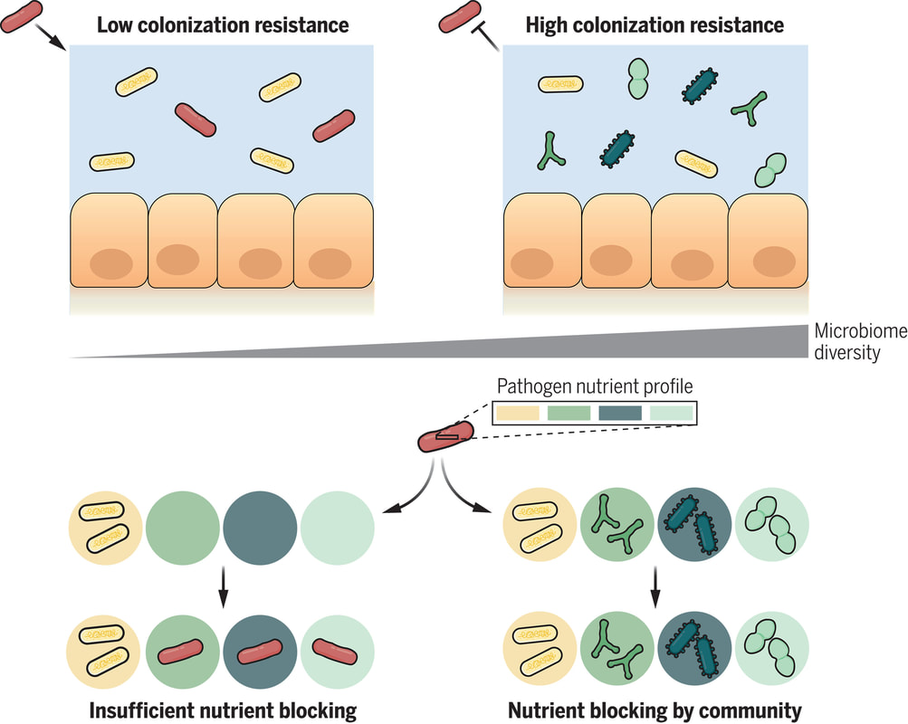
While certain plastics may be deemed safer, it's still advisable to minimize plastic use whenever possible. Consider alternatives like glass, metal, or bamboo, which are safer for both your health and the environment. By reducing your reliance on single-use plastics and opting for reusable, durable items, you can play a significant role in decreasing plastic pollution and its impact on the planet. SolutionsA 2020 review in Earth-Science Reviews identified microplastics in air pollution as potentially the largest contributor to microplastic contamination worldwide, affecting even remote regions like the Arctic and the vast expanses of our oceans. The pervasive nature of microplastics in the atmosphere is alarming, as these particles are not only inhaled but also deposited on land and water surfaces through precipitation, leading to widespread environmental and health impacts. However, there are steps individuals can take to mitigate their exposure to microplastics and reduce their environmental footprint:
Reducing plastic consumption and waste generation is an effective strategy. Simple steps like using reusable shopping bags, using your own coffee mug when getting coffee to go, avoiding plastic-wrapped dry cleaning, bringing drinking water from home in glass water bottles instead of buying bottled water, and store foods in glassware or mason jars instead of plastic bags. You can also take your own leftover container to restaurants, which can significantly cut down the amount of plastic that ends up in landfills and oceans, thereby decreasing the microplastic contamination in our food and water. Strategies such as these will help to reduce the amount of plastic that can migrate into your food. Plastic is all around us and can be extremely difficult to avoid. But if you start looking around, you may find many areas of your life where you can eliminate the use of plastic and replace the it with something inert that won’t harm the environment and your health. Given that adults may ingest thousands of microplastics annually through water consumption alone, it is advisable to minimize the use of plastic water bottles. Opting for a non-plastic water container, like one made from stainless steel or copper, can significantly reduce this exposure. Additionally, experts recommend avoiding microwaving food in plastic containers or placing them in the dishwasher, as heat can cause more plastic to leach into food, and release into the environment. These changes, while seemingly small, can collectively make a significant difference in reducing microplastic pollution and protecting both human health and the environment. In the battle against plastic pollution, both businesses and individuals play crucial roles. One initiative that stands out is the B Corporation movement. B Corporations are businesses committed to reducing global waste and promoting fair hiring and manufacturing practices across their supply chains. These companies actively work to minimize the use of materials that generate microplastics, making them leaders in sustainability. When shopping, look for the B Corporation logo—a "B" encircled—to support companies that adhere to these eco-friendly standards. On an individual level, protecting yourself from airborne microplastics is becoming increasingly important. Microplastic particles in the air, though often larger than typical pollutants like PM10 and PM2.5, still pose significant health risks. Thankfully, these larger particles are easier to capture with a high-performance air purifier. While many air purifiers can only trap smaller pollutants, high-performance models with centrifugal fans are specifically designed to capture even large and heavy microplastics. These purifiers filter out particles as small as 0.003 microns, which is far smaller than the tiniest microplastics. Consider using a personal air purifier in spaces where microplastics are likely to accumulate, such as bedrooms or workspaces, where they can be emitted from clothing, appliances, and containers. Additionally, a car air purifier can help filter out microplastics from tire and brake wear, which can infiltrate your vehicle's interior, especially in high-traffic areas. By choosing B Corporation products and investing in effective air purification, you can significantly reduce your environmental impact and protect your health from the dangers of microplastic pollution. DetoxificationEmerging research suggests that sweating, whether through exercise or sauna use, may play a role in detoxifying the body from accrued microplastics. A 2022 study detected microplastic particles such as polyethylene, PET, and polymers from sportswear in sweat collected after exercise, indicating that perspiration could aid in the elimination of these particles alongside other toxins like pesticides, flame retardants, and bisphenol-A. This adds to a growing body of evidence showing that sweating can facilitate the excretion of heavy metals, petrochemicals, and other pollutants. As with other toxins, microparticle content in sweat could indicate efficacy of interventions promoting clearance. Given the increasing prevalence of microplastics in our environment, inducing sweat through regular sauna use or exercise could offer a simple and accessible detoxification method to help reduce the body's burden of microplastics. However, more research is needed to understand the full impact of repeated sweating on microplastic levels in the body. Additionally, regulatory limits specific to nanoplastics in food and drinks could help safeguard public health given the unprecedented exposure uncovered by advanced microscopy techniques. After all, "seeing" the risk is the first step toward safety. referencesThe human gut is teeming with a diverse array of bacteria collectively known as the gut microbiota. Among its many functions, one of the most vital is colonization resistance—the ability to prevent harmful pathogens from taking up residence in the gut and causing disease. However, understanding which microbiota communities are protective and which allow pathogens to thrive has long been a challenge. In a groundbreaking study led by Spragge et al., researchers shed light on the complex dynamics of gut microbiota and their role in colonization resistance against two significant bacterial pathogens: Klebsiella pneumoniae and Salmonella enterica serovar Typhimurium. Their findings, published in Science, unveil the critical importance of microbiome diversity in safeguarding against pathogenic invasion. Traditionally, it was believed that certain individual bacterial species might confer colonization resistance. However, Spragge et al. discovered that the true protective power lies in the collective diversity of the microbiota. They conducted meticulous experiments both in vitro and in gnotobiotic mice (mice that have been raised in a controlled environment where the microbial composition of their gut is precisely known and controlled), evaluating the ability of single bacterial species and increasingly diverse microbiota communities to resist pathogen colonization. Surprisingly, the researchers found that single species alone provided limited protection against the pathogens. It was only when these species were combined into diverse communities consisting of up to 50 different species that colonization resistance was significantly enhanced. This underscores the importance of ecological diversity in promoting gut health. Moreover, the study identified certain key species within these diverse communities that played a pivotal role in bolstering colonization resistance, even though they offered little protection on their own. These key species acted by consuming nutrients required by the pathogens, thereby depriving them of essential resources for growth and establishment in the host. Importantly, Spragge et al. demonstrated that microbiome diversity not only increases the probability of protection against pathogens but also enhances the overlap in nutrient utilization profiles between the microbiota community and the pathogen. This nutrient blocking mechanism serves as a potent defense strategy against pathogenic invasion. The implications of these findings are profound. They provide compelling evidence for the health benefits of a diverse gut microbiome and offer insights into the rational design of pathogen-resistant microbiota communities. By harnessing the protective power of microbiome diversity, we may pave the way for innovative strategies to combat infectious diseases and promote overall gut health. In conclusion, Spragge et al.'s study unveils the intricate interplay between microbiome diversity and colonization resistance, highlighting the collective strength of diverse bacterial communities in defending against pathogenic threats. This research not only expands our understanding of gut microbiota dynamics but also holds promise for the development of novel therapeutics aimed at fortifying the body's natural defenses against infections. referencesSpragge, Frances, et al. “Microbiome Diversity Protects against Pathogens by Nutrient Blocking.” Science, vol. 382, no. 6676, 15 Dec. 2023, https://doi.org/10.1126/science.adj3502.
This article challenges the conventional understanding of heart disease, particularly the widely accepted theory that attributes its cause primarily to events occurring in the coronary arteries. Instead, a paradigm shift is proposed, contending that a deeper understanding of heart disease, encompassing angina, unstable angina, and myocardial infarction (heart attack), necessitates a focus on events within the myocardium, the muscular tissue of the heart. Over the past decades, the prevailing belief in the coronary artery theory has led to costly surgical interventions, widespread medication use with questionable benefits, and dietary recommendations that may exacerbate rather than alleviate the problem. By delving into the precise pathophysiological events that underlie heart attacks, we can uncover alternative approaches to prevention and treatment, such as adopting a "Nourishing Traditions"-style diet and utilizing safe and affordable medicines like g-strophanthin. Furthermore, this shift in perspective prompts us to confront broader issues, including the impact of modern lifestyles on human health, the need for a new medical paradigm, and the importance of ecological consciousness. Ultimately, reexamining the root causes of heart disease offers a pathway to addressing this pervasive health challenge and forging a healthier future for all. The information is summarized based on the work of Dr. Thomas Cowan, vice president of the Physicians Association for Anthroposophical Medicine and is a founding board member of the Weston A. Price Foundation. During his career he has studied and written about many subjects in medicine. These include nutrition, homeopathy, anthroposophical medicine, and herbal medicine. Challenging the Conventional model: Revisiting the Causes of Heart AttacksThe traditional understanding of heart attacks, largely centered on arterial blockage due to plaque buildup, has faced challenges in recent years. Initially, it was believed that blockages in the major coronary arteries led to oxygen deficiency in the heart, causing chest pain (angina) and eventually progressing to a heart attack. This simplistic view prompted invasive procedures like angioplasty, stents, and coronary bypass surgery as standard treatments. However, clinical observations and research findings have cast doubts on this approach. Anecdotal evidence (admittedly low quality evidence) from a trial in rural Alabama revealed surprising outcomes among individuals with single artery blockages. Contrary to expectations, less than 10% of those who experienced heart attacks did so in the region of the heart supplied by the blocked artery. Similarly, a comprehensive study conducted by the Mayo Clinic highlighted the limited efficacy of bypass surgery in preventing future heart attacks. While the procedure offered relief from chest pain, it did not significantly reduce the risk of subsequent heart events, except in high-risk patients. Contrary to popular belief, blockages exceeding 90% are often compensated for by collateral blood vessels, which develop over time to ensure uninterrupted blood flow to the heart. This extensive network of collateral vessels serves as a natural bypass system, mitigating the impact of arterial blockages on blood circulation. However, diagnostic procedures like coronary angiograms, which rely on injecting heavy dye into the arteries, often fail to accurately assess the extent of blockages and the true blood flow in the heart. As a result, many patients undergo invasive treatments such as bypass surgery, stents, or angioplasty based on misleading information about the severity of their arterial blockages. Moreover, studies have shown that these procedures provide minimal benefit, if any, to patients, particularly those with minimally symptomatic blockages exceeding 90%. Despite the widespread use of these interventions, their efficacy in restoring blood flow and preventing heart attacks remains questionable. These revelations underscore the need for a reevaluation of conventional treatment strategies and a deeper exploration of the underlying mechanisms behind heart attacks. Rather than focusing solely on arterial blockages, a more holistic approach that considers factors beyond plaque buildup may offer greater insights into the prevention and management of heart disease. Beyond the Coronary Artery TheoryThe prevailing focus in cardiology has long been on the stable, progressing plaque within the coronary arteries, deemed responsible for heart attacks. However, recent insights challenge this notion, redirecting attention to the unpredictable nature of unstable plaques. Unlike their calcified counterparts, unstable plaques are soft and prone to rapid evolution, abruptly occluding arteries and triggering downstream oxygen deficits, angina, and ischemia. These vulnerable plaques are believed to be a blend of inflammatory buildup and low-density lipoprotein (LDL), the primary targets of statin drugs. Consequently, the widespread adoption of statin therapy is advocated as a preventive measure against heart attacks, fueled by angiogram studies purportedly showcasing the prevalence of unstable plaques as the leading cause of myocardial infarctions (MIs). Yet, autopsies and pathology studies present a different narrative. Thrombosis, deemed crucial in precipitating MIs, is found in only a fraction of cases upon meticulous examination. Furthermore, measurements of myocardial oxygen levels during MIs reveal no discernible deficit, challenging the conventional understanding of ischemia as the primary mechanism. While thrombosis does occur in conjunction with MIs, its occurrence in less than half of cases underscores the inadequacy of attributing MIs solely to arterial blockages. The timing of thrombosis, often post-MI, begs the question: what precipitated the event in the first place? These inconsistencies underscore the limitations of existing theories surrounding coronary artery involvement in MIs. As the spotlight shifts away from stable plaques, a pressing question emerges: What truly underlies the genesis of heart attacks? Unveiling the Autonomic Symphony: The Heart's Harmonious BalanceAn accurate understanding of myocardial ischemia necessitates consideration of the primary risk factors associated with heart disease, including gender, diabetes, smoking, and chronic psychological stress. Curiously, none of these risk factors directly implicate coronary artery pathology; instead, they impact capillary health or exert indirect effects. Over the past five decades, key medications in cardiology, such as beta-blockers, nitrates, aspirin, and statins, have demonstrated some benefits for heart patients. However, their mechanisms of action must be scrutinized within a comprehensive theory of myocardial ischemia. A groundbreaking revelation in heart disease prevention and treatment stems from the autonomic nervous system's role in ischemia genesis, as illuminated by heart-rate variability monitoring. The autonomic nervous system comprises two branches—the sympathetic and parasympathetic—responsible for regulating physiological responses. Imbalance between these branches emerges as a significant contributor to heart disease. Studies reveal a notable reduction in parasympathetic activity among patients with ischemic heart disease, particularly preceding ischemic events triggered by physical or emotional stressors. Conversely, abrupt increases in sympathetic activity rarely culminate in ischemia without antecedent parasympathetic decline. Notably, women exhibit stronger vagal activity than men, potentially influencing sex-based disparities in MI incidence. Multiple risk factors, including hypertension, smoking, diabetes, and stress, diminish parasympathetic activity, underscoring the pivotal role of the regenerative nervous system in heart health. Conversely, pharmacological interventions like nitrates, aspirin, and statins stimulate parasympathetic mediators, promoting ANS balance. In essence, while traditional risk factors and interventions influence plaque and stenosis development, their paramount impact lies in restoring ANS equilibrium. Thus, understanding the sequence of events leading to myocardial infarction demands a deeper exploration of autonomic nervous system dynamics. The Underlying pathophysiology of Myocardial IschemiaIn the vast majority of cases, the pathology leading to myocardial infarction (MI) begins with a decreased tonic activity of the parasympathetic nervous system (rest and digest), often exacerbated by physical or emotional stressors. This reduction prompts an increase in sympathetic nervous system activity, triggering heightened adrenaline production and directing myocardial cells to break down glucose via aerobic glycolysis, rather than their preferred fuel source of ketones and fatty acids (often explaining why patients report feeling tired before a MI). Remarkably, despite these metabolic shifts, no change in blood flow, as measured by the myocardial cell oxygen level (pO2), occurs. The shift towards glycolysis results in a surge of lactic acid production within myocardial cells, a phenomenon observed in nearly all MIs. This surge, coupled with localized tissue acidosis, impedes calcium entry into cells, compromising their contractility. Consequently, localized edema ensues, leading to hypokinesis—the hallmark of ischemic disease—and eventual tissue necrosis characteristic of an MI. Moreover, the ensuing tissue edema alters arterial hemodynamics, escalating sheer pressure and exacerbating plaque instability. This process elucidates the rupture of unstable plaques and their role in exacerbating arterial blockage during critical, acute scenarios. This explanation accounts for all the observable phenomena associated with heart disease. Understanding the etiology of heart disease holds profound implications beyond academic curiosity. It informs therapeutic strategies aimed at preserving parasympathetic activity, fostering holistic approaches to heart health, and challenging prevailing "civilized" industrial lifestyles. Central to this paradigm shift is the recognition of the vital role played by g-strophanthin—a hormone derived from the strophanthus plant. G-strophanthin is an endogenous hormone made in the adrenal cortex from cholesterol, whose production is inhibited by statin drugs, that does two things that are crucial for heart health and are done by no other medicine. G-strophanthin uniquely stimulates the production of acetylcholine, the primary neurotransmitter of the parasympathetic nervous system, while also converting lactic acid—the metabolic poison implicated in ischemic processes—into pyruvate, a preferred myocardial cell fuel. Perhaps this “magic” is why Chinese medicine practitioners say that the kidneys (i.e., adrenals, where ouabain is made) nourish the heart. Embracing this understanding not only guides therapeutic interventions but also underscores the imperative of dietary modifications. A diet abundant in healthful fats and fat-soluble nutrients, while low in processed carbohydrates and sugars, emerges as a cornerstone of heart health—a departure from the industrialized diets synonymous with modern civilization. In essence, unraveling the metabolic symphony orchestrating myocardial ischemia offers a transformative lens through which to perceive heart disease, fostering a holistic approach that transcends conventional paradigms and embraces the profound interconnectedness of mind, body, and environment. referencesGiorgio Baroldi. The Etiopathogenesis of Coronary Heart Disease. CRC Press EBooks, Informa, 20 Jan. 2004. Accessed 29 Mar. 2024.
Sroka K. On the genesis of myocardial ischemia. Z Kardiol. 2004 Oct;93(10):768-83. doi: 10.1007/s00392-004-0137-6. PMID: 15492892. Helfant, R. H., et al. “Coronary Heart Disease. Differential Hemodynamic, Metabolic, and Electrocardiographic Effects in Subjects with and without Angina Pectoris during Atrial Pacing.” Circulation, vol. 42, no. 4, 1 Oct. 1970, pp. 601–610, www.ncbi.nlm.nih.gov/pubmed/11993303., https://doi.org/10.1161/01.cir.42.4.601. Takase, B., Kurita, A., Noritake, M., Uehata, A., Maruyama, T., Nagayoshi, H., ... & Nakamura, H. (1992). Heart rate variability in patients with diabetes mellitus, ischemic heart disease, and congestive heart failure. Journal of electrocardiology, 25(2), 79-88. Sroka, K., Peimann, C. J., & Seevers, H. (1997). Heart rate variability in myocardial ischemia during daily life. Journal of electrocardiology, 30(1), 45-56. Scheuer, J., & Brachfeld, N. (1966). Coronary insufficiency: relations between hemodynamic, electrical, and biochemical parameters. Circulation Research, 18(2), 178-189. Schmid, P. G., Greif, B. J., Lund, D. D., & Roskoski Jr, R. O. B. E. R. T. (1978). Regional choline acetyltransferase activity in the guinea pig heart. Circulation Research, 42(5), 657-660. Katz, A. M. (1971). Effects of ischemia on the cardiac contractile proteins. Cardiology, 56(1-6), 276-283. Manunta, Paolo, et al. “Endogenous Ouabain in Cardiovascular Function and Disease.” Journal of Hypertension, vol. 27, no. 1, 1 Jan. 2009, pp. 9–18, journals.lww.com/jhypertension/Abstract/2009/01000/Endogenous_ouabain_in_cardiovascular_function_and.3.aspx, https://doi.org/10.1097/HJH.0b013e32831cf2c6. Doepp, Manfred. “May Strophanthin Be a Valuable Cardiac Drug ? .” American Journal of Medical and Clinical Research & Reviews, vol. 2, no. 9, 15 Sept. 2023, pp. 1–6, ajmcrr.com/index.php/pub/article/view/75/74, https://doi.org/10.58372/2835-6276.1069. Accessed 29 Mar. 2024. Thayer, J. F., Yamamoto, S. S., & Brosschot, J. F. (2010). The relationship of autonomic imbalance, heart rate variability and cardiovascular disease risk factors. International journal of cardiology, 141(2), 122-131. Recent breakthrough studies have shone a light on the intriguing link between our microbiome – the diverse community of microorganisms residing in our gut and mouth – and the secret to a longer, healthier life. Scientists have long suspected that our genes, environment, and internal factors like the microbiome play a role in determining how long we live, but the specifics remained elusive. Now, thanks to cutting-edge research, we're getting closer to unraveling the mysteries of longevity. In this groundbreaking exploration, scientists employed a sophisticated approach called Mendelian randomization (MR) to delve into the intricate relationships between the human microbiome and longevity. By analyzing genetic data from large cohorts, they uncovered some compelling associations that shed light on the microbial players in the quest for a longer life. The Gut Chronicles: Microbial Superstars and CulpritsThe gut microbiome, a bustling metropolis of bacteria, has been a focal point in the quest for longevity. The study identified certain gut microbes as potential champions in the battle against aging. Microbial heroes like Coriobacteriaceae, Oxalobacter, and the probiotic Lactobacillus amylovorus were found to be positively linked to increased odds of longevity. On the flip side, a few gut microbes emerged as potential antagonists, with names like Fusobacterium nucleatum, Coprococcus, Streptococcus, Lactobacillus, and Neisseria negatively associated with longevity. These microbial foes might have a role in determining how gracefully we age. Oral Health: More Than Just a Pretty SmileThe study didn't stop at the gut; it extended its gaze to the oral microbiome, a less-explored but equally important realm. The findings suggested a fascinating connection between the oral microbiome and longevity. Specific oral bacteria were identified as potential influencers in the longevity game. Interestingly, the research hinted at a lower gut microbial diversity among centenarians (diversity appears to lower with age), but no significant difference in their oral microbiota. This finding underscores the importance of tracking the movements of these beneficial microbes across different parts of the body for a longer and healthier life. Decoding the Genetic Blueprint for LongevityThe study leveraged Mendelian randomization to unravel the causality between the microbiome and longevity. This approach, using genetic variants as tools, allowed scientists to explore the potential causal links between specific microbial features and the length of our lives. The bidirectional analyses provided a wealth of information, not only pinpointing specific microbes associated with longevity but also revealing the microbial preferences of genetically longevous individuals. For instance, genetic predisposition to longevity correlated with a higher abundance of Prevotella and a lower abundance of Bacteroides, suggesting a potential link between dietary choices and a longer life. Microbes and Diseases: Unraveling the We The study didn't just stop at longevity; it ventured into the realm of diseases. Certain microbes associated with longevity were found to have correlations with specific diseases. For example, Coriobacteriaceae, linked to longevity, was significantly reduced in patients with heart failure, suggesting a potential protective role against cardiovascular diseases. This "microbiota—disease—longevity" axis provides a nuanced understanding of how our microbial companions might influence not only our lifespan but also our susceptibility to various health conditions. What's Next in the Quest for a Longer LifeWhile the study opens exciting new avenues, there are some limitations to consider. The identified causalities didn't all reach statistical significance due to the vast number of microbial features tested. However, the robustness of the findings was supported by the replication of several identified causal links in independent datasets. Moving forward, researchers aim to collect more comprehensive individual-level data, including microbiome profiles, genetics, socio-economic factors, behaviors, and environmental influences. This holistic approach will help tease apart the individual contributions of these factors to longevity. In conclusion, this pioneering study, using Mendelian randomization, has provided us with a roadmap to explore the intricate connections between our microbiome and the quest for a longer, healthier life. As we unlock the secrets hidden in our genes and microbes, we inch closer to personalized approaches for healthy aging and interventions that could extend our time on this planet. referencesLiu, Xiaomin, et al. “Mendelian Randomization Analyses Reveal Causal Relationships between the Human Microbiome and Longevity.” Scientific Reports, vol. 13, no. 1, 29 Mar. 2023, p. 5127, www.nature.com/articles/s41598-023-31115-8, https://doi.org/10.1038/s41598-023-31115-8.
In an update to its 2007 scientific statement, the American Heart Association (AHA) emphasizes the significant and multifaceted benefits of resistance training (RT) on cardiovascular health. Contrary to the misconception that RT solely enhances muscle mass and strength, the statement highlights the favorable physiological and clinical effects of this form of exercise on cardiovascular disease (CVD) and associated risk factors. The scientific statement aims to provide comprehensive insights into the impact of RT, either alone or in combination with aerobic training, on traditional and nontraditional CVD risk factors. More is not always betterEpidemiological evidence suggests that RT is associated with a lower risk of all-cause mortality and CVD morbidity and mortality. Adults who participate in RT have ≈15% lower risk of all-cause mortality and 17% lower risk of CVD, compared with adults who report no RT. Approximately 30 to 60 minutes per week of RT is associated with the maximum risk reduction for all-cause mortality and incident CVD. Notice this "U" shape in the curve when examining the relationship between RT and morbidity and mortality. This curve suggests that some RT is clearly beneficial, but has the volume of RT increases past a certain point the benefits drop and it becomes harmful. The concept of a "biphasic response" is fundamental to understanding hormesis. It describes the characteristic dose-response relationship observed in hormetic processes, where a substance or stressor elicits opposite effects at low and high doses. The response can be visualized as a U-shaped or J-shaped curve, illustrating the beneficial effects at low doses and potential harm at higher doses. Benefits of RT on Traditional CVD Risk FactorsThe AHA's scientific statement underscores the positive influence of RT on traditional CVD risk factors, including blood pressure (BP), glycemia, lipid profiles, and body composition. Numerous studies indicate that engaging in RT is associated with reduced resting BP, improved glycemic control, and favorable alterations in lipid profiles, contributing to a lower risk of all-cause mortality and CVD morbidity. Despite recommendations suggesting 2 days per week of RT, only 28% of U.S. adults adhere to this guideline, highlighting the need for increased awareness and promotion. RT and resting blood pressureRT has demonstrated the ability to reduce resting BP across diverse populations, with notable benefits observed in individuals with prehypertension and hypertension. The mechanisms behind these benefits include enhancements in endothelial function, vasodilatory capacity, and vascular conductance. The reductions in BP achieved through RT are comparable to those achieved with antihypertensive medications. RT and GlycemiaRT shows promise in improving glycemia and insulin resistance, leading to a lower incidence of diabetes. The evidence suggests a nonlinear dose-response association, with up to 60 minutes per week of RT associated with the maximum risk reduction for diabetes. RT and Lipid ProfilesWhile the effect on lipid profiles is modest, RT results in favorable changes in high-density lipoprotein cholesterol, total cholesterol, and triglycerides. These improvements are more pronounced in older adults and those with elevated cardiometabolic risk. Rt, Body composition, and weightRT positively influences body composition by increasing lean body mass and reducing body fat percentage. It is particularly effective in overweight or obese individuals, contributing to increased metabolic rate and mitigating weight gain over time. Benefits of RT on Nontraditional CVD Risk FactorsIn addition to traditional risk factors, the scientific statement highlights the potential mechanisms by which RT positively affects nontraditional CVD risk factors. These include increased cardiorespiratory fitness, improved endothelial function, and potential benefits for sleep quality, psychological health, and well-being. The AHA's updated scientific statement reinforces the pivotal role of resistance training in cardiovascular health, providing a comprehensive overview of its impact on both traditional and nontraditional risk factors. As the evidence supporting RT's benefits continues to grow, the statement serves as a valuable resource for clinicians and public health professionals, offering practical strategies for promoting and prescribing resistance training to enhance cardiovascular health in diverse populations. ReferencesPaluch, Amanda E, et al. “Resistance Exercise Training in Individuals with and without Cardiovascular Disease: 2023 Update: A Scientific Statement from the American Heart Association.” Circulation, 7 Dec. 2023, https://doi.org/10.1161/cir.0000000000001189. Accessed 11 Dec. 2023.
Momma H, Kawakami R, Honda T, Sawada SS. Muscle-strengthening activities are associated with lower risk and mortality in major non-communicable diseases: a systematic review and meta-analysis of cohort studies. Br J Sports Med. 2022 Jul;56(13):755-763. doi: 10.1136/bjsports-2021-105061. Epub 2022 Feb 28. PMID: 35228201; PMCID: PMC9209691. Nasal Breathing: A Breath of Fresh Air for Cardiovascular Wellness – Insights from New Research2/4/2024 The leading cause of death in the United States is cardiovascular disease, and the risk of cardiovascular issues can be predicted by factors such as blood pressure, heart rate variability, blood pressure variability, and cardiac vagal baroreflex sensitivity. The interplay between the cardiovascular and respiratory systems is highlighted, with a particular emphasis on how respiration affects key prognostic cardiovascular variables. This study explores the impact of nasal breathing compared to oral breathing on cardiovascular health in young adults. Nasal breathing is associated with humidification, warming, and filtration of inhaled air, potentially leading to bronchodilation and improved breathing efficiency. While past research has shown nasal breathing to have positive effects on resting metabolic demands, its influence on cardiovascular markers is not well-understood. The primary hypothesis is that nasal breathing, as opposed to oral breathing, will result in decreased blood pressure, improved heart rate variability, reduced blood pressure variability, and increased cardiac vagal baroreflex sensitivity at rest. The study aims to contribute to the understanding of how breathing patterns influence prognostic cardiovascular variables, aligning with the broader interest in the impact of breathing pace and training on cardiovascular health. The secondary hypothesis focuses on the effects of nasal breathing during submaximal exercise. The expectation is that nasal breathing, by attenuating the ventilatory response and metabolic demands, will lead to reduced blood pressure responses, improved heart rate variability, and decreased blood pressure variability during exercise. This aspect is particularly relevant due to the association between elevated exercise blood pressure and the risk of developing hypertension and cardiovascular disease. FindingsThe study findings are summarized, focusing on the impact of nasal vs. oral breathing on physiological and subjective variables at rest and during exercise. At rest, nasal breathing is associated with lower mean and diastolic blood pressure, improved heart rate variability metrics, reduced LF/HF ratio, and lower ratings of perceived exertion (RPE) and breathlessness (RPB). However, it increased systolic blood pressure average real variability. During submaximal exercise, differences between nasal and oral breathing were observed for RPB, suggesting a modest effect on reducing breathlessness during acute exercise. The discussion delves into the potential clinical significance of these findings, particularly the reduction in diastolic blood pressure during nasal breathing at rest. The study suggests a greater parasympathetic to sympathetic dominance during nasal breathing, indicated by changes in frequency-domain metrics of heart rate variability. Although nasal breathing did not significantly affect beat-to-beat blood pressure variability, there is speculation about potential connections between respiratory variables and blood pressure changes, emphasizing the need for further investigation. The study notes that the impact of nasal breathing on cardiovascular variables may have implications for various populations and suggests avenues for future research, including examining nasal breathing's effects on blood pressure over longer durations, both at rest and during activities like exercise. The discussion also touches on the potential benefits of interventions like mouth-taping overnight, emphasizing the importance of considering nasal breathing in the context of broader health outcomes. In summary, the study highlights the potential benefits of nasal breathing, with improvements in various cardiovascular and subjective measures at rest. While the effects during exercise are more modest, the findings contribute to understanding the nuanced relationship between respiratory patterns and cardiovascular health. referencesWatso, Joseph C., et al. “Acute Nasal Breathing Lowers Diastolic Blood Pressure and Increases Parasympathetic Contributions to Heart Rate Variability in Young Adults.” American Journal of Physiology. Regulatory, Integrative and Comparative Physiology, vol. 325, no. 6, 1 Dec. 2023, pp. R797–R808, pubmed.ncbi.nlm.nih.gov/37867476/, https://doi.org/10.1152/ajpregu.00148.2023.
|
The Awareness domain contains research, news, information, observations, and ideas at the level of self in an effort to intellectualize health concepts.
The Lifestyle domain builds off intellectual concepts and offers practical applications.
Taking care of yourself is at the core of the other domains because the others depend on your health and wellness.
Archives
September 2024
Categories
All
|